Animal Anatomy Diagrams: Name Each Part, Then Print It
Line drawings of animals and their organs, with a numbered ring on every part and two sheets to print.
100 pages
Horses, dogs and other whole animals 28
Skeletons, muscles and organs drawn inside the outline of the standing animal, starting with the 33 bones of the horse. An alligator and an armadillo join them, and a dog skull beside a horse skull.
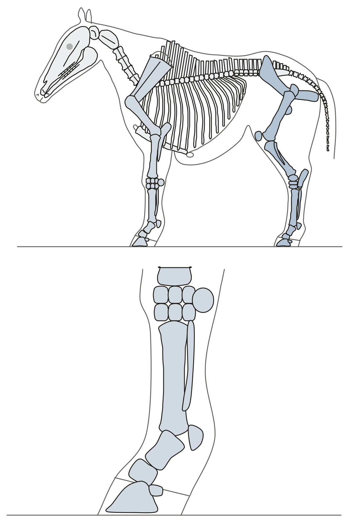 Horse Skeletal Anatomy QuizA horse skeleton from the left side with 33 numbered bones: 24 large ones, and 9 from the knee down on a close view. Textbook and horseman's names both count. Tarsal bones, horse · Skull, horse · Distal sesamoid bone, horse
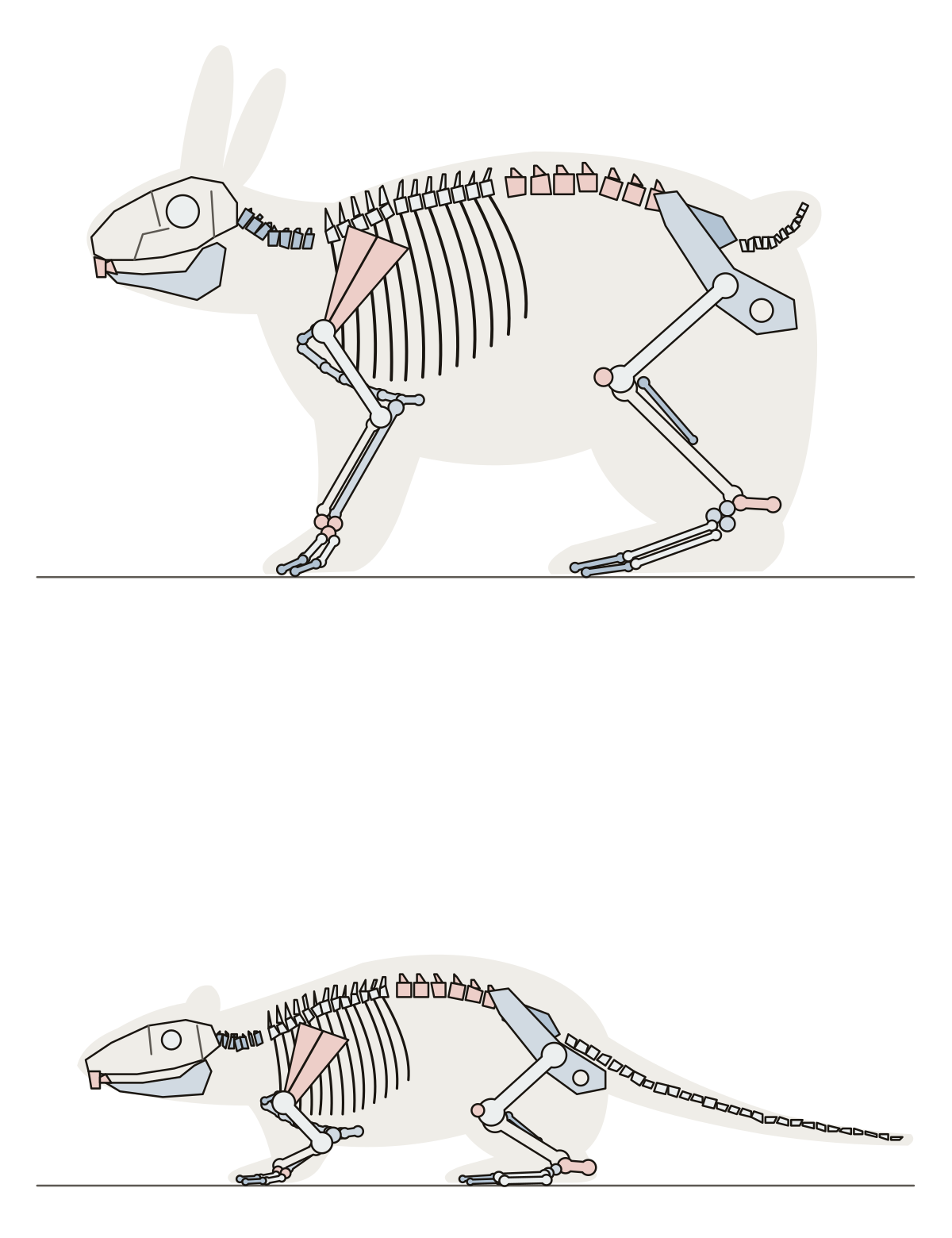 Labeled Rabbit Skeleton and Rat SkeletonA rabbit skeleton and a rat skeleton from the side, each inside the outline of the animal: 26 bones on the rabbit, 12 on the rat.
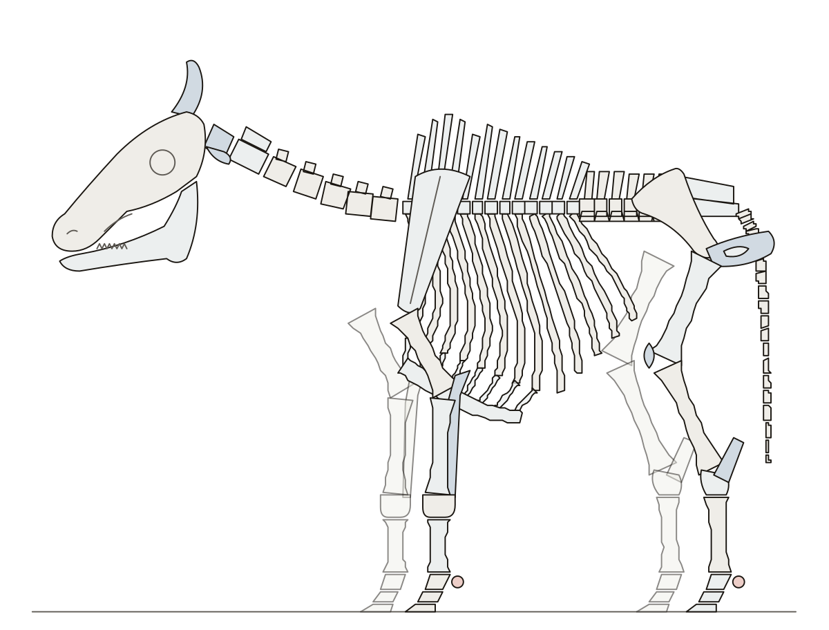 Cow Skeleton LabeledA skeleton of a cow with labeling: skull, horn core, backbone region by region, ribs, both limbs down to the cannon bones, dewclaws and phalanges.
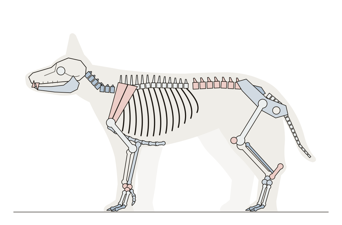 Labeled Dog SkeletonThe skeleton of a standing dog from the side, inside the outline of the animal, with its bones named from the skull to the tail.
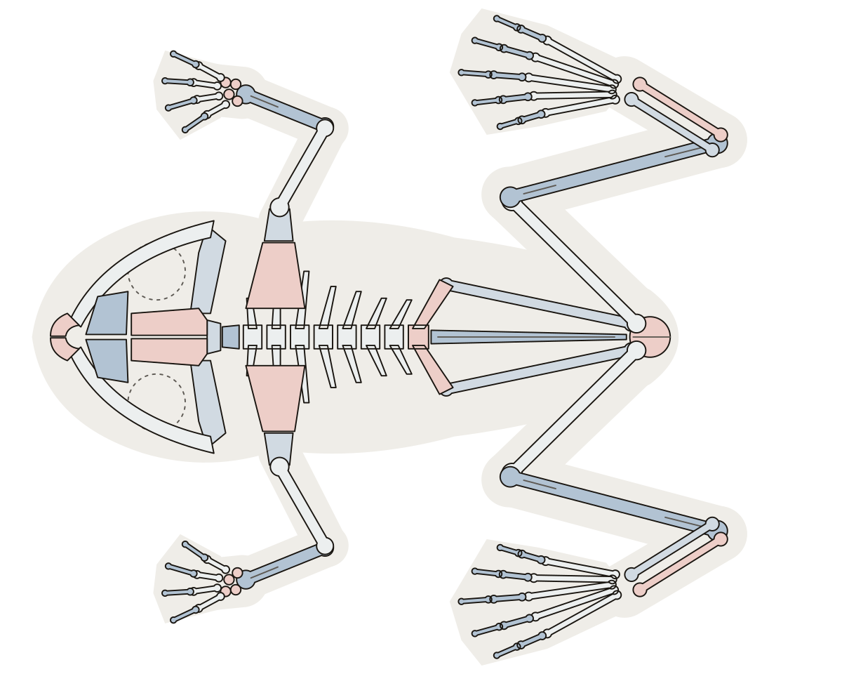 Frog Skeleton LabeledThe skeleton of a frog from above, inside the outline of the sitting animal: skull, short spine, urostyle, hips and folded legs.
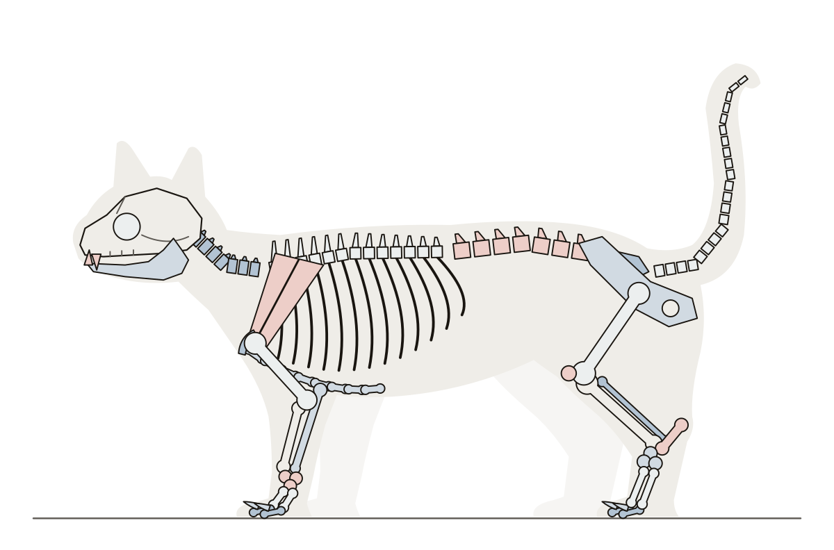 Cat Skeleton LabeledA standing cat from the side with its tail up and its skeleton drawn inside: the bones named from the skull to the tip of the tail.
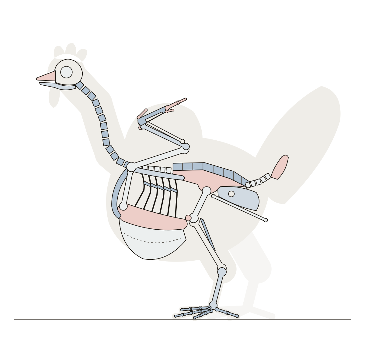 Labeled Bird SkeletonThe skeleton of a standing chicken from the side: skull and beak, long neck, wishbone, keel, wing, fused hips, tail and leg.
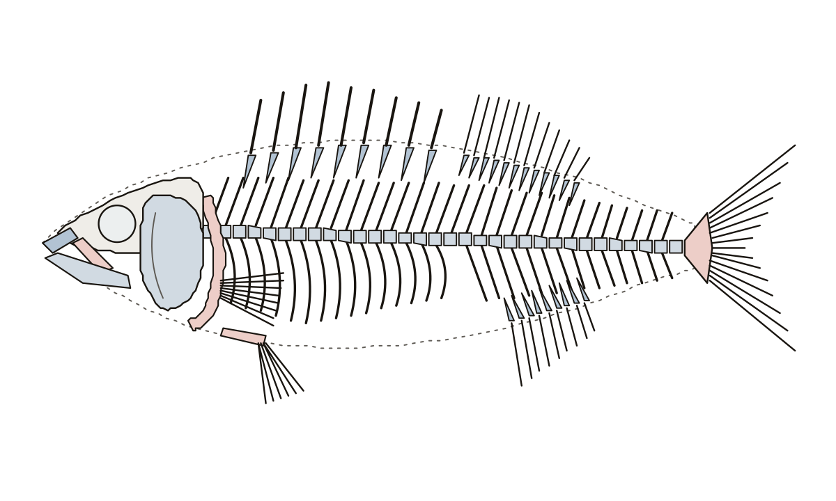 Fish Skeleton LabeledThe skeleton of a perch from the side: skull, jaws, gill cover, backbone, neural and hemal spines, ribs, fin spines, soft rays and the hypural plate.
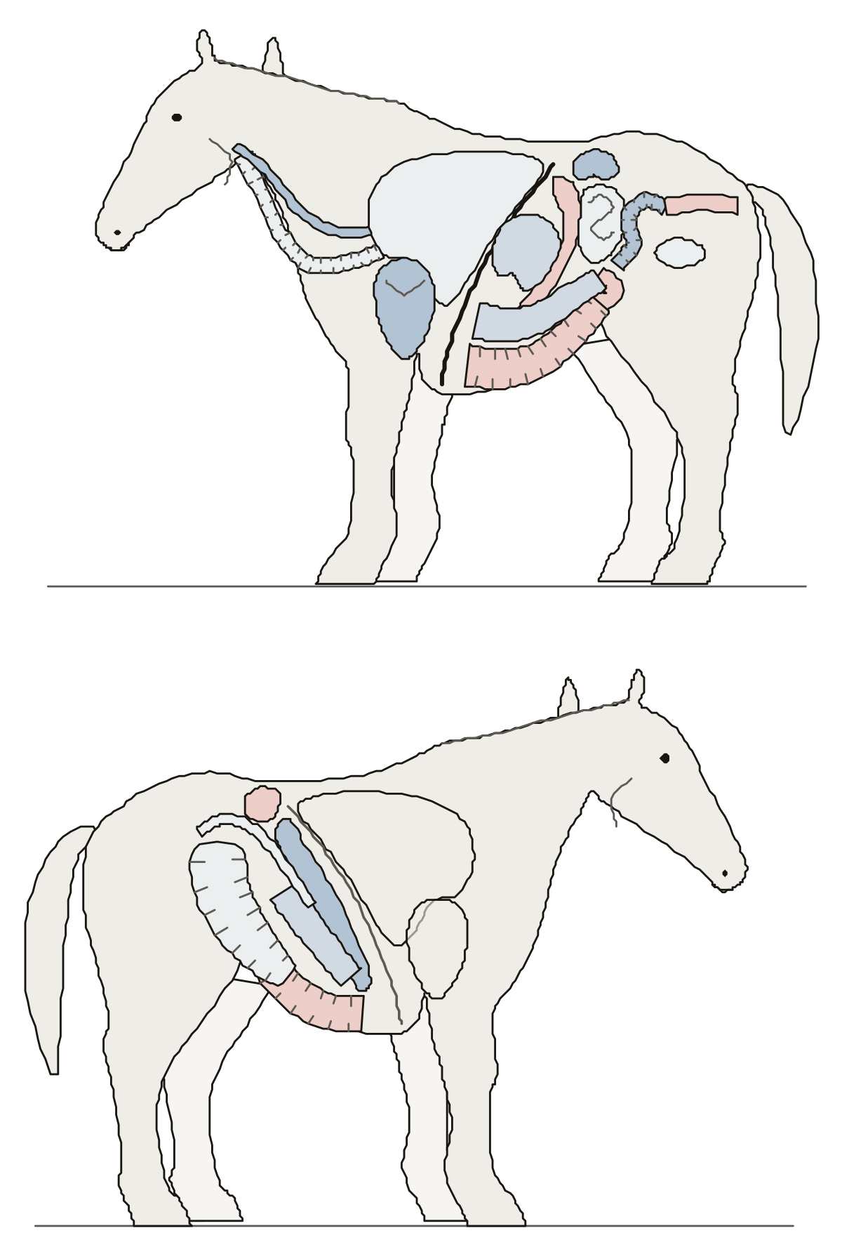 Horse Anatomy QuizA standing horse from each side with its organs inside: heart, lungs, diaphragm, stomach, spleen and kidneys, then the liver, the cecum and the four parts of the large colon.
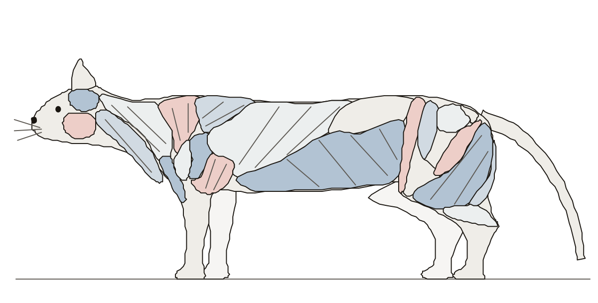 Cat Muscles QuizA standing cat from the side with its surface muscles as patches: masseter, the three trapezius muscles, the deltoids, latissimus dorsi, sartorius, biceps femoris and more.
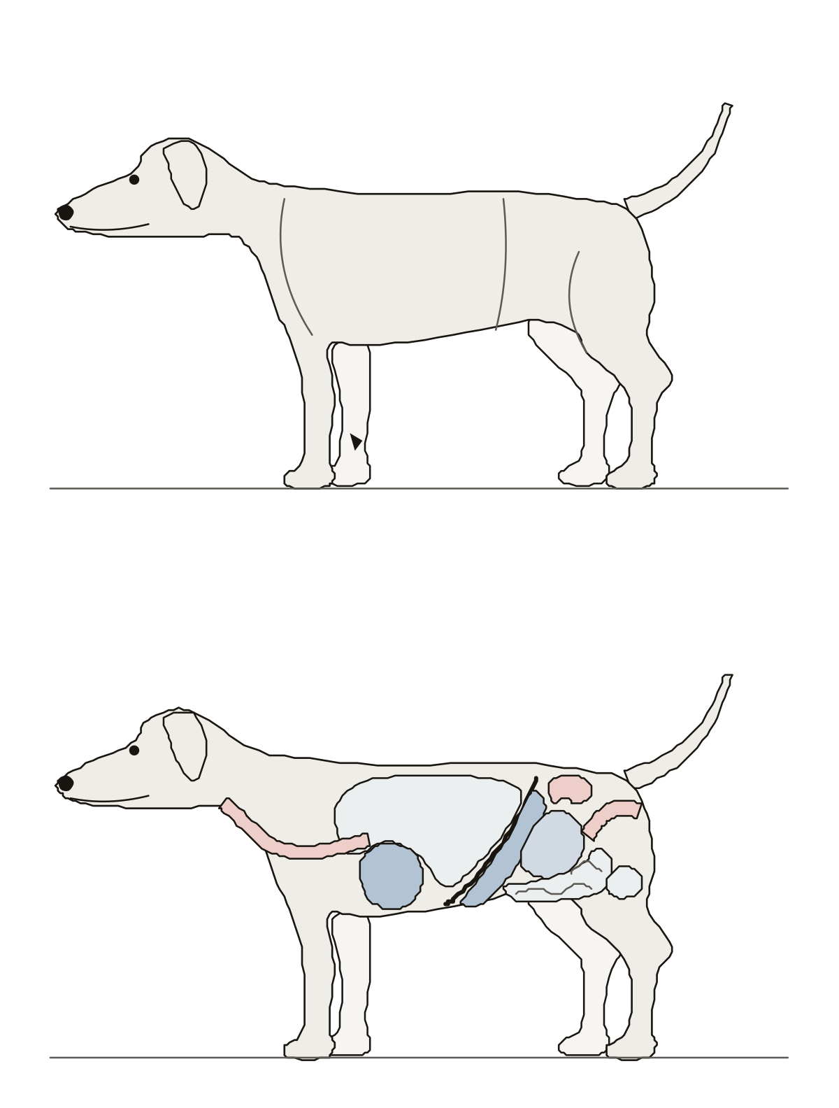 Dog Anatomy QuizA standing dog in two views: muzzle, stop, withers, loin, croup, stifle and hock on the outside, then lungs, heart, liver, stomach, intestines and kidney inside.
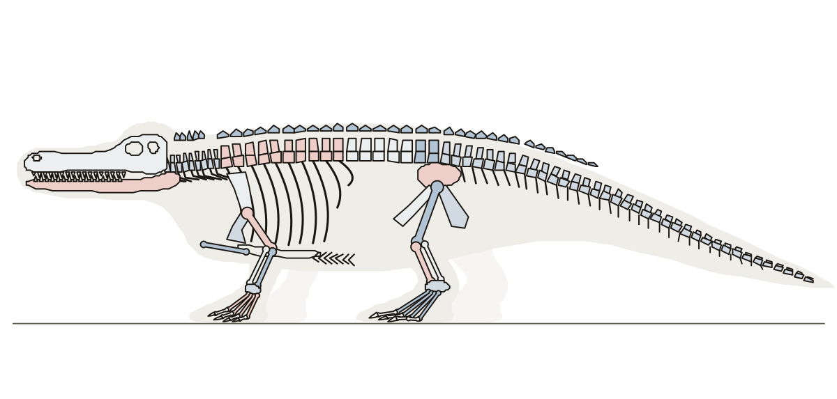 Alligator Skeleton LabeledA walking alligator from the side with its skeleton drawn inside: the bones named from the long skull to the tail, with the belly ribs and the plates of the back.
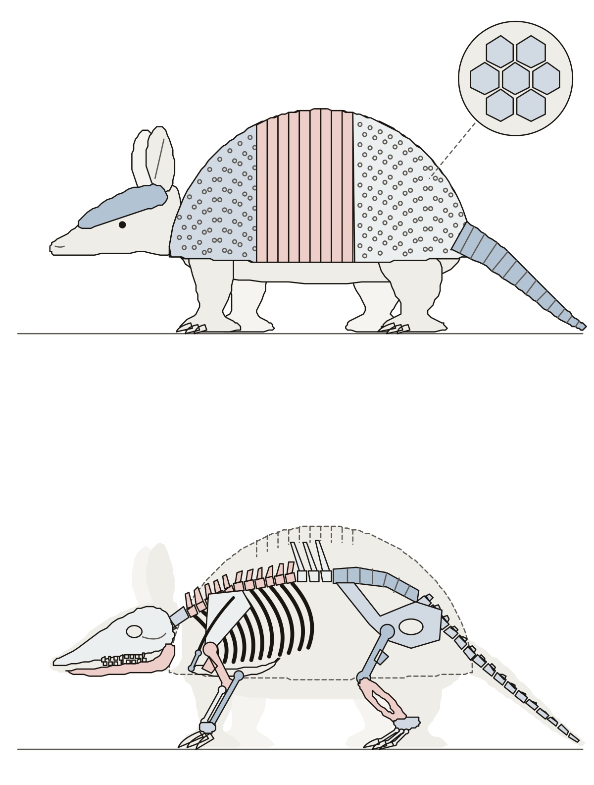 Armadillo Skeleton LabeledA nine-banded armadillo from the side: head shield, shoulder shield, bands, pelvic shield and tail rings, then the skeleton under the shell.
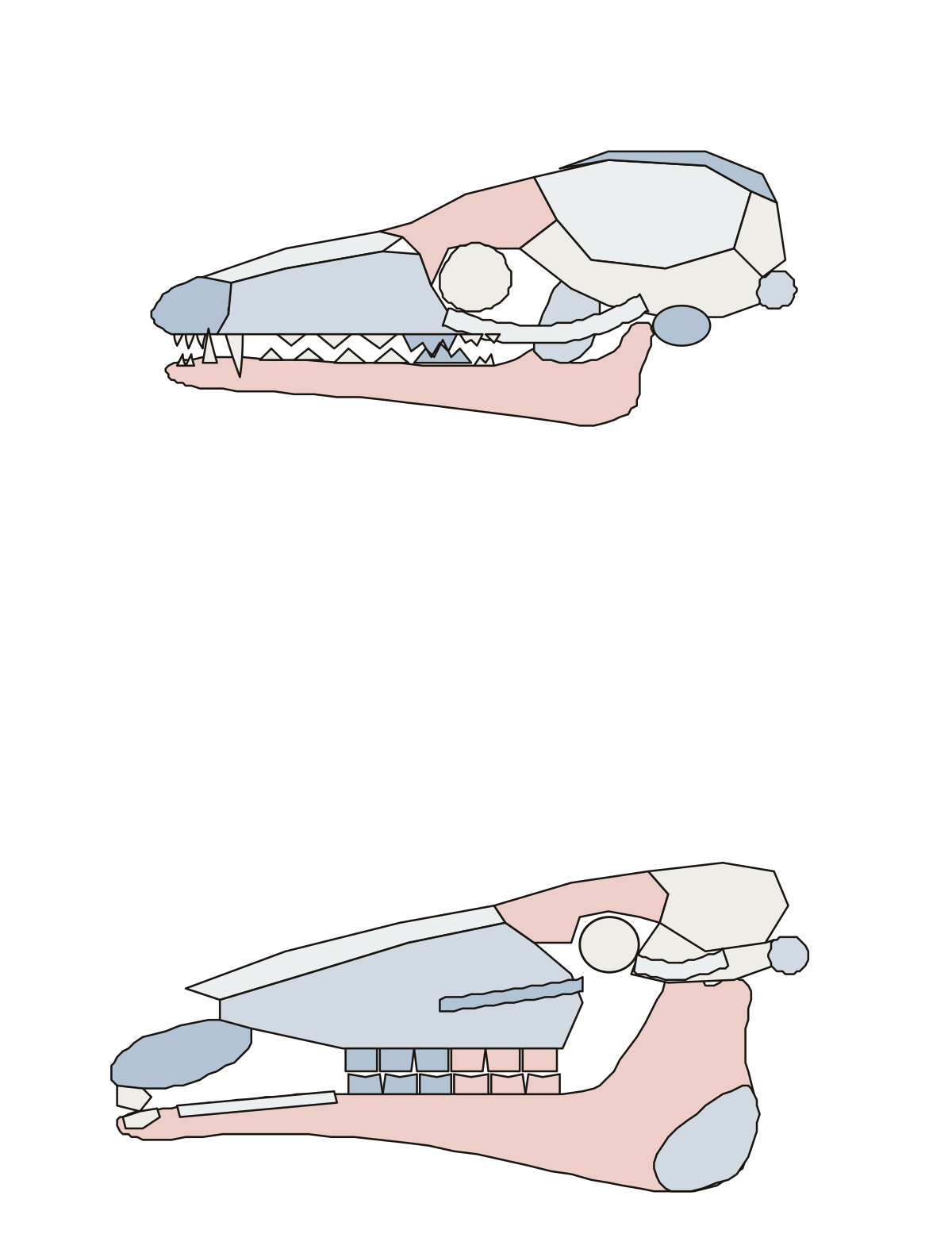 Animal Skull Anatomy QuizA dog skull and a horse skull from the side: jaw bones, eye socket, cheek bone and teeth, asked one part at a time.
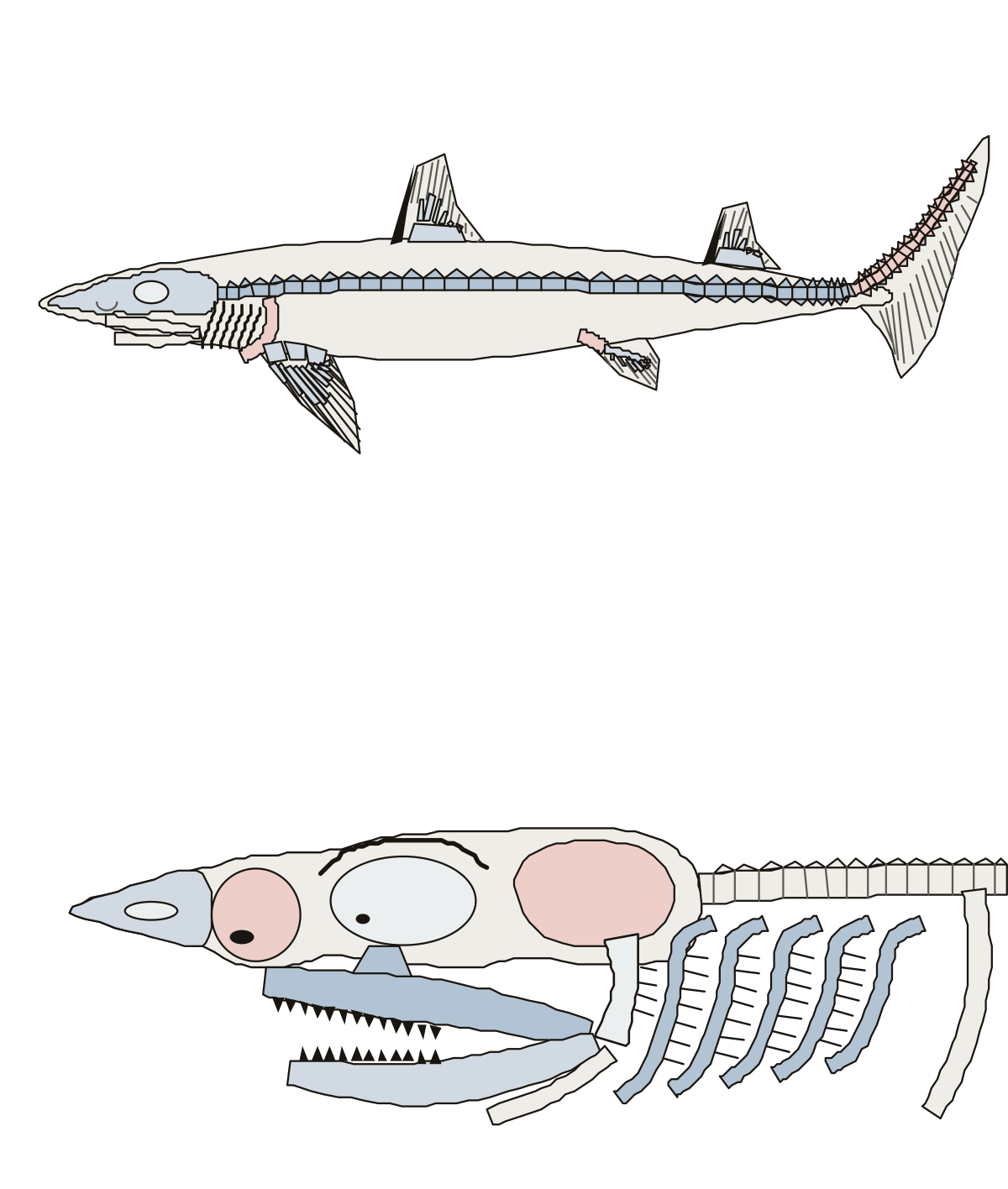 Dogfish Shark Skeleton LabeledA spiny dogfish from the side: chondrocranium, vertebral column, girdles, fin cartilages, fin rays and fin spines; then the head with its jaws, hyomandibular and gill arches.
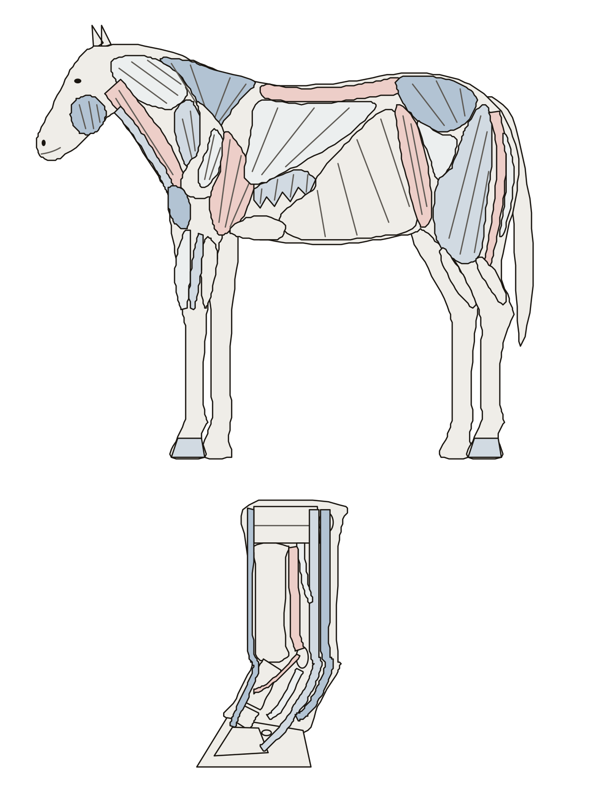 Horse Muscle Anatomy QuizA standing horse from the side with its surface muscles as patches, from the masseter to the semitendinosus, then the lower leg with its flexor tendons, suspensory ligament and check ligament.
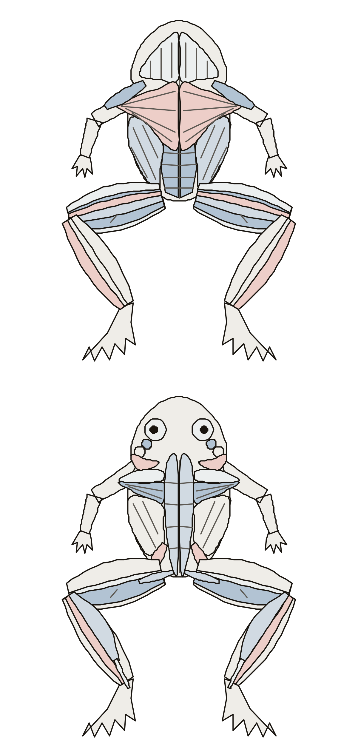 Frog Muscle QuizA frog from below and from above with its muscles as patches: mylohyoid, pectoralis, rectus abdominis, sartorius, the gracilis muscles, triceps femoris, gastrocnemius and more.
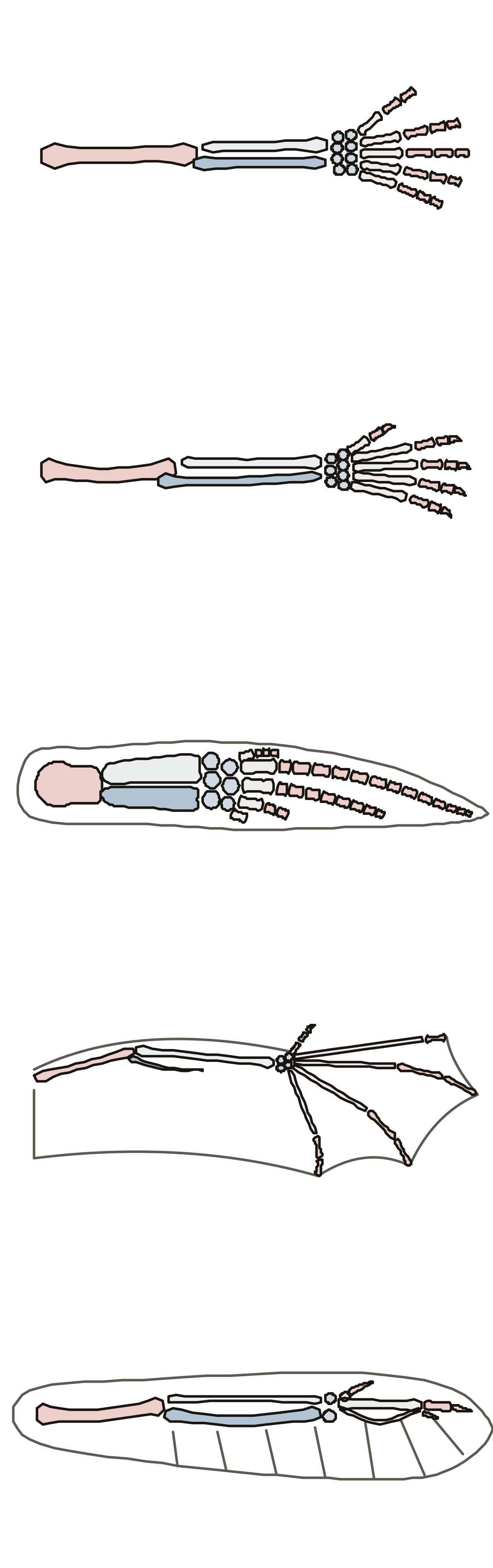 Comparative Anatomy QuizFive forelimbs, one view each: human arm, cat foreleg, whale flipper, bat wing and bird wing, with the humerus, radius, ulna, carpals, metacarpals and phalanges asked on each.
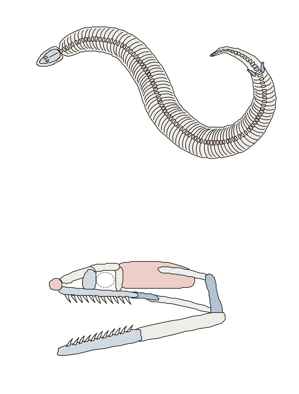 Labeled Snake Skeleton DiagramA python's skeleton in two views: skull, rib-bearing vertebrae, pelvic spurs and tail; then the skull with quadrate, supratemporal and jaw bones.
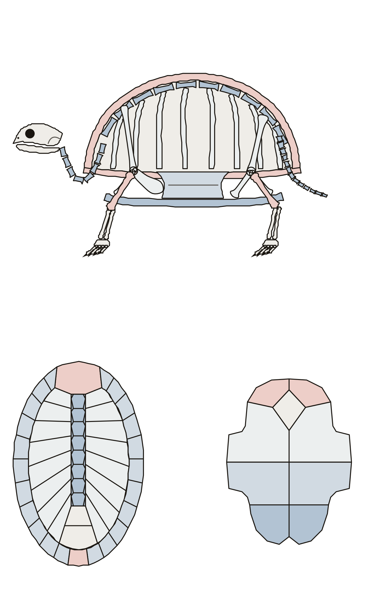 Turtle Skeleton DiagramA turtle skeleton in two views: skull, neck, ribs, shoulder girdle, pelvis and legs inside the shell, then the bones of the carapace and the plastron. Hyoplastron, turtle · Plastron, turtle · Cervical vertebrae, turtle
- Blank Dog Skeleton DiagramThe dog skeleton drawing with nothing written on it, to print and fill in: a standing dog from the left side, with its answer key.
- Unlabelled Frog Muscle DiagramsThe frog muscle drawings with nothing written on them, to print and fill in: the frog from below, the frog from above, and the answer key.
- Skeleton Diagram of a Frog Without LabelsThe frog skeleton drawing with nothing written on it, to print and fill in: the whole skeleton from above, skull to toes, with its answer key.
- Cat Skeleton Diagram Not LabeledThe cat skeleton drawing with no names on it, to print and fill in: 27 bones from the canine teeth to the tail, with its answer key.
- Dogfish Shark Skeleton Diagram UnlabeledThe dogfish skeleton drawing with nothing written on it, to print and fill in: the whole fish above, the head and gill arches below, with its answer key.
- Cat Muscles Diagram UnlabeledThe cat muscles drawing with nothing written on it, to print and fill in: twenty lines from the jaw to the calf, and an answer key.
- Label the Armadillo SkeletonAn armadillo's skeleton as a worksheet: eighteen numbered parts on the bones of a nine-banded armadillo, skull to tail, a line for each name, and a key.
- Label the Snake SkeletonA snake's skeleton as a worksheet: five numbered bones on a python seen from above, eleven on its skull from the side, a line for each name, and a key.
Hearts, eyes and kidneys 21
One organ per page, from animals a school or college lab often uses: sheep, pig, cow, frog, dog, fish, chicken and octopus.
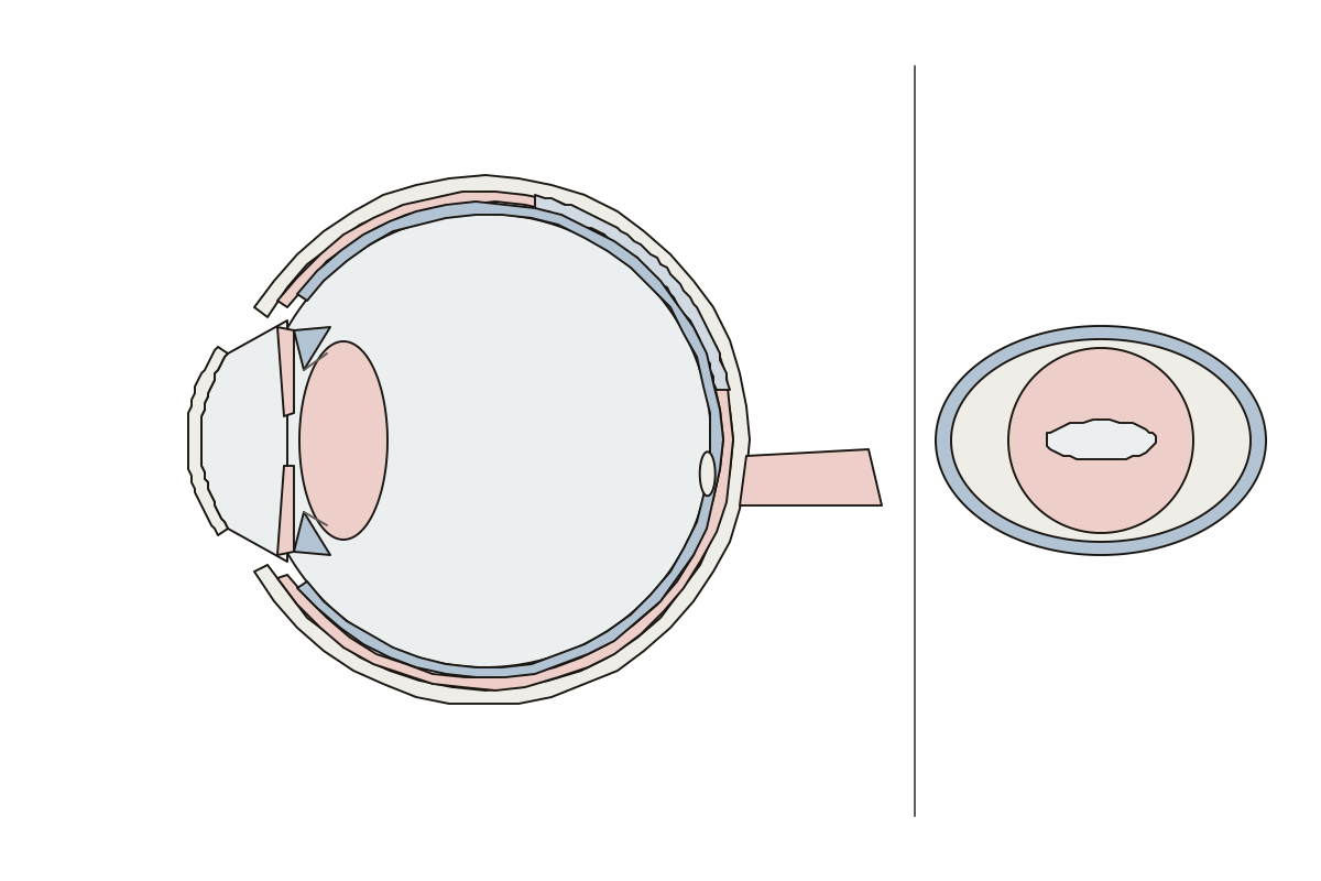 Cow Eye DiagramA cow's eye cut across and from the front: cornea, iris, pupil, lens, retina, tapetum lucidum, choroid, sclera, optic disc and optic nerve. Sclera, cow · Choroid, cow · Ciliary body, cow
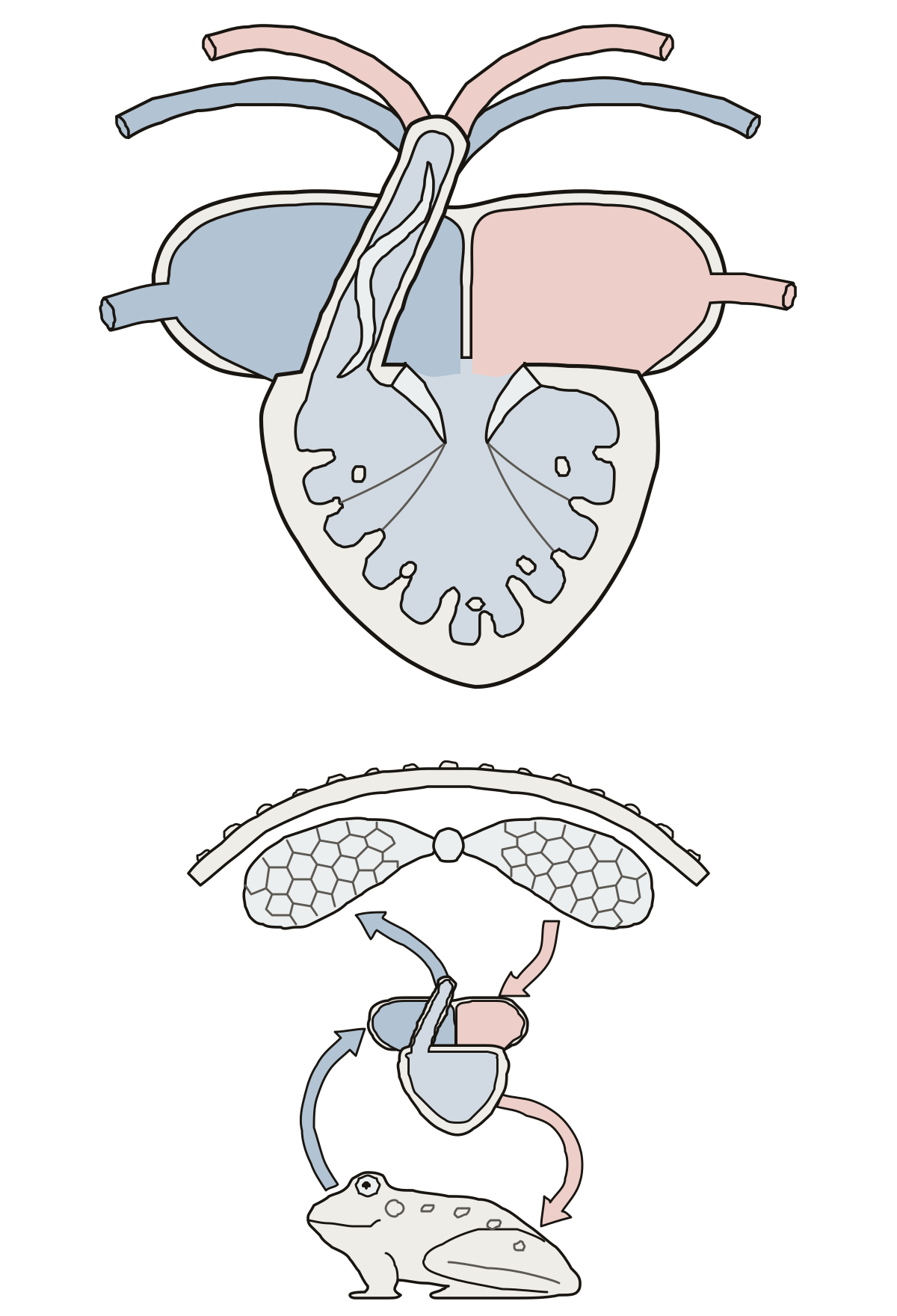 Frog Heart DiagramA frog's heart: 8 numbered parts, from the two atria and single ventricle to the spiral valve, aorta, pulmonary artery and the two circuits. Spiral valve · Pulmocutaneous circuit · Right atrium
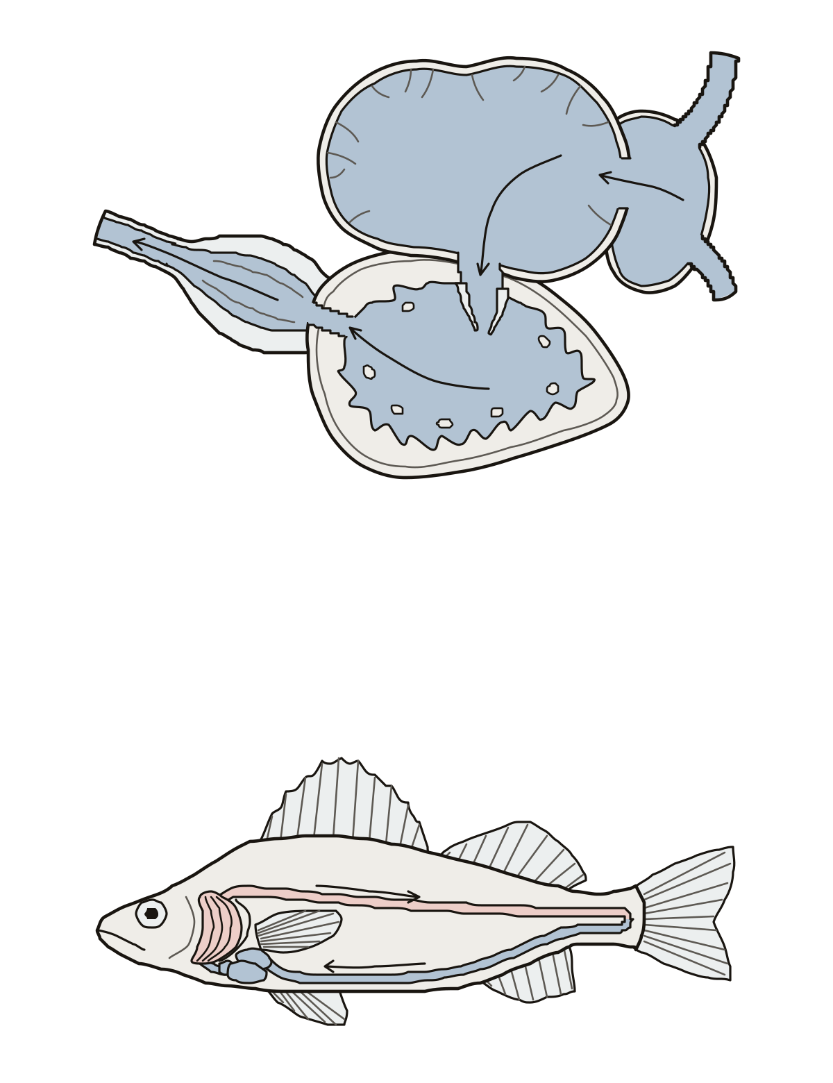 Fish Heart DiagramA fish heart: 9 numbered parts, from the sinus venosus, atrium and ventricle to the bulbus arteriosus, ventral aorta, gills and dorsal aorta. Ventral aorta · Atrium · Sinus venosus
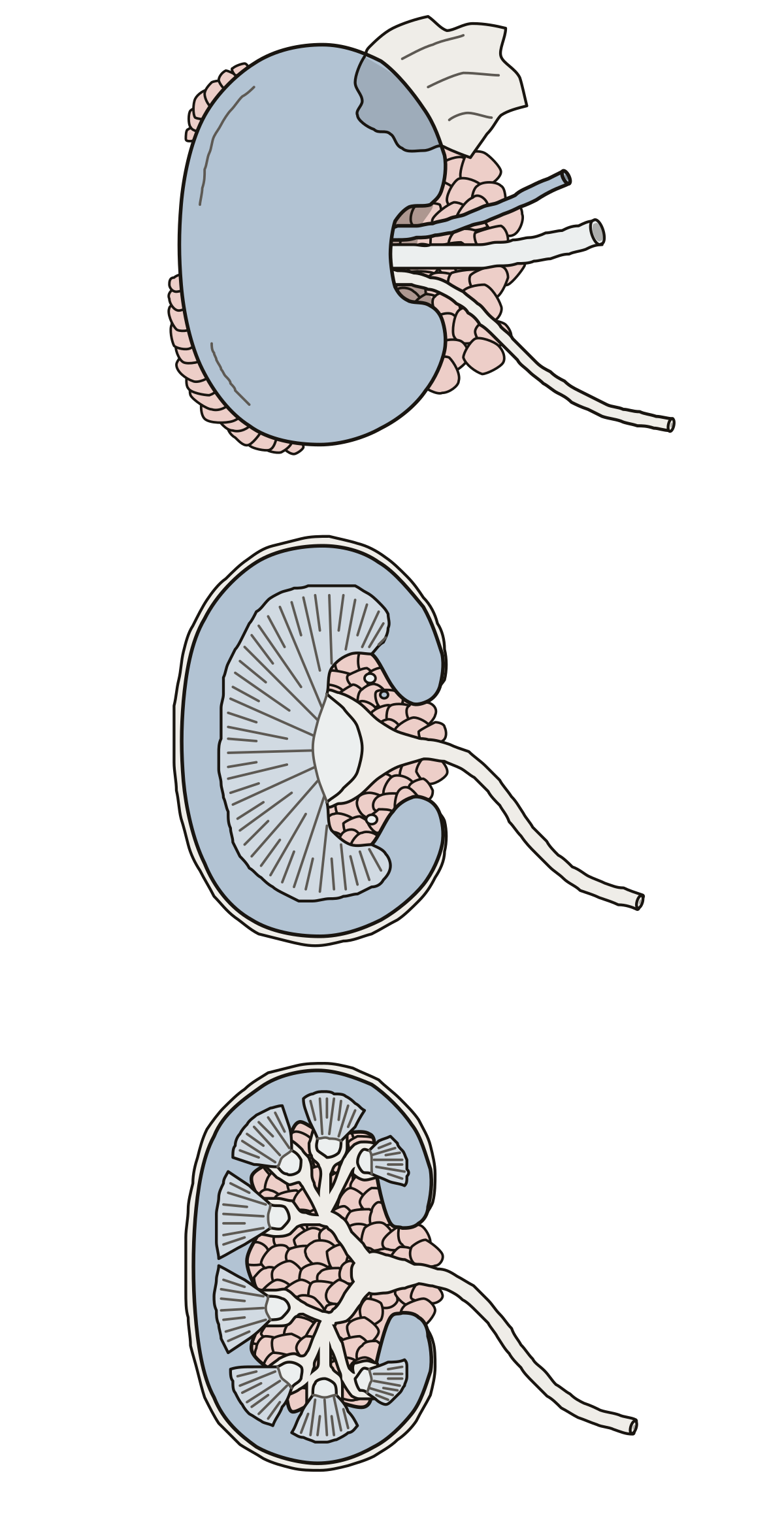 Kidney Dissection LabeledA sheep kidney outside and in section, and a pig kidney in section: fat, capsule, hilum, vessels, ureter, cortex, medulla, renal crest, pelvis, calyces.
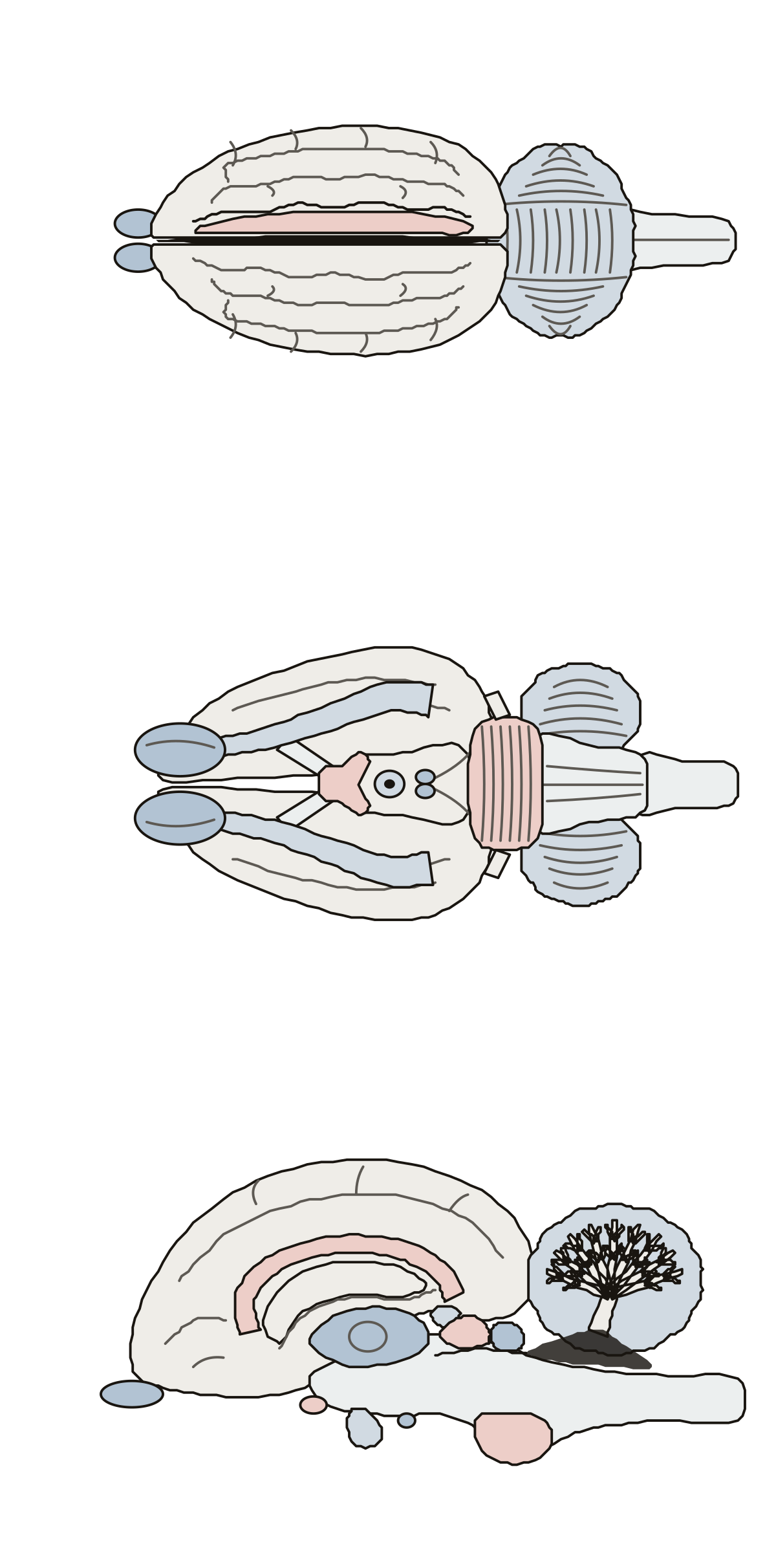 Sheep Brain DiagramA sheep brain in three views: cerebrum, cerebellum, olfactory bulbs, optic chiasm, pons, medulla, corpus callosum, thalamus, colliculi and arbor vitae.
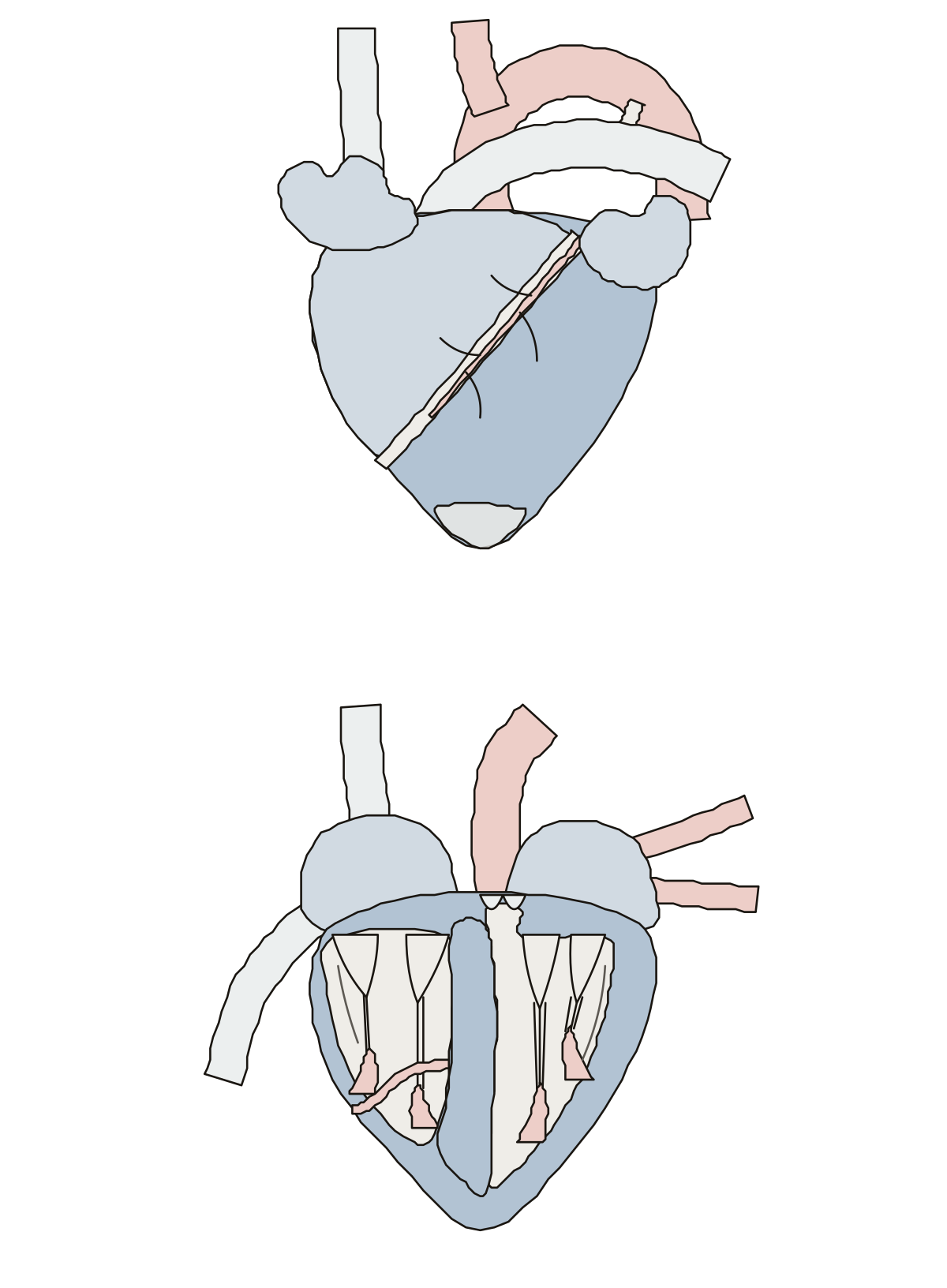 Sheep Heart DiagramA sheep heart outside and in frontal section: auricles, ventricles, aorta, brachiocephalic trunk, pulmonary trunk, venae cavae, valves, cords and septum.
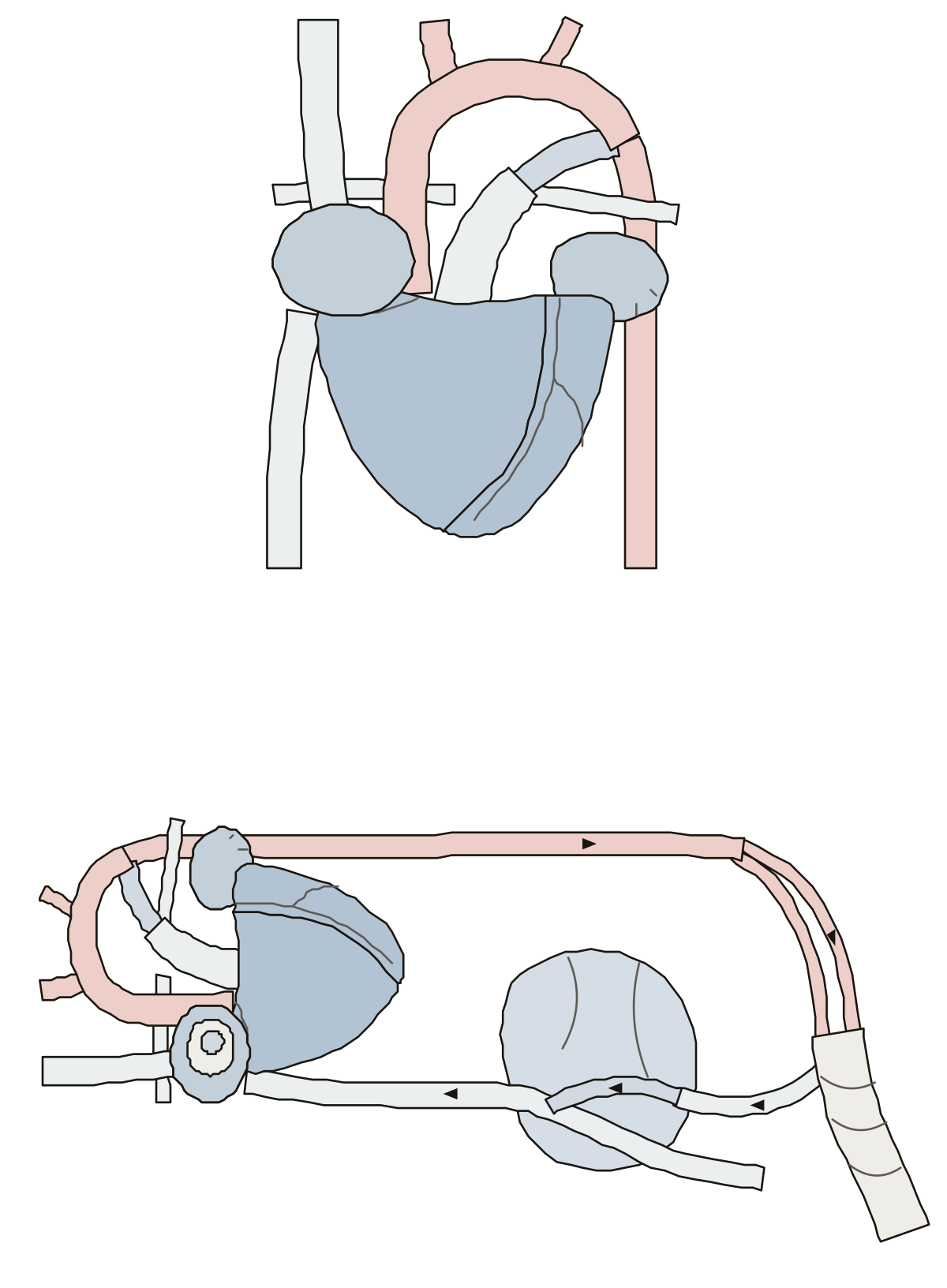 Fetal Pig Heart DiagramA fetal pig's heart with its vessels, then the route before birth: foramen ovale, ductus arteriosus, ductus venosus, and the umbilical vein and arteries.
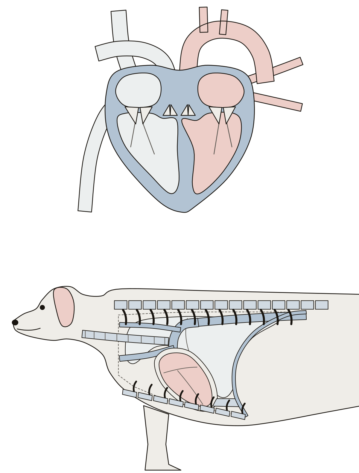 Dog Heart DiagramA dog's heart opened to show its four chambers, four valves and great vessels, then the heart in place in the chest against the ribs, sternum and diaphragm.
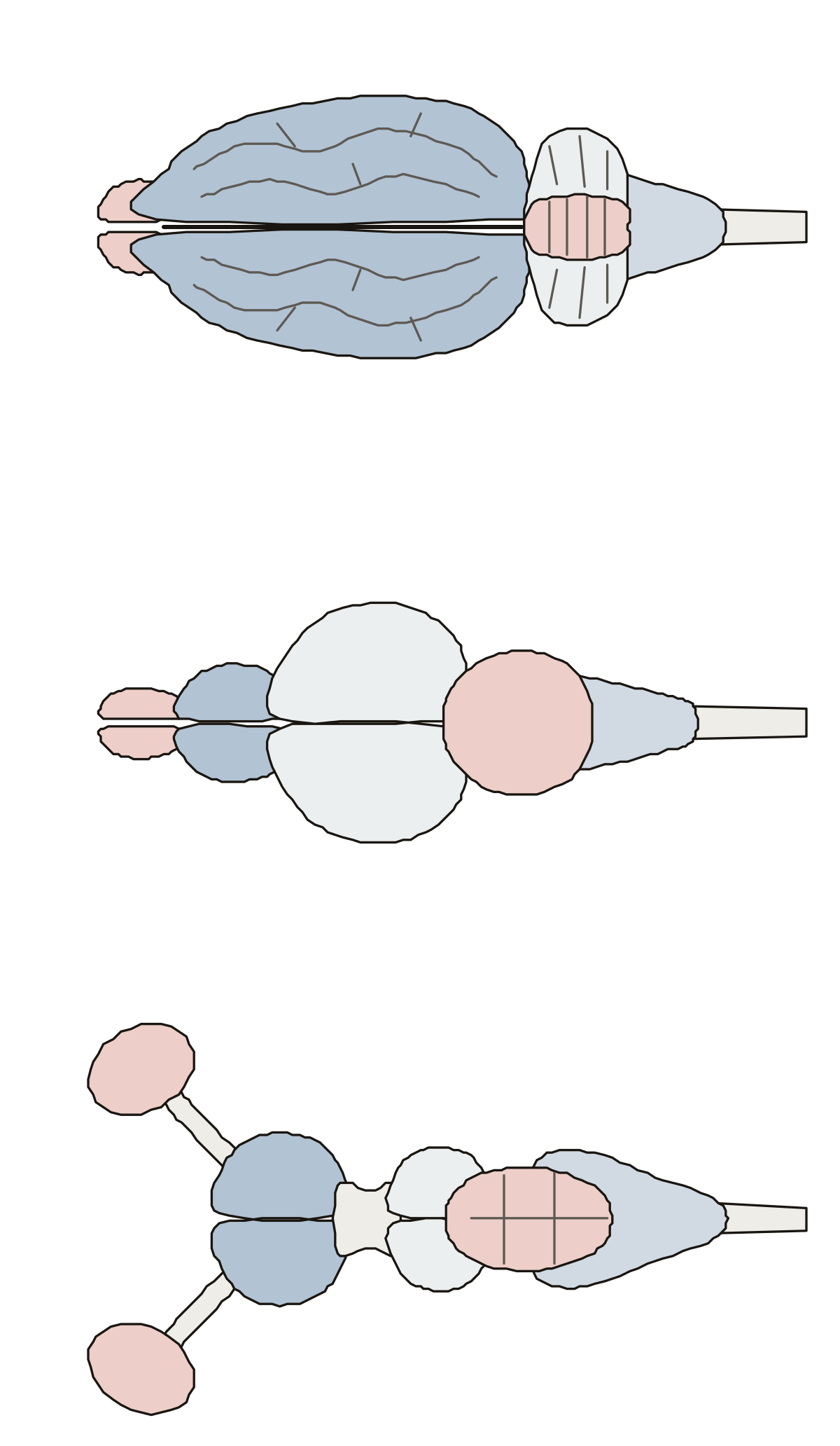 Pig Brain DiagramThree brains from above: a pig's with its large folded cerebrum, a fish's with its large optic lobes, and a dogfish shark's with its large olfactory bulbs.
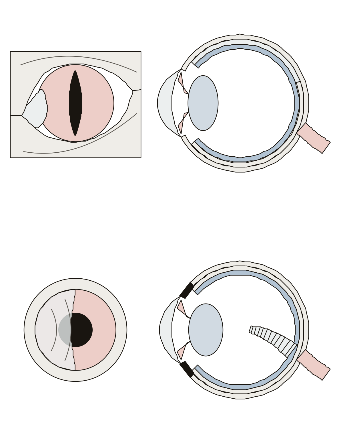 Cat Eye DiagramA cat's eye with its slit pupil, third eyelid and tapetum lucidum, and a bird's eye with its nictitating membrane, scleral ring and pecten, each from the front and cut through.
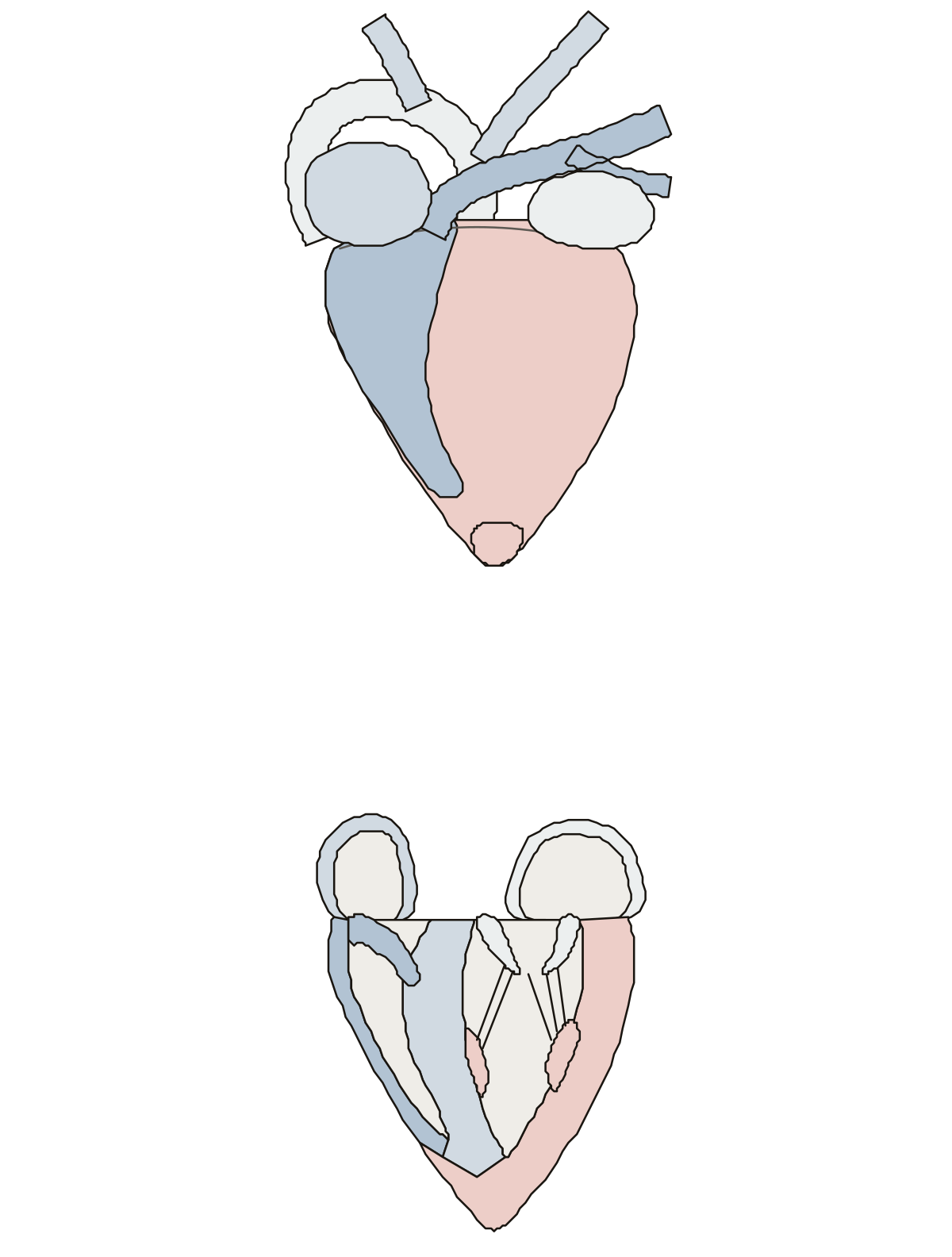 Bird Heart DiagramA chicken's heart from the breast side: four chambers, the aorta arching to the right and its two branches; then the ventricles cut open.
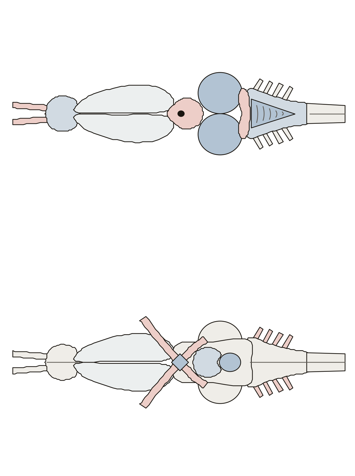 Frog Brain LabeledA frog's brain in two views: olfactory lobes, cerebral hemispheres, diencephalon, pineal body, optic lobes, cerebellum and medulla from above; optic chiasm, infundibulum and pituitary body from below.
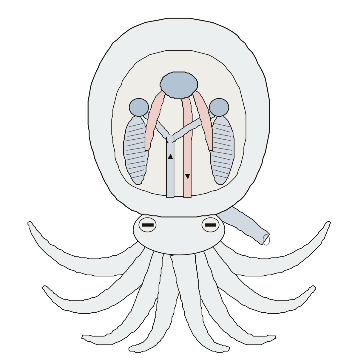 Octopus Hearts DiagramAn octopus with a window in its mantle: the systemic heart, two branchial hearts, gills, atria, aorta and vena cava, with the mantle and siphon.
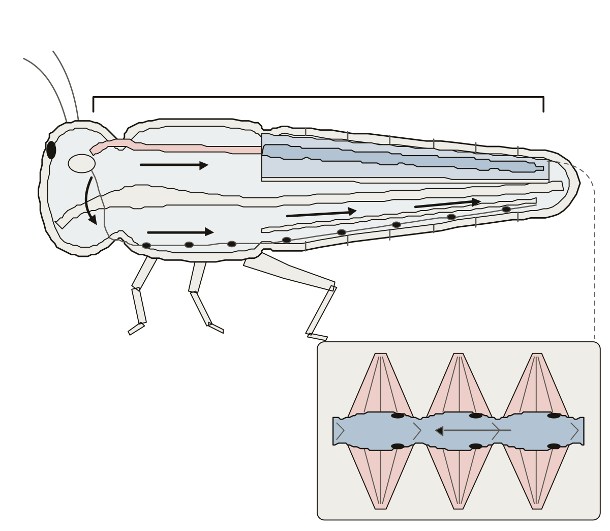 Insect Heart DiagramAn insect cut lengthwise: the dorsal vessel with its heart and aorta, the ostia and alary muscles, the two diaphragms, the pericardial sinus and the hemocoel.
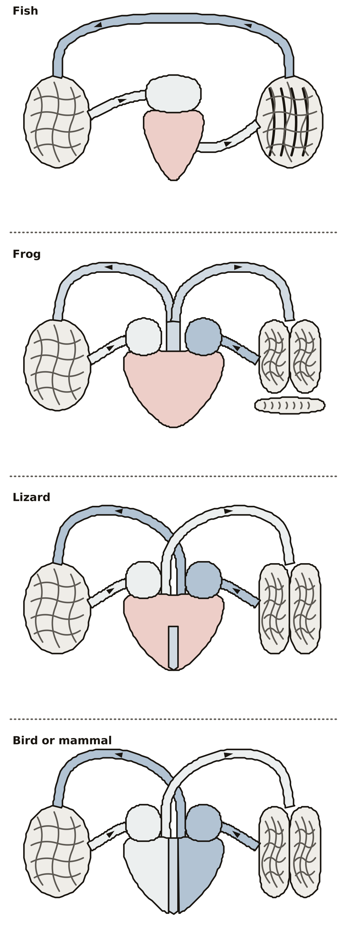 Evolution of Heart in Vertebrates DiagramThe hearts of a fish, a frog, a lizard and a bird or mammal on one chart: one atrium or two, one ventricle or two, one circuit or two.
- Cow Eye Diagram UnlabeledThe cow eye drawing with nothing written on it, to print and fill in: the eyeball cut across and the eye from the front, with its answer key. Vitreous humor, cow · Aqueous humor, cow · Optic nerve, cow
- Frog Brain Anatomy QuizA quiz from the first question on a frog's brain: find the hemispheres, optic lobes, cerebellum and medulla from above, then the optic chiasm, infundibulum and pituitary body from below.
- Dog Heart Anatomy Not LabeledThe dog heart drawing with nothing written on it, to print and fill in: chambers, valves and great vessels, then the heart in the chest, with its answer key.
- Bird Heart Diagram BlankThe bird heart drawing with nothing written on it, to print and fill in: eight parts on the whole heart, seven on the cut heart, with its answer key.
- Animal Eye Anatomy QuizA quiz from the first screen on a cat's eye and a bird's eye, each from the front and cut through: tap the pupil, third eyelid and tapetum lucidum, then the pecten, scleral ring and nictitating membrane.
- Label the Frog Brain DiagramA frog's brain as a worksheet: ten numbered parts on the view from above, five on the underside, a line for each name, and a key.
Digestion and breathing, animal by animal 11
The same body system in different animals: a pig's simple stomach, a cow's rumen, a rabbit's cecum, a bird's gizzard and a fish's gills.
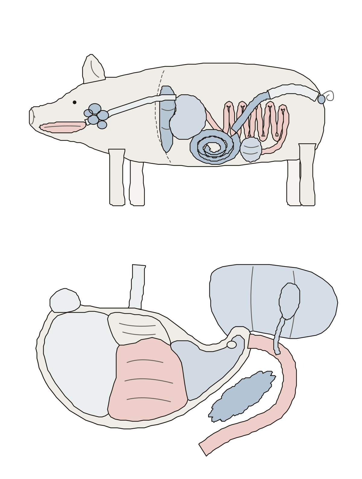 Pig Digestive System DiagramA pig's digestive tract from the side: mouth, esophagus, stomach, liver, small intestine, cecum, spiral colon, rectum; then the stomach's four regions.
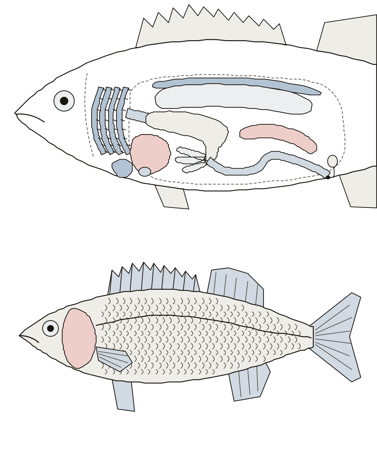 Fish Digestive System DiagramA bony fish, the perch, with its side opened: esophagus, stomach, pyloric ceca, intestine, liver, swim bladder, kidney, gills and heart; then the fins outside.
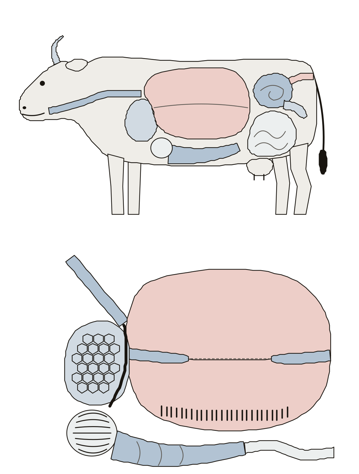 Cattle Digestive System DiagramA cow's digestive tract from the side: esophagus, rumen, reticulum, omasum, abomasum and intestines; then the stomach opened to show its sacs, pillars and linings.
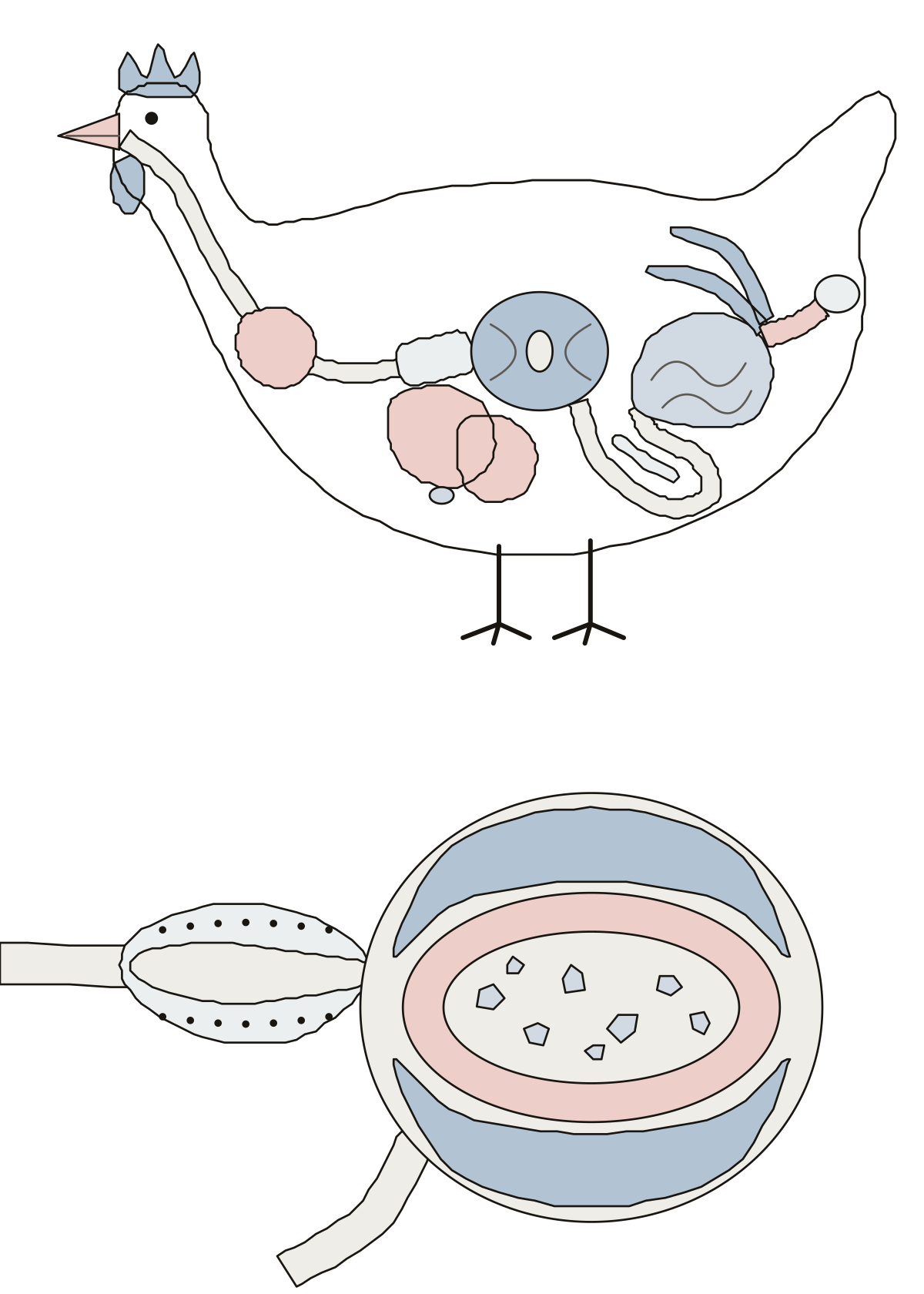 Bird Digestive System DiagramA chicken's digestive tract drawn inside the bird: beak, esophagus, crop, proventriculus, gizzard, liver, duodenal loop, intestines, ceca and cloaca.
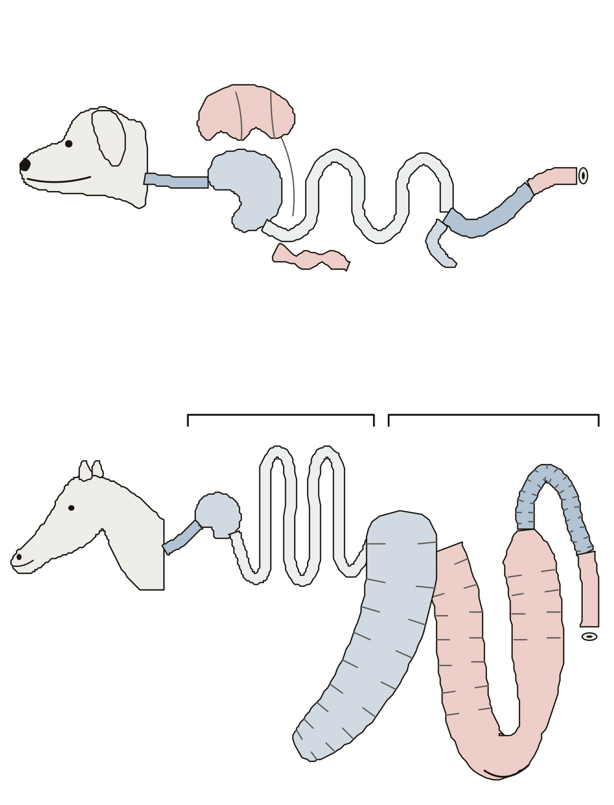 Monogastric Digestive System DiagramThe single-stomach tract of a dog and of a horse, laid out in order: stomach, small intestine, cecum, colon and rectum, with the horse's huge hindgut.
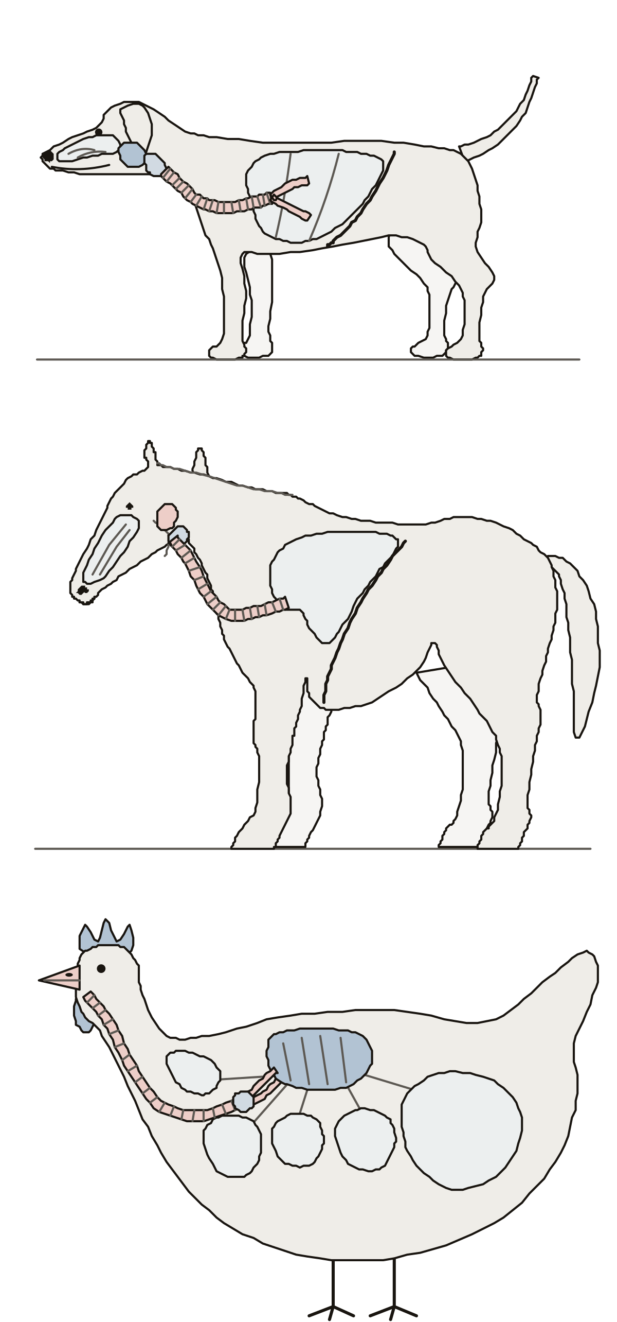 Dog Respiratory System DiagramThe airways of a dog from nostrils to diaphragm, the same on a horse with its guttural pouch, and a chicken with its syrinx, small lung and five kinds of air sac.
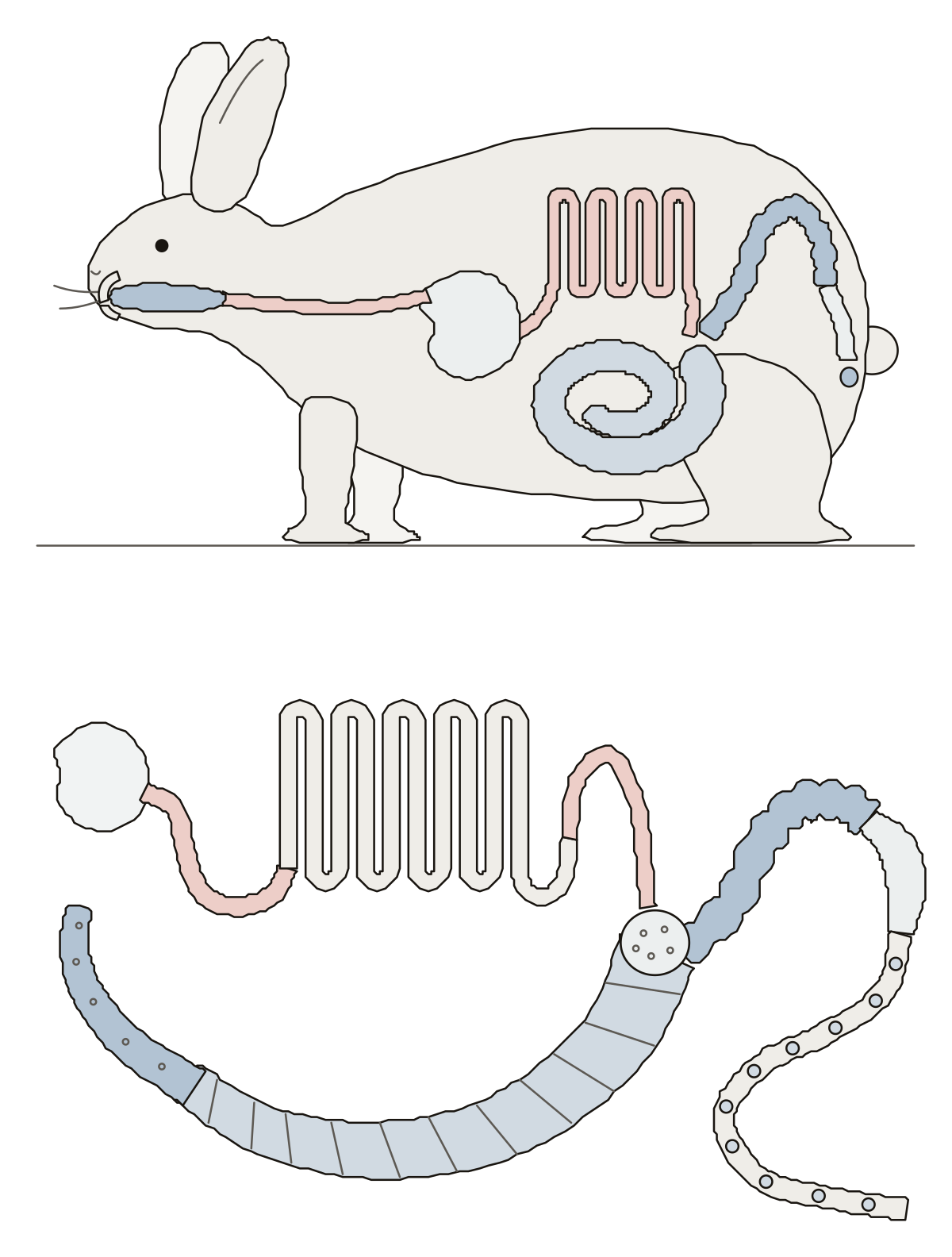 Rabbit Digestive System DiagramA rabbit from the side with its tract inside, mouth to anus; then the small intestine, the round bulb, the appendix and the colon in its parts.
- Bird Digestive System No LabelThe bird digestive system drawing with nothing written on it, to print and fill in: the tract inside a hen above, the gizzard cut open below, with its answer key.
- Label the Rabbit Digestive SystemA rabbit's digestive system as a worksheet: eight numbered parts inside the rabbit, eight on the gut laid out flat, a line for each name, and a key.
- Horse Digestive System Diagram to LabelA horse's digestive tract as a worksheet: nine numbered parts from the stomach to the rectum, a line for each name, and a key.
- Label Cattle Digestive SystemA cow's digestive system as a worksheet: ten numbered parts from the mouth through the four stomach compartments to the rectum, a line for each name, and a key.
Lab specimens, part by part 40
Biology lab specimens as line drawings to learn before lab day: earthworm, crayfish, squid, mussel, clam, jellyfish, sponge, frog, shark, rat, cat and fetal pig, and the life cycle of Obelia.
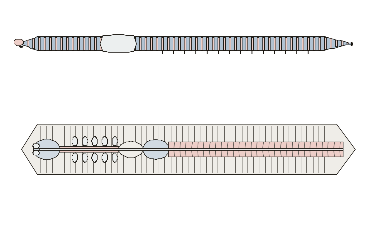 Earthworm Diagram LabeledAn earthworm outside and opened along the back: 14 parts to name. Crop · Clitellum · Pharynx
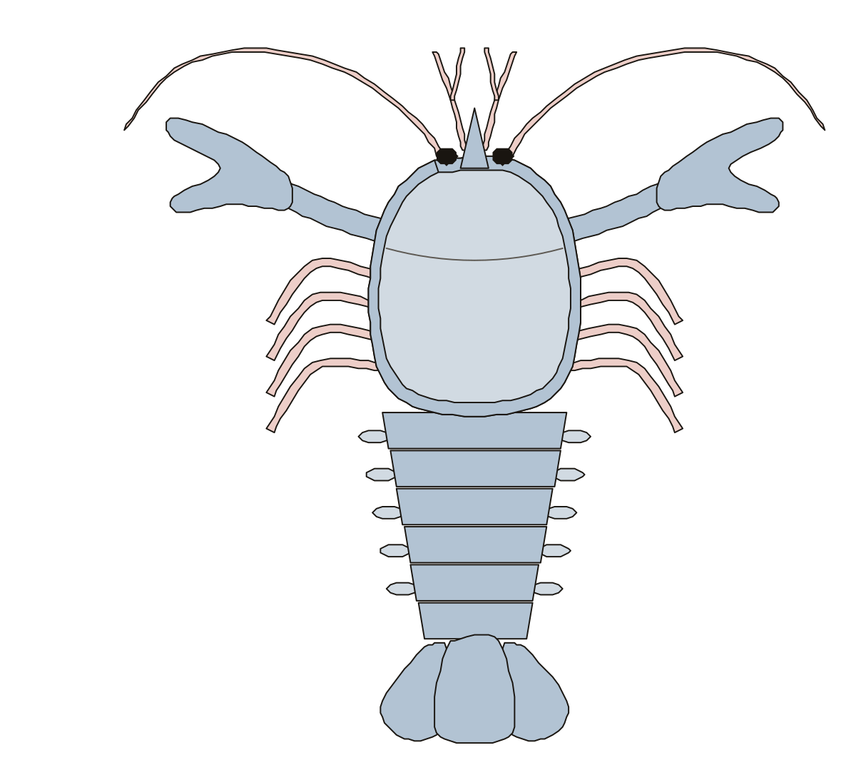 Crayfish Diagram LabeledA crayfish from above, head to tail fan: 12 parts to name. Uropods · Swimmerets · Antennules
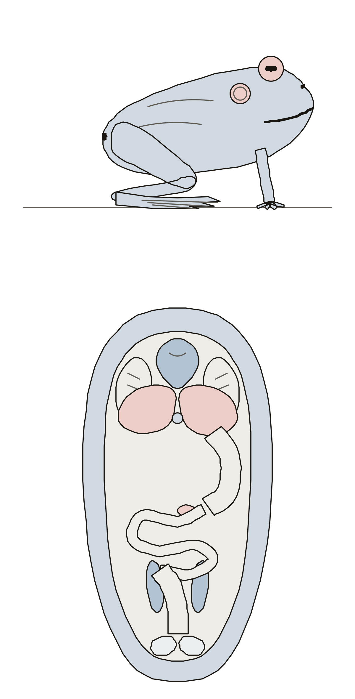 Frog Diagram LabeledA frog outside and dissected: eye, eardrum, nostril, mouth, limbs, webbed foot, cloaca, heart, lungs, liver, gall bladder, stomach, gut, kidneys, bladder. Pancreas · Small intestine · Kidneys
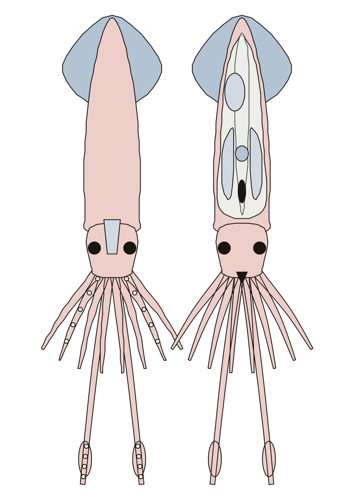 Squid Diagram LabeledA squid: 14 numbered parts, from the mantle, fins, funnel, arms and tentacles to the pen, gills, heart, ink sac and beak inside. Mantle · Fins · Funnel
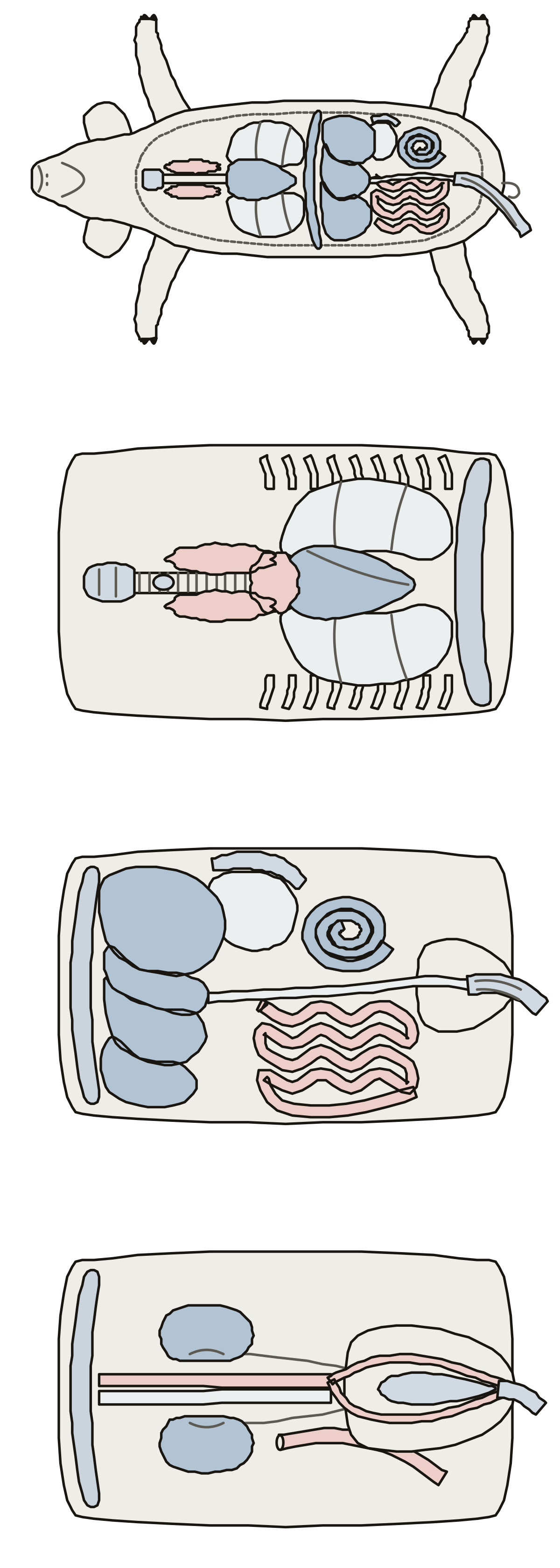 Fetal Pig Diagram Labeled AnswersA fetal pig opened in four views: lungs, heart, diaphragm, liver, thymus, thyroid, trachea, stomach, spleen, spiral colon, kidneys, bladder and the cord's vessels.
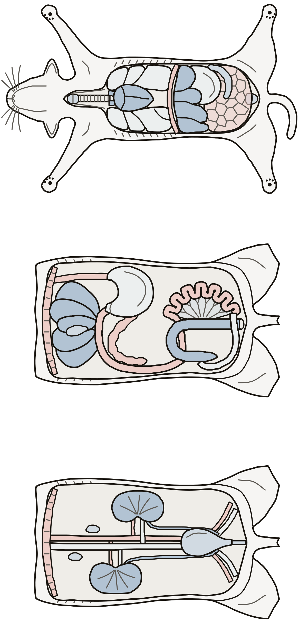 Cat Digestive System DiagramA cat opened in three views: trachea, lungs, liver, stomach, spleen and omentum; duodenum, jejunum, ileum, cecum and colon; kidneys, ureters, bladder and urethra.
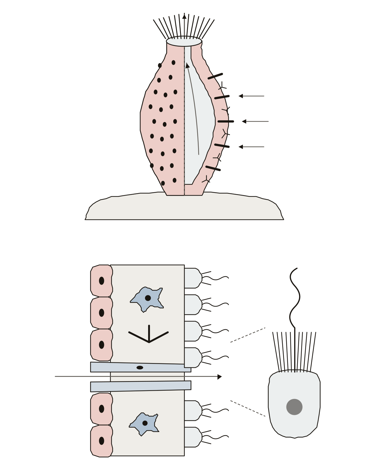 Parts of a SpongeA simple sponge, the water animal, cut open: osculum, pores, spongocoel and spicules; then its body wall with collar cells and the other cell types.
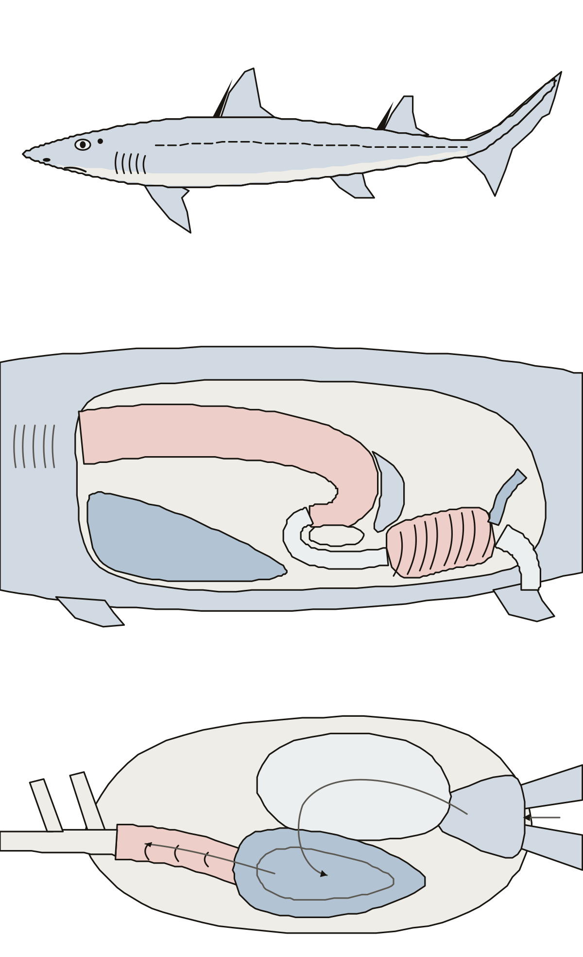 Shark Diagram LabeledA spiny dogfish shark in three views: fins, gill slits and spiracle outside; stomach, liver and spiral valve inside; and the four chambers of the heart.
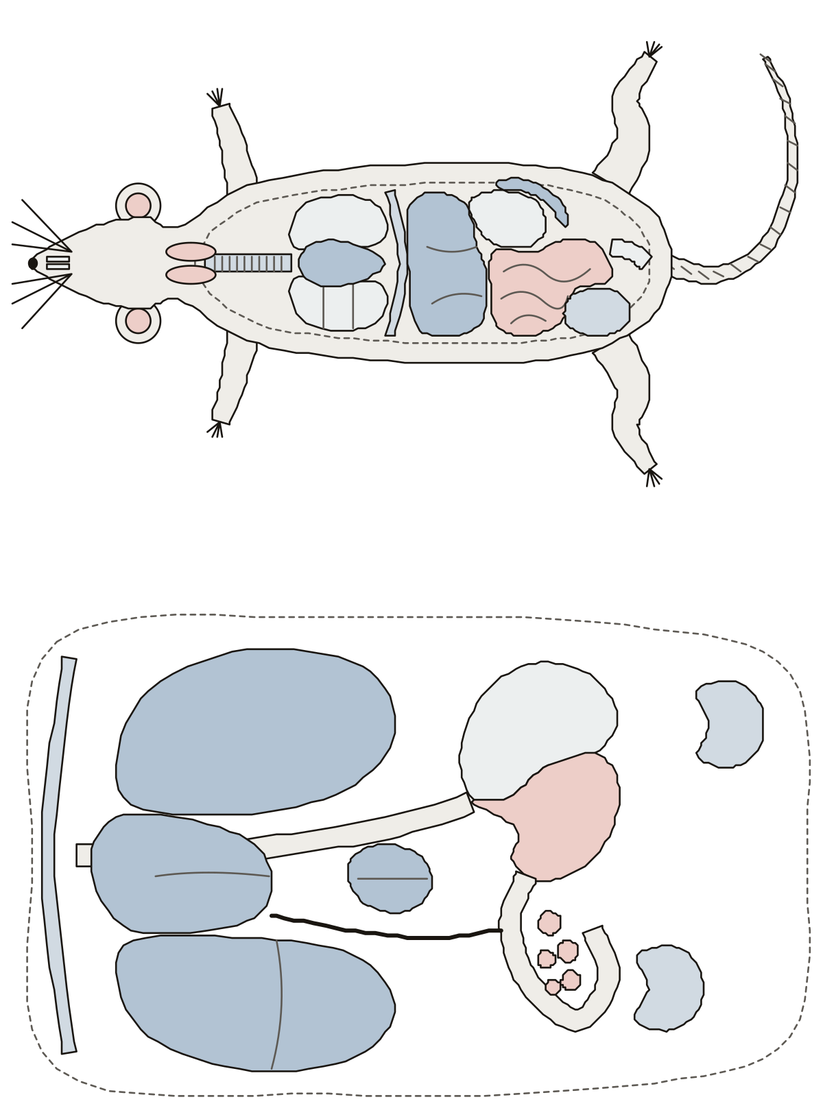 Rat Diagram LabeledA rat on its back, opened: trachea, lungs, heart, liver, stomach, spleen, intestines and cecum; then the four liver lobes, the two-part stomach and the kidneys.
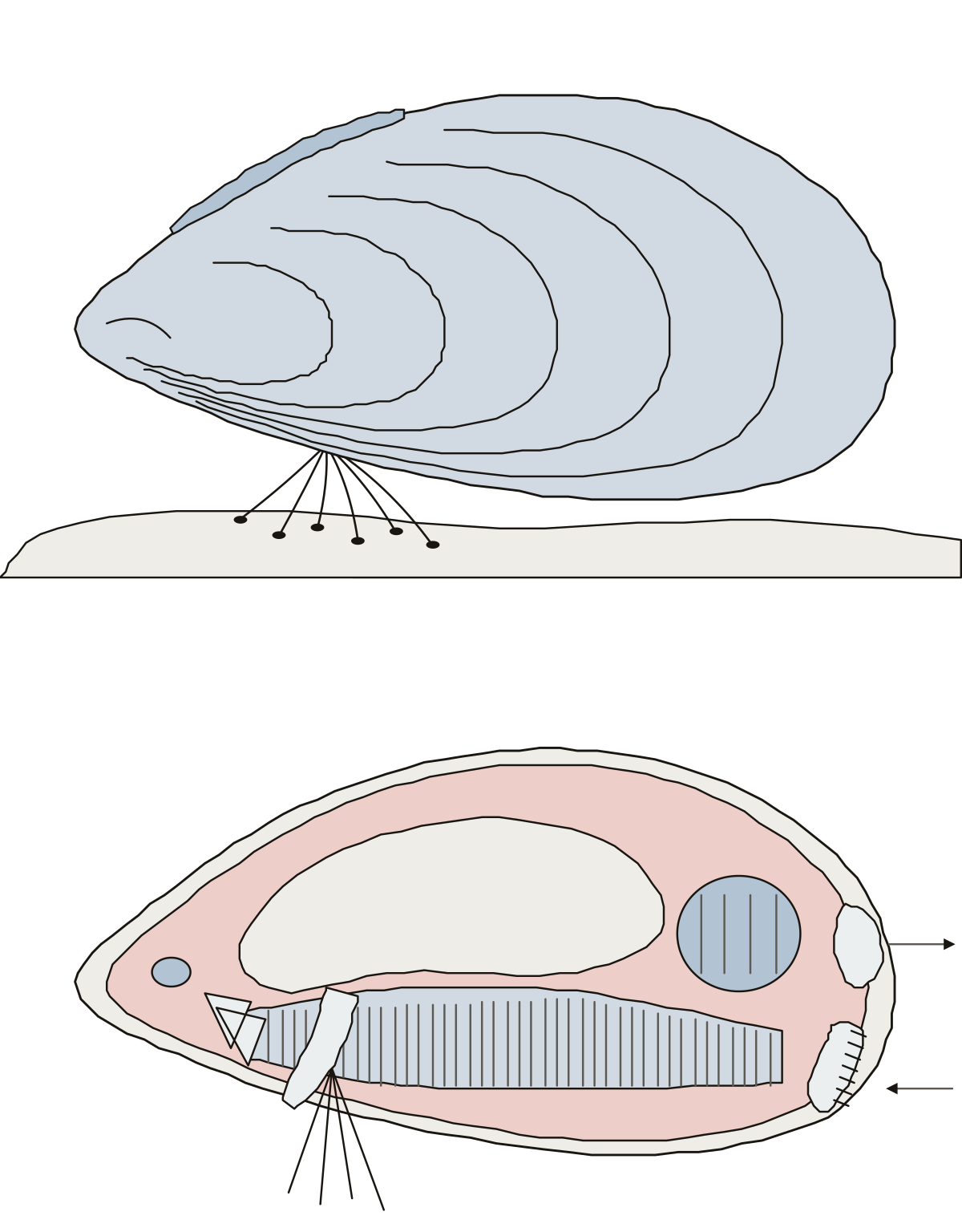 Parts of a MusselA sea mussel on its rock: valve, umbo, growth lines, hinge ligament and byssal threads; then the mantle, gills, foot, adductor muscles, palps and siphons inside.
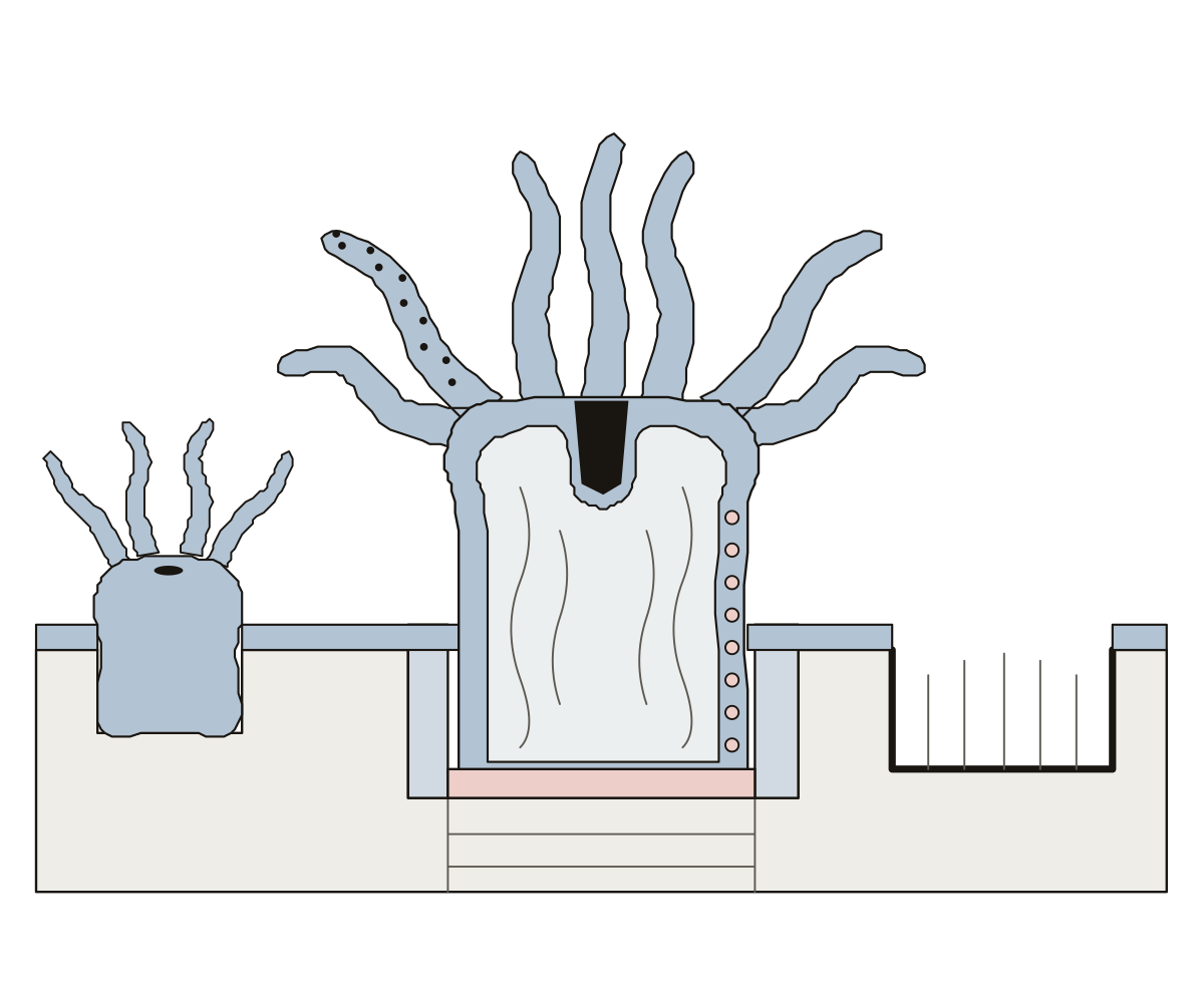 What Are the Parts of a Coral?A coral polyp cut in half: tentacles, mouth, stomach, stinging cells and zooxanthellae, with the calyx, basal plate, coenosarc and skeleton it builds.
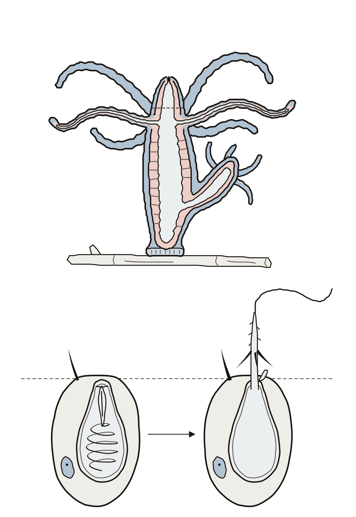 Parts of a HydraA hydra cut lengthwise: tentacles, hypostome, mouth, ectoderm, endoderm, mesoglea, bud and basal disc; then a stinging cell with its nematocyst, cnidocil, thread and barbs.
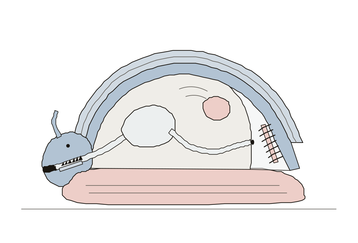 Parts of a MolluscA simple mollusk cut lengthwise: shell, mantle, mantle cavity, gills, foot, head, mouth, radula, visceral mass, stomach, heart and anus.
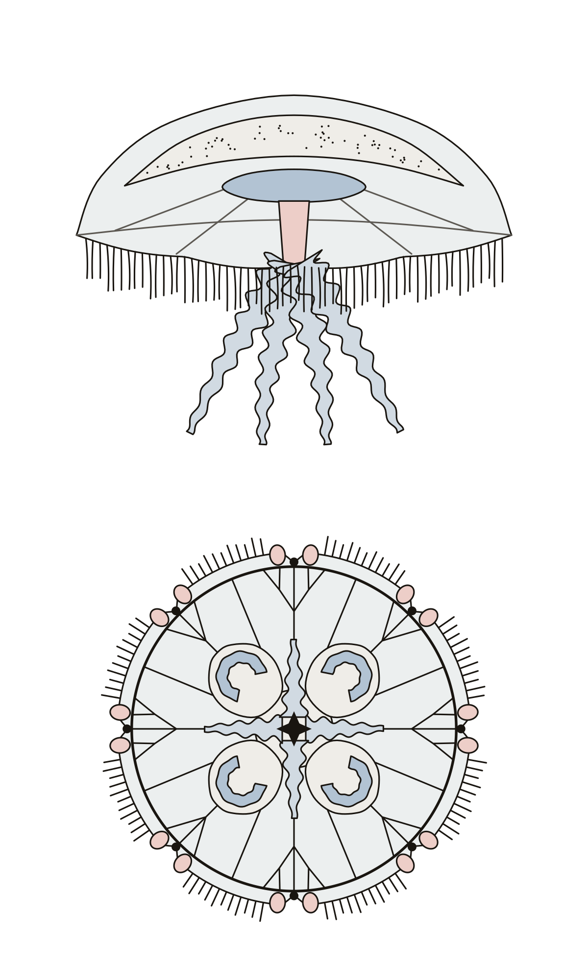 Body Parts of a JellyfishA moon jellyfish in two views: bell, mesoglea, stomach, manubrium, oral arms and tentacles from the side; mouth, gonads, gastric pouches, canals, rhopalia and lappets from below.
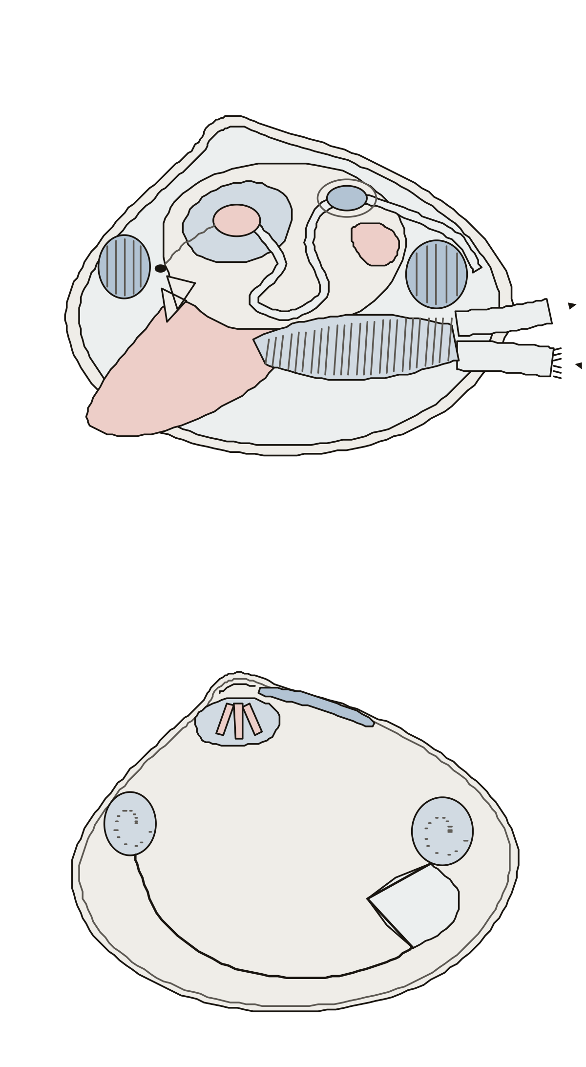 Body Parts of a ClamA hard-shell clam in two views: mantle, foot, gills, siphons, adductor muscles, stomach, intestine, heart and kidney; then the umbo, hinge teeth, muscle scars, pallial line and pallial sinus.
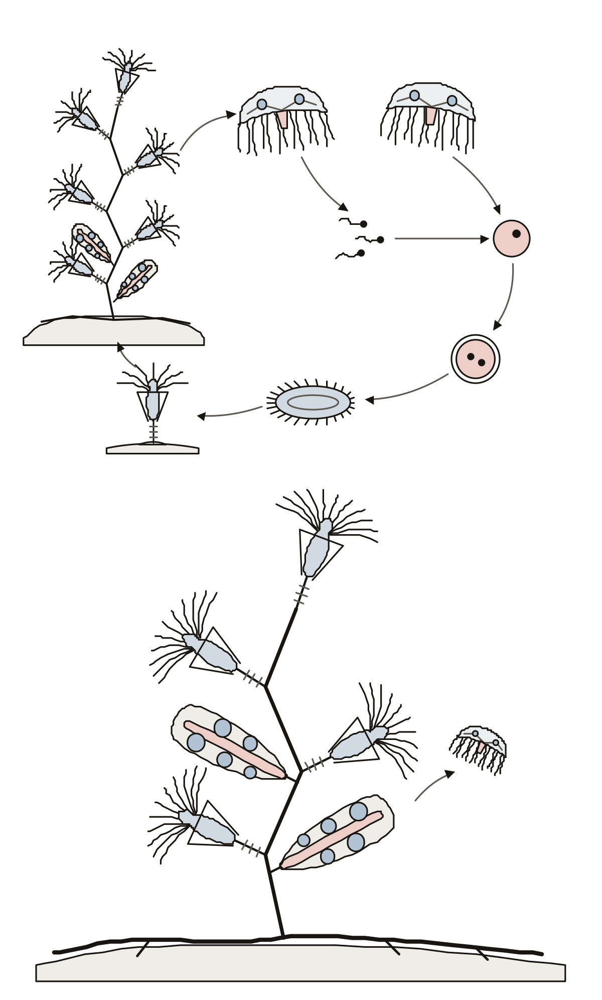 Label the Life Cycle of ObeliaThe life cycle of Obelia: colony, medusa, sperm, egg, fertilized egg, planula and young polyp in a ring, then the hydranths, blastostyles, medusa buds and hydrorhiza of the colony.
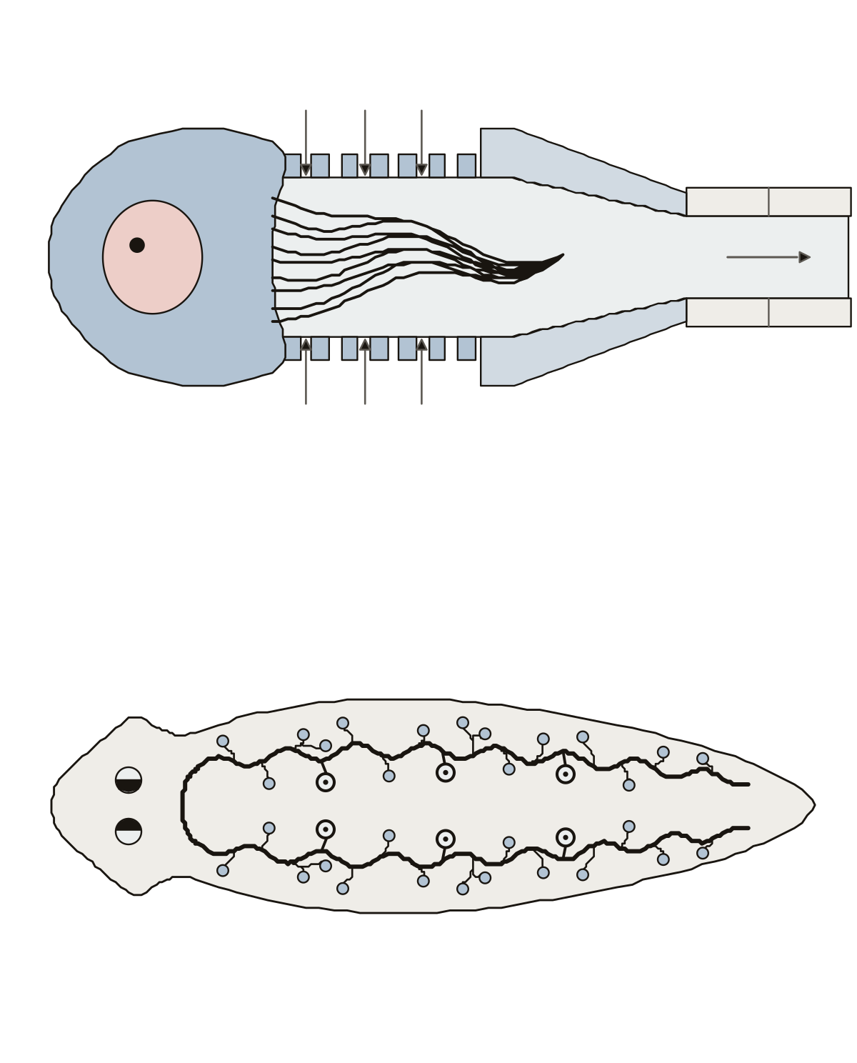 Diagram of a Flame CellA flame cell cut along its length: nucleus, cilia, fenestrations, tube cell, lumen and tubule; then a planarian with its excretory canals, branches and pores.
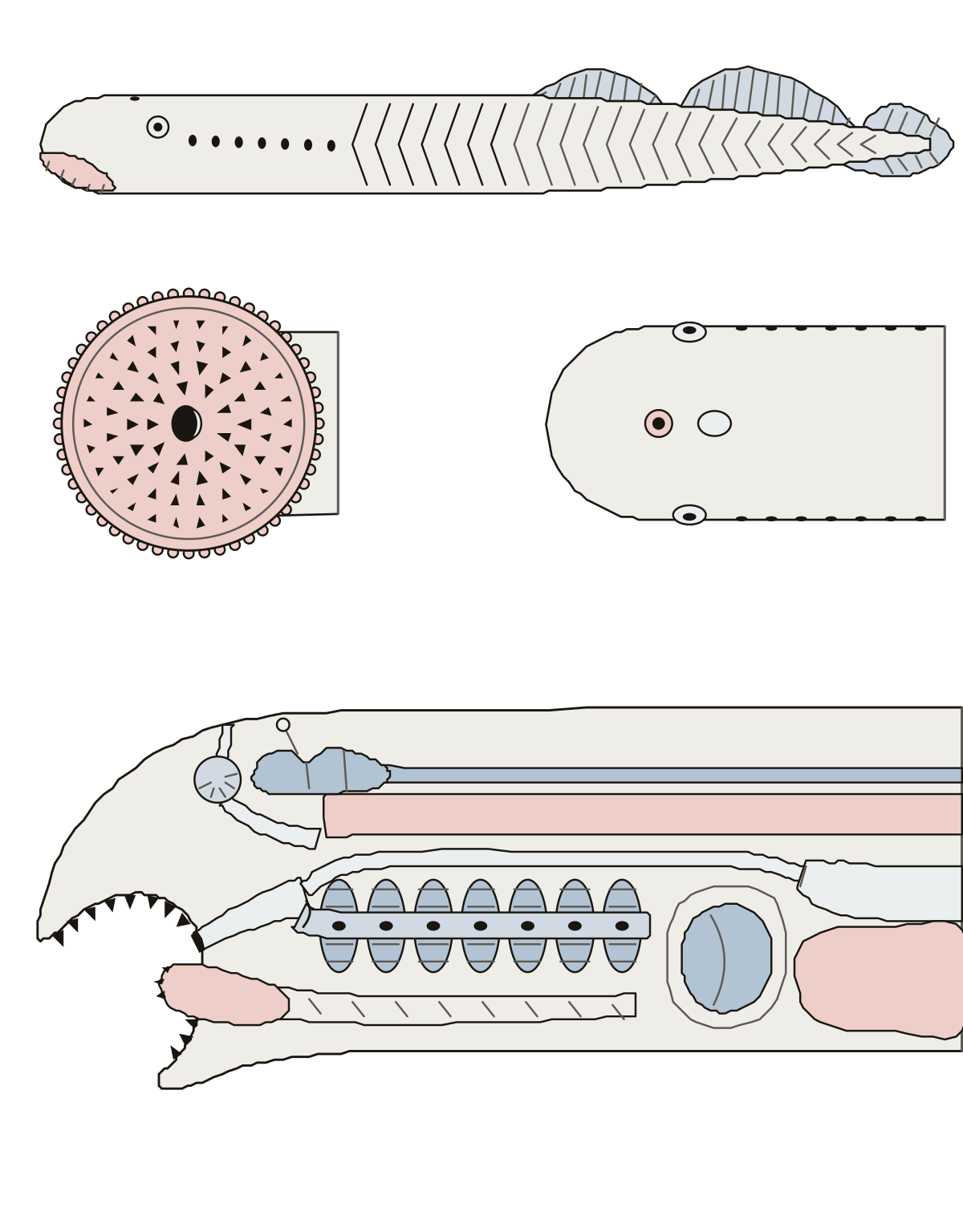 Lamprey Anatomy QuizA sea lamprey in two views: the oral disc, single nostril, seven gill openings and fins outside; the notochord, gill pouches, respiratory tube and heart inside.
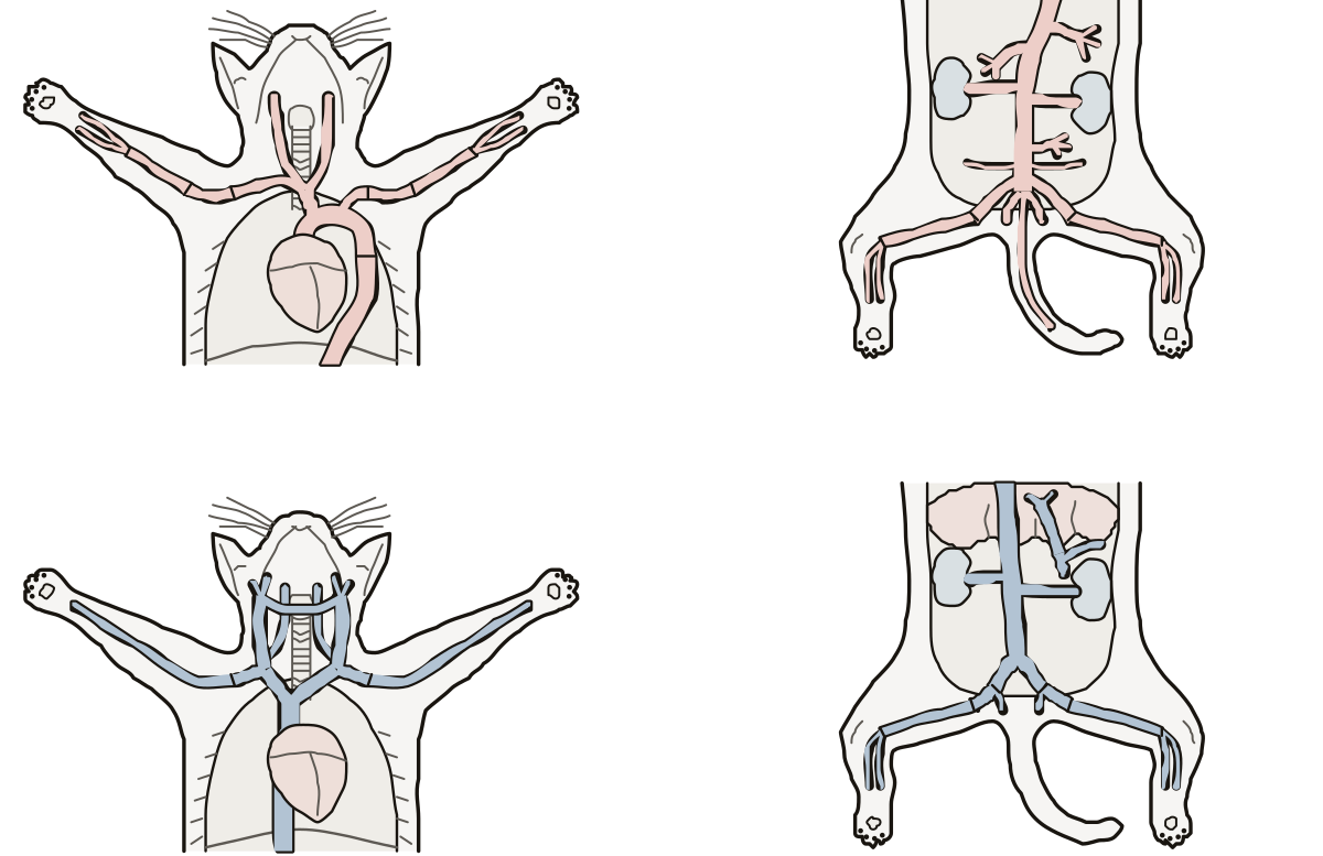 Cat Blood Vessels Labeling QuizA cat from the front in four views: the arteries from the aortic arch to the caudal artery of the tail, then the veins from the external jugular to the femoral vein.
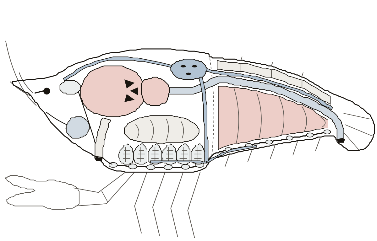 Crayfish Internal Anatomy QuizA crayfish from the side with the body wall drawn away: the food path, the heart and its arteries, gills, green gland, brain, nerve cord and tail muscles. Digestive gland, crayfish · Ventral nerve cord, crayfish · Sternal artery, crayfish
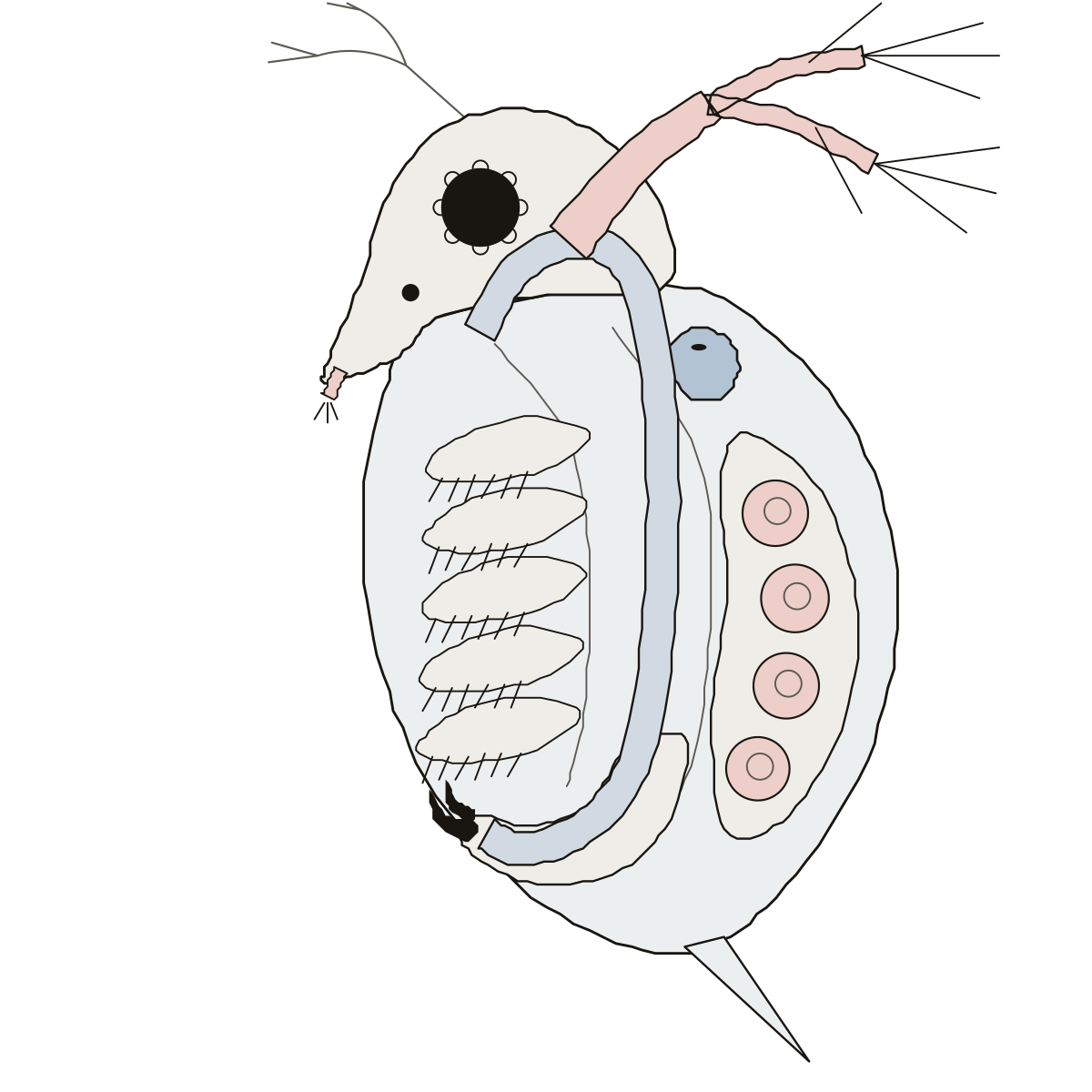 Daphnia Heart DiagramA Daphnia from the side: heart, compound eye, ocellus, antennae, carapace, intestine, brood chamber with eggs, legs, postabdomen and claws. Antennae, Daphnia · Ocellus, Daphnia · Brood chamber, Daphnia
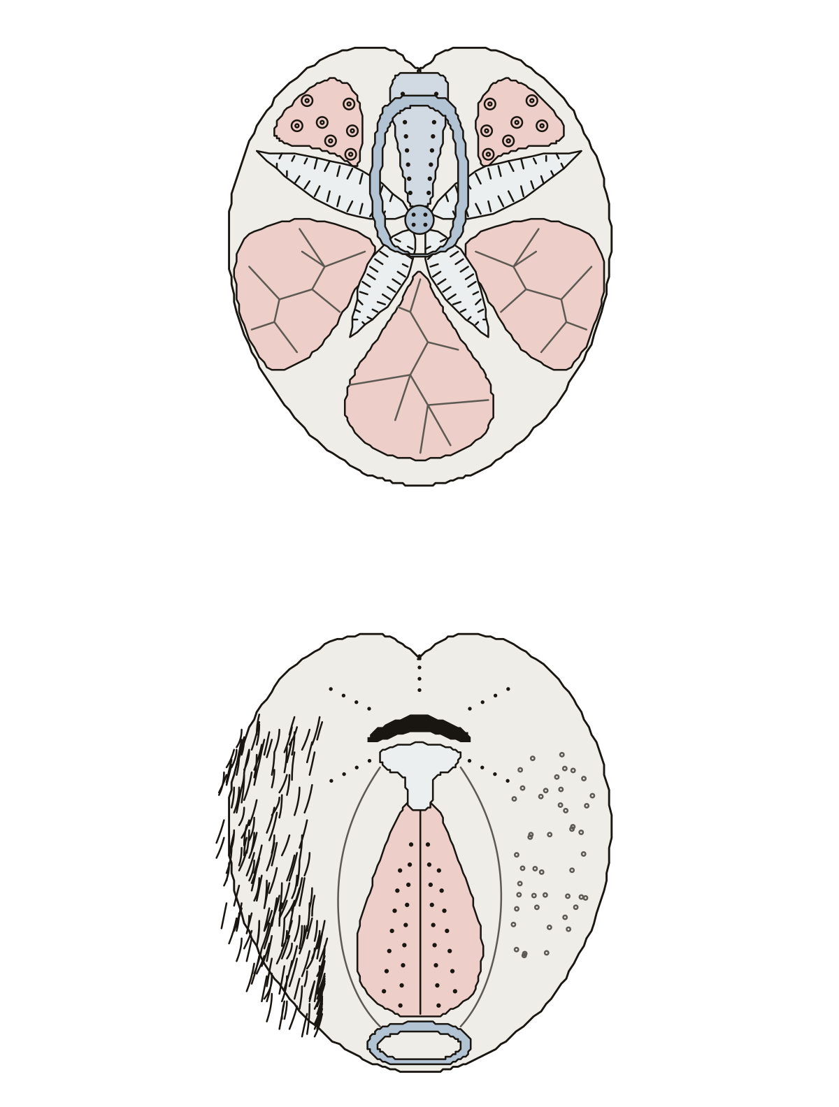 Heart Urchin DiagramThe test of a heart urchin in two views: petals, front groove and apical disc on top; mouth, labrum, plastron and fasciole underneath.
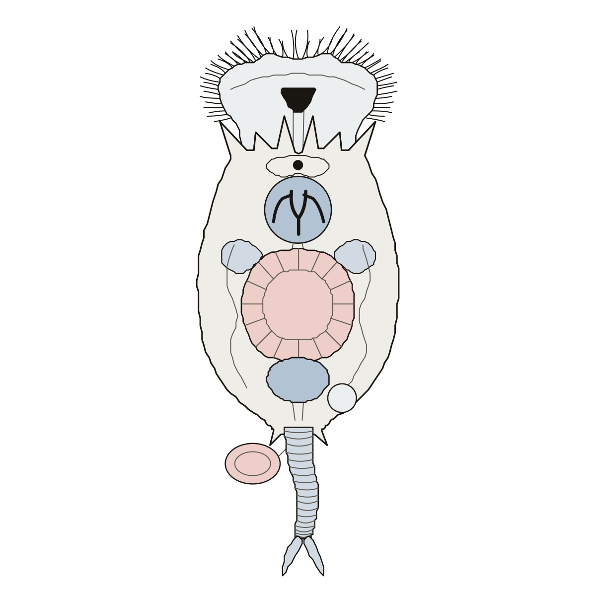 Brachionus LabeledA Brachionus rotifer from above: corona, cilia, mouth, mastax, trophi, lorica and spines, stomach, gastric glands, intestine, bladder, eyespot, foot and toes. Corona, rotifer · Intestine, rotifer · Mastax, rotifer
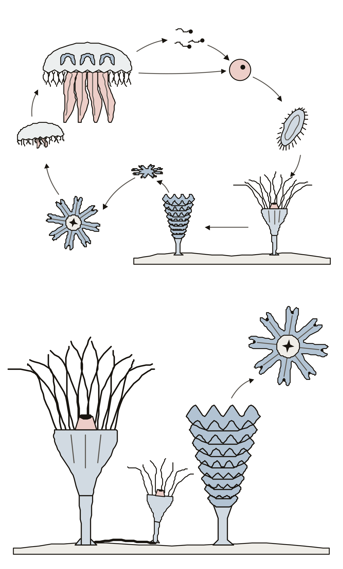 Jellyfish Life Cycle DiagramThe life cycle of the moon jelly, Aurelia: medusa, egg, sperm, planula, scyphistoma, strobila and ephyra in a ring, then the polyp with its tentacles, mouth, stalk and bud. Mouth, moon jelly · Young medusa, moon jelly · Adult medusa, moon jelly
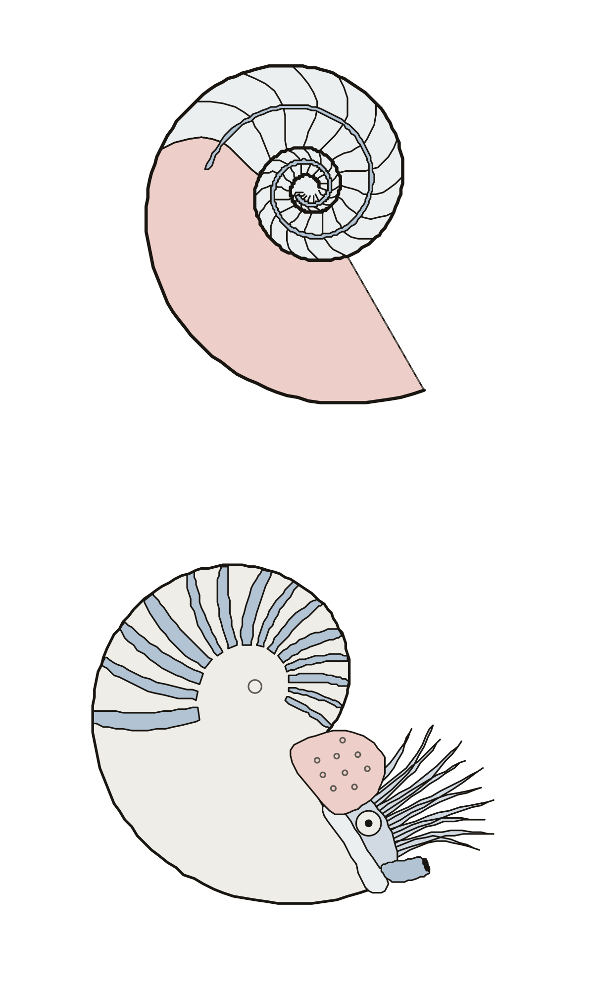 Nautilus Shell LabeledA chambered nautilus in two views: the chambers, septa, siphuncle, body chamber and opening of the cut shell, then the hood, tentacles, eye, funnel and mantle. Tentacles, nautilus · Siphuncle, nautilus · Funnel, nautilus
- Mollusk Diagram QuizA quiz from the first screen on a simple mollusk cut lengthwise: tap the shell, mantle, foot, head and mouth, then the radula, visceral mass, stomach, heart, mantle cavity, gills and anus.
- Frog Diagram No LabelsThe frog drawing with nothing written on it, to print and fill in: a sitting frog from the side and a frog opened from below, with its answer key. Nostril · Bladder · Eye
- Crayfish Diagram UnlabeledThe crayfish drawing with nothing written on it, to print and fill in: the external anatomy from above, feelers to tail fan, with its answer key. Antennae · Carapace · Cephalothorax
- Sponge Diagram UnlabeledThe sponge drawing with nothing written on it, to print and fill in: the whole sponge cut open and its body wall enlarged, with its answer key.
- Unlabelled Rat Dissection DiagramThe rat drawing with nothing written on it, to print and fill in: the whole rat opened on its back and its liver, stomach and kidneys enlarged, with its answer key.
- Mussel Diagram UnlabeledThe mussel drawing with nothing written on it, to print and fill in: the shell on its rock above, the opened mussel below, with its answer key.
- Hydra Unlabelled DiagramsThe hydra drawing with nothing written on it, to print and fill in: the animal cut lengthwise above, one stinging cell below, with its answer key.
- Jellyfish Anatomy QuizA quiz from the first screen on a moon jellyfish: tap the bell, tentacles, oral arms, stomach, mesoglea and manubrium from the side, then the mouth, gonads, pouches, canals, rhopalia and lappets from below.
- Jellyfish Anatomy Diagram UnlabeledThe jellyfish drawing with nothing written on it, to print as a worksheet: six parts from the side, seven from below, with its answer key.
- Mussel Anatomy QuizA quiz from the first screen on a sea mussel: tap the valve, umbo, growth lines, hinge ligament and byssal threads, then the mantle, gills, foot, palps, adductor muscles and siphons inside.
- Coral Polyp Diagram to LabelA coral polyp as a worksheet: ten numbered parts on a stony coral cut down through one polyp, tentacles to skeleton, a line for each name, and a key.
- Clam Labeled Diagram BlankThe clam drawing with nothing written on it, to print and fill in: thirteen lines on the body, seven inside the empty shell, and an answer key.
- Label Mollusk Body Plan DiagramThe mollusk body plan as a worksheet: twelve numbered parts on a simple mollusk cut lengthwise, a line for each name, and a key.
- Daphnia Image LabelingA Daphnia as a worksheet: thirteen numbered parts on a water flea seen from the side, heart to claws, a line for each name, and a key. Head, Daphnia · Carapace, Daphnia · Claws, Daphnia
- Label the Heart UrchinA heart urchin's test as a worksheet: seven numbered parts from above, five on the underside, a line for each name, and a key.
This set is for the animal side of a biology, agriculture or veterinary science course. It starts with the skeleton of the horse, and pages on other animals, single organs and the specimens of a school dissection lab are added one drawing at a time.
More about these tools
Each page is one calm line drawing with a numbered ring on every part. A ring lights up and you choose or type its name, or you get a name and tap the part. Missed parts come back when the round ends.
Use a page the evening before a lab practical or a horse club knowledge contest, so the names are familiar before you meet the real thing. Teachers print the blank drawing as a worksheet and the labeled one as its key. Names were checked against a veterinary anatomy text on the date each page shows.
Keep exploring
- Animal body-part diagrams (94)
- Blank US maps (61)
- Blank maps (159)
- Bone labeling quizzes (49)
- Capitals quizzes (11)
- Car and engine part diagrams (54)
- Cell and organelle diagrams (63)
- Clothing, shoes and jewelry (45)
- Computer, phone and camera diagrams (43)
- Anatomy concept maps (1)
- Country map quizzes (28)
- DNA, cell division and cell processes (53)
- Science Quizzes
- All quizzes