DNA and Cell Division Diagrams: Label Each Figure, Then Print It
One textbook figure per page, a numbered blank on every part, and a blank copy and key to print.
53 pages
DNA and how it is read 21
The double helix and its building blocks, then the machinery that copies DNA, transcribes it and translates the message into protein, and a gel that sorts DNA pieces by size. An amino acid and the kinds of mutation follow.
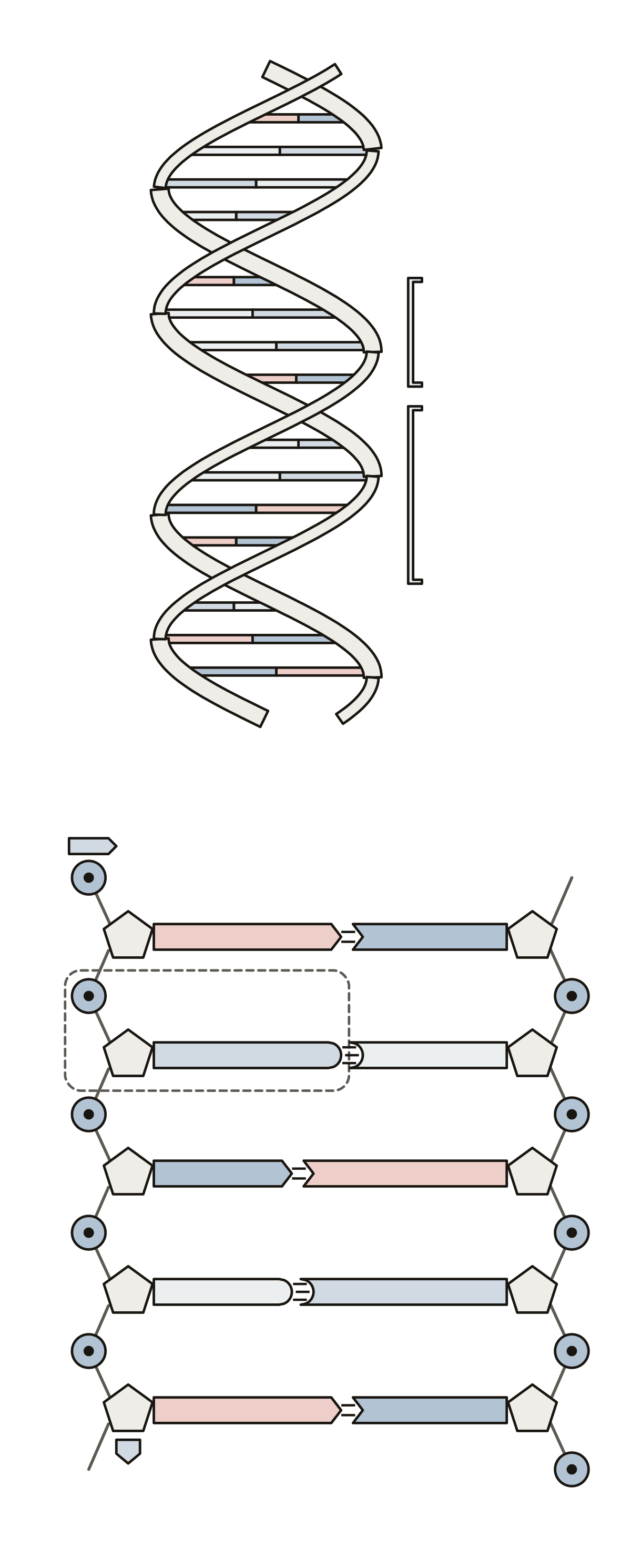 DNA Diagram LabeledThe DNA double helix and a flattened ladder: 14 numbered parts, from the sugar-phosphate backbone and grooves to adenine, thymine, guanine and cytosine.
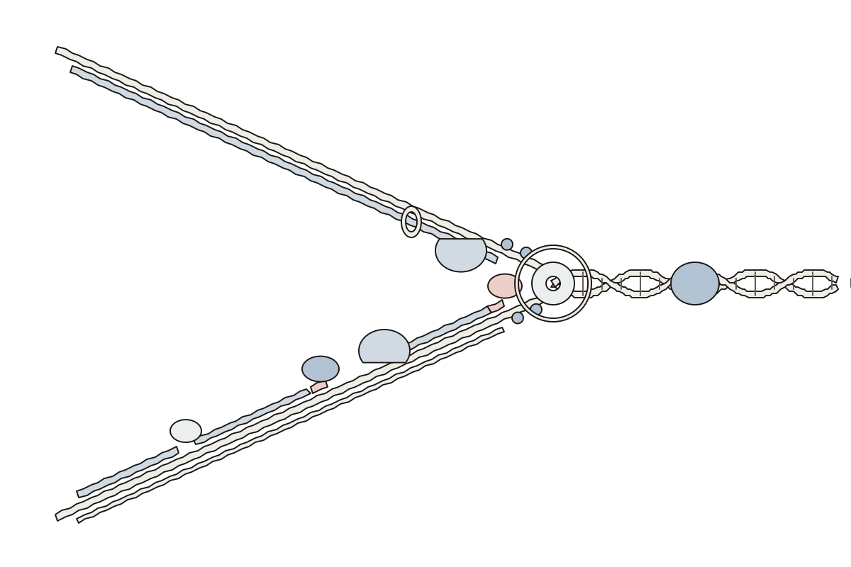 DNA Replication Diagram LabeledA DNA replication fork: 14 numbered parts, from helicase, topoisomerase and primase to the leading strand, lagging strand, Okazaki fragments and ligase.
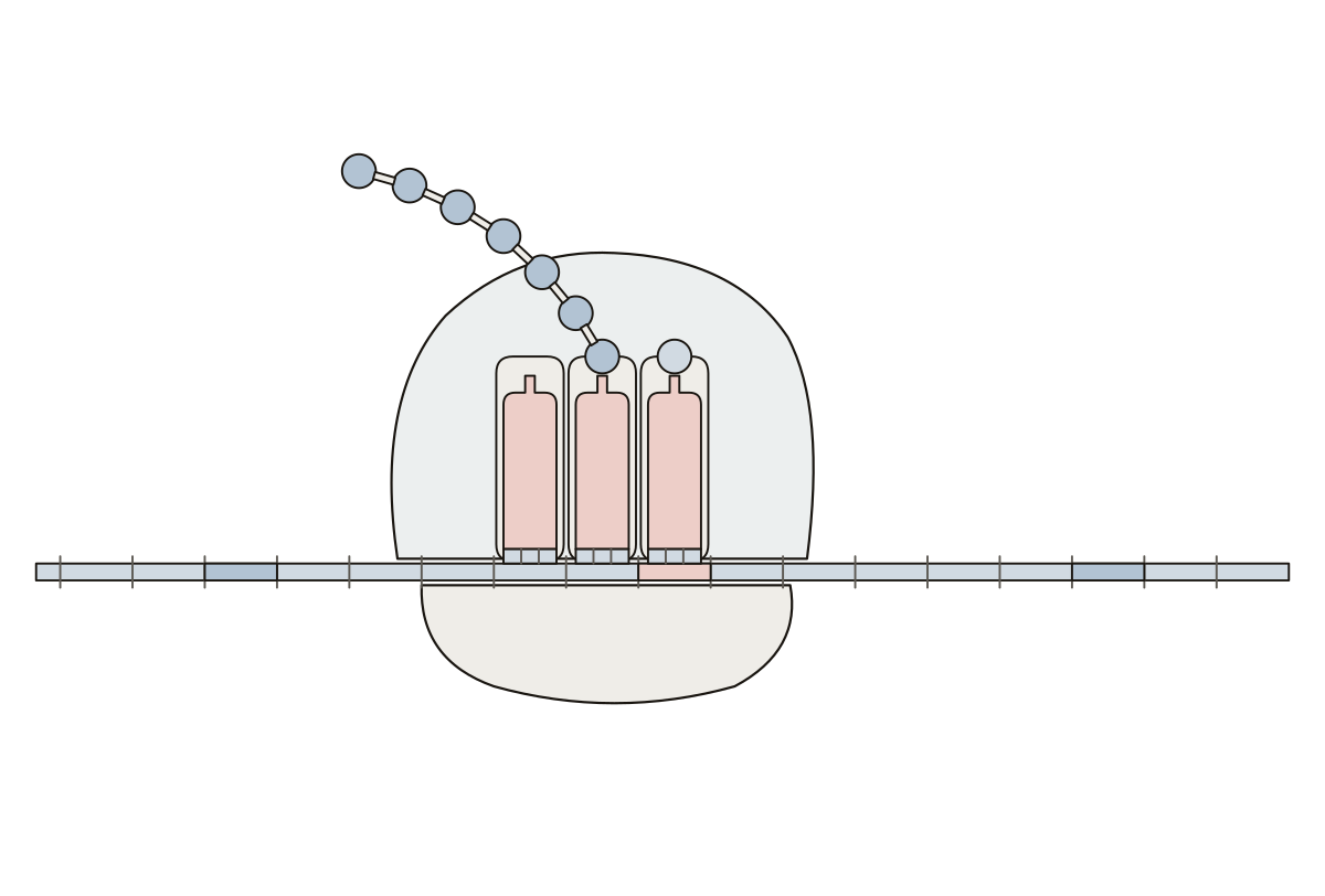 Translation Diagram LabeledTranslation at the ribosome: 14 numbered parts, from the mRNA, codon and anticodon to the A, P and E sites and the growing polypeptide chain.
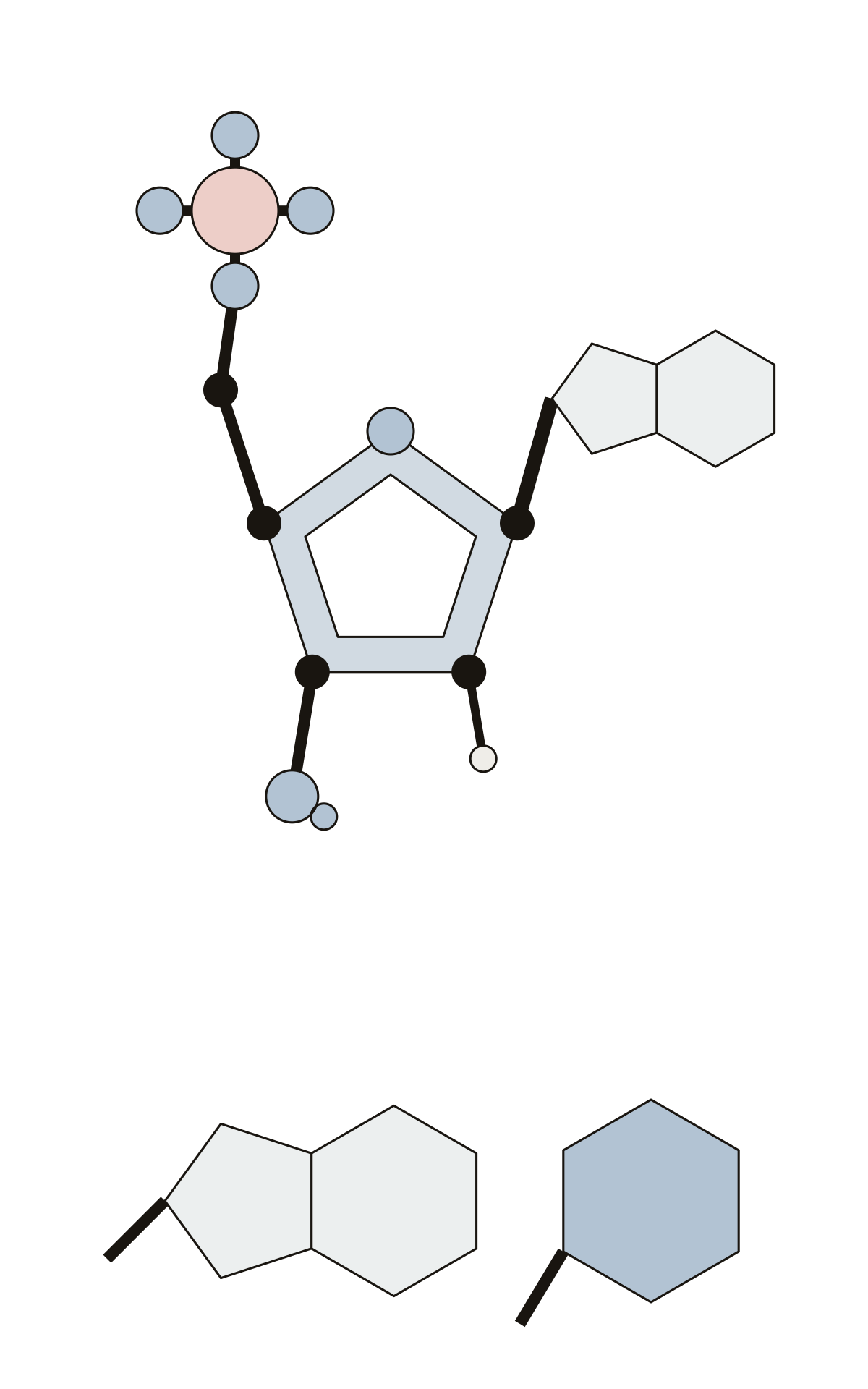 Parts of a NucleotideOne DNA nucleotide: 12 numbered parts, from the phosphate group, sugar and base to the 1′ to 5′ carbons, the glycosidic bond, purines and pyrimidines. 4′ carbon · 5′ carbon · Pentose sugar
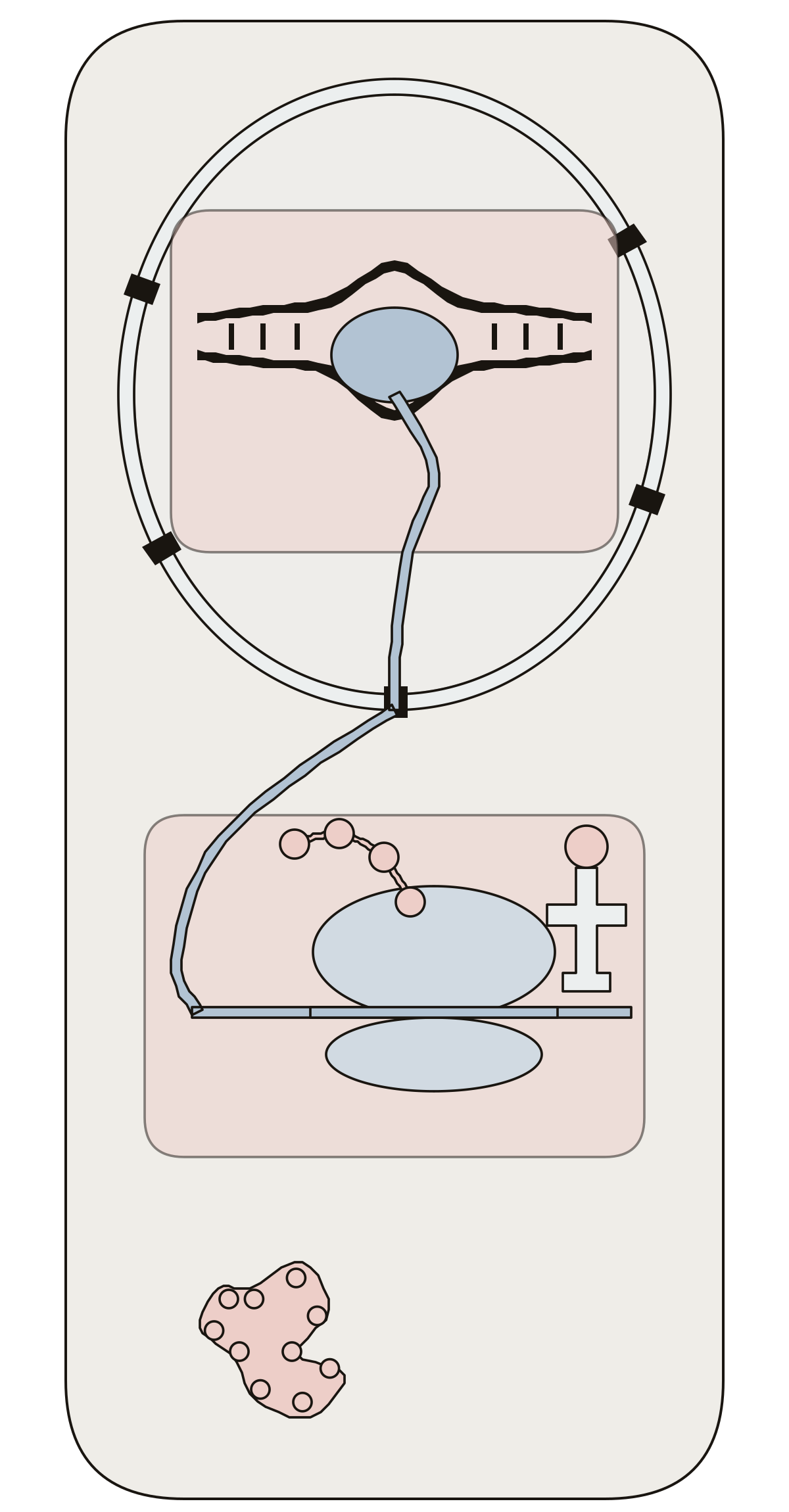 Protein Synthesis Diagram LabeledOne cell from DNA to protein: 13 numbered parts, from DNA, RNA polymerase and mRNA to the nuclear pore, ribosome, tRNA, amino acid and protein. Nuclear pore · Polypeptide chain · mRNA
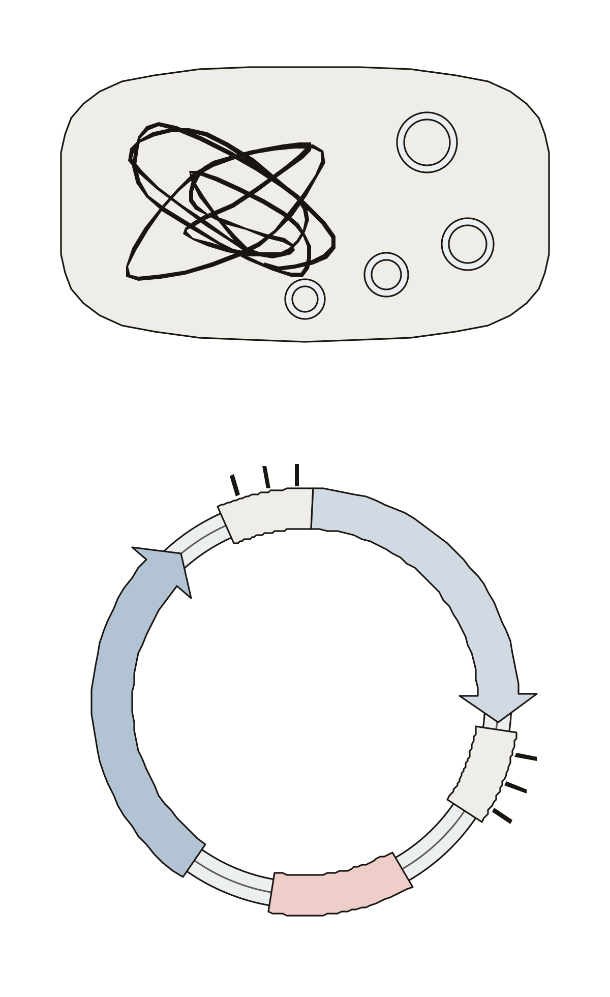 Parts of a PlasmidA plasmid in a bacterium and as a map: 7 numbered parts, from the chromosome and plasmid to the origin of replication, resistance gene and cloning site. Chromosome · Restriction sites · Multiple cloning site
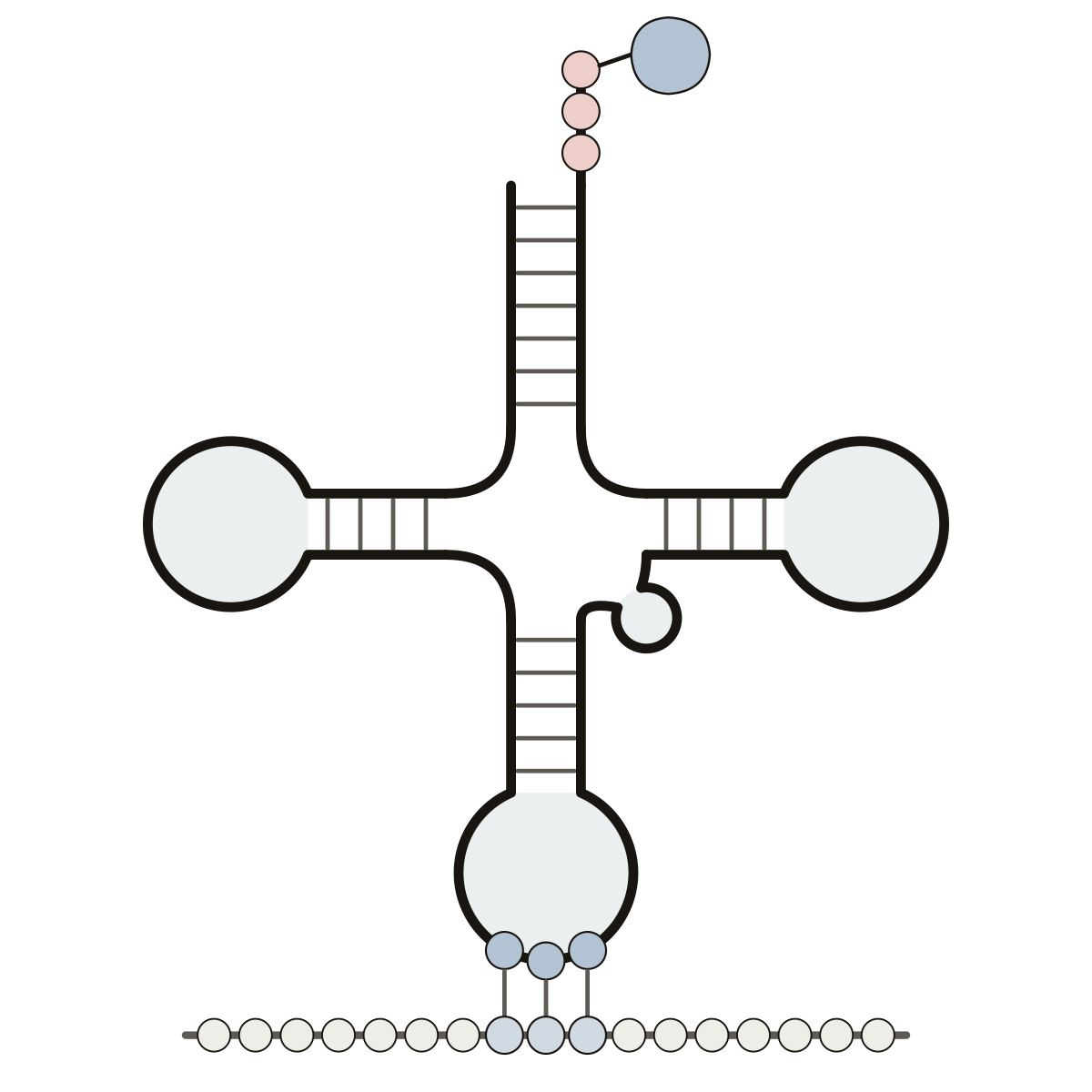 Transfer RNA Diagram (tRNA)Acceptor stem, CCA tail, amino acid, D loop, anticodon loop, anticodon, variable loop and TΨC loop, with the codon on the mRNA.
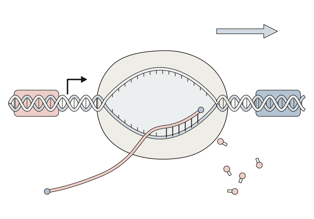 Transcription Labeled DiagramRNA polymerase copying a gene: promoter, start site, template and coding strands, transcription bubble, RNA transcript and terminator.
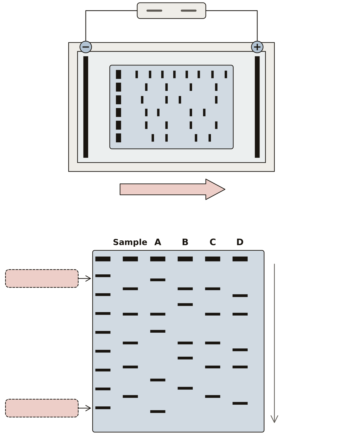 Genetic Fingerprinting WorksheetGel, wells, buffer, tank, electrodes, bands and the DNA ladder, then a finished gel to read for a match.
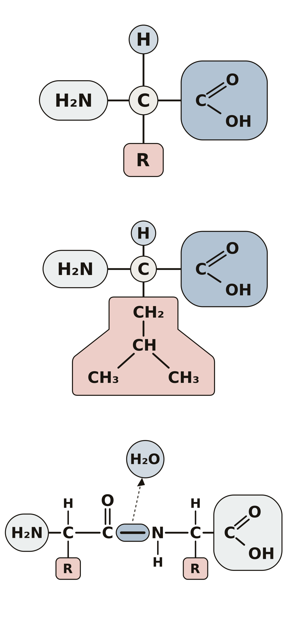 Amino Acid Structure DiagramAlpha carbon, amino group, carboxyl group, hydrogen atom and side chain, then leucine, the peptide bond and the two ends of a chain.
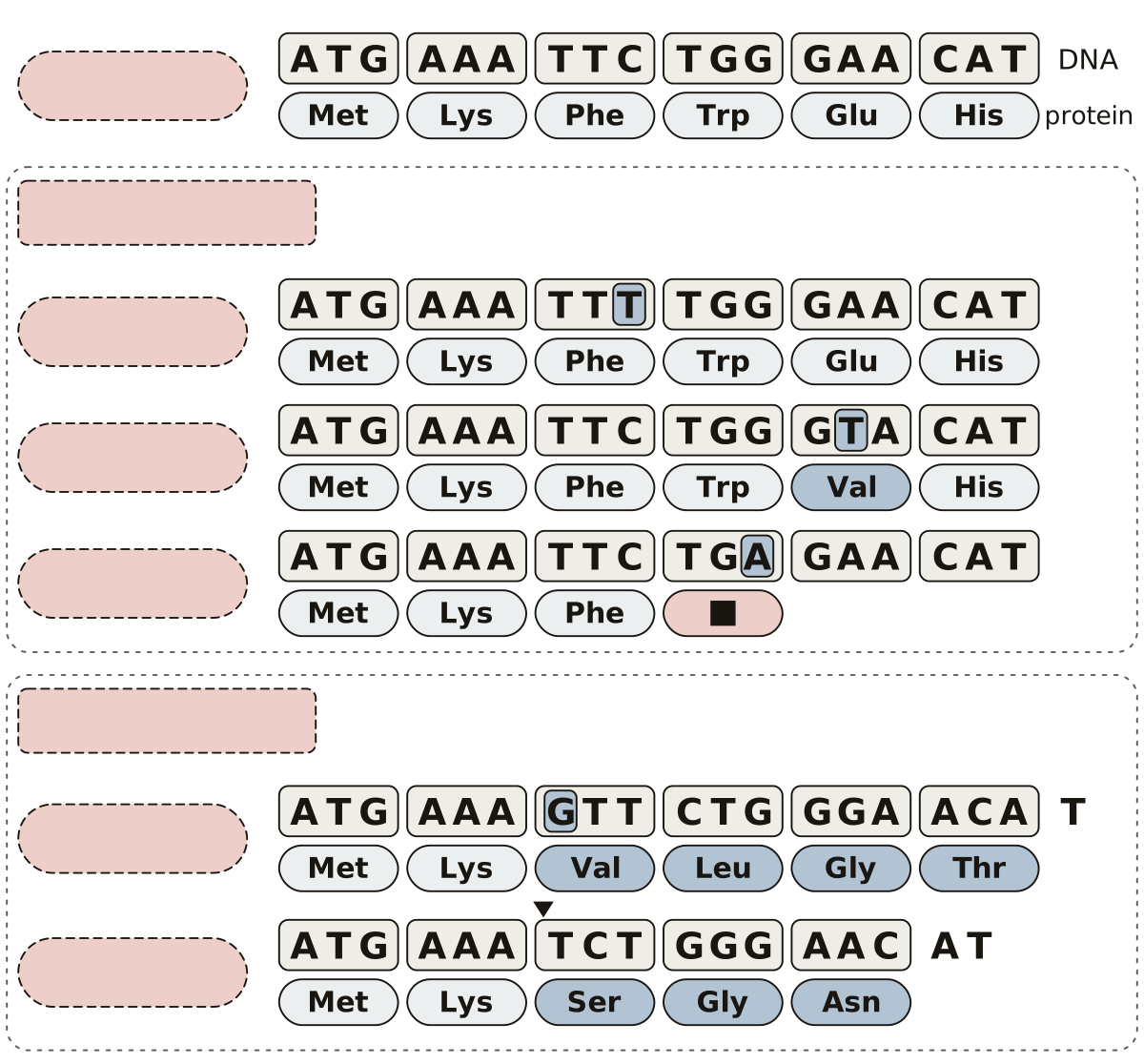 Types of Mutations DiagramSubstitution, silent, missense and nonsense mutations, insertion, deletion, frameshift and a premature stop codon, on one DNA sequence.
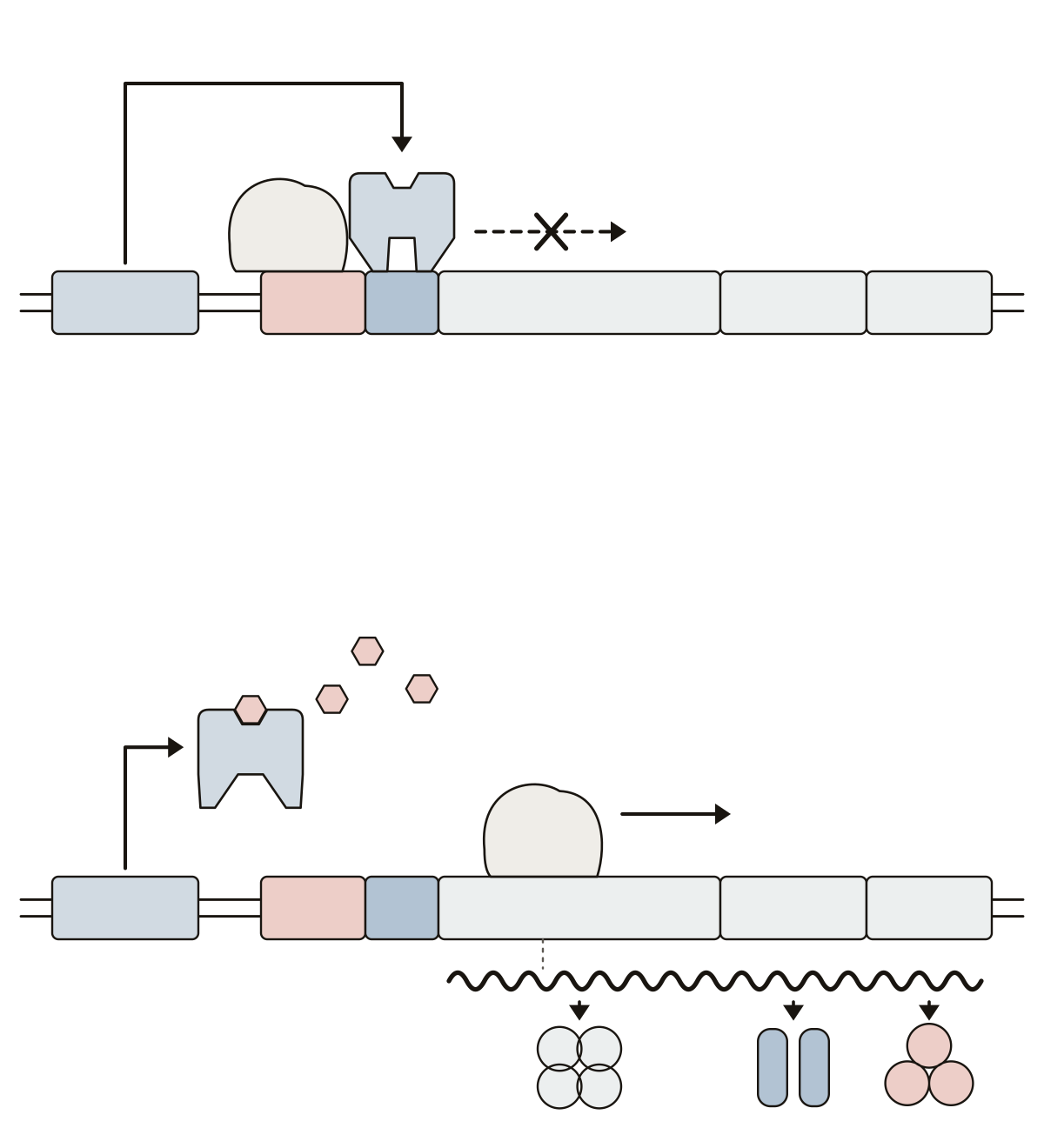 Lac Operon DiagramlacI, promoter, operator, lacZ, lacY and lacA with the repressor and RNA polymerase; then allolactose, the mRNA and the three enzymes it codes for.
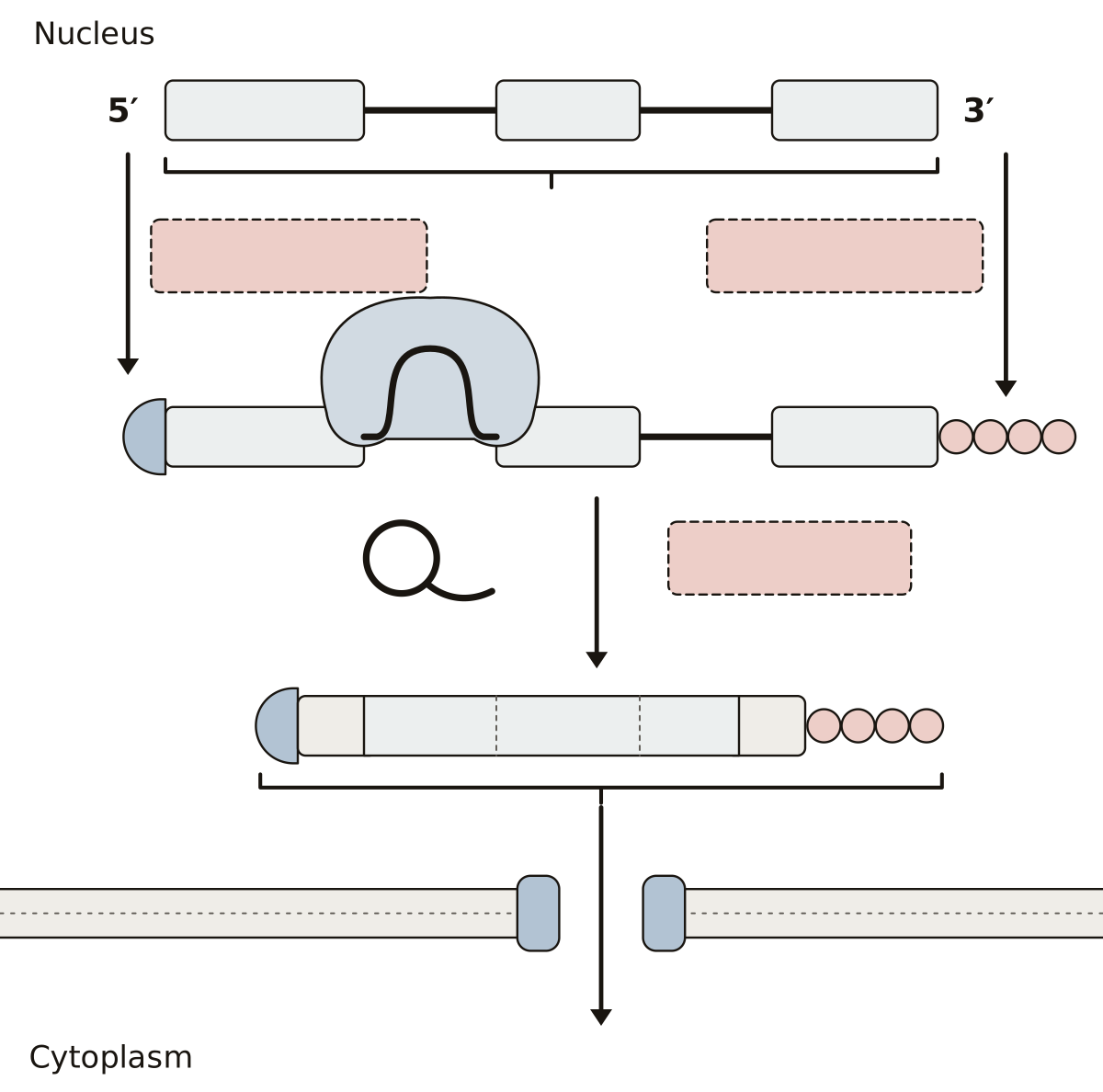 Pre-mRNA Processing DiagramPre-mRNA, exons, introns, the 5′ cap, the poly-A tail, the spliceosome and the lariat, then the mature mRNA with its untranslated regions and coding sequence.
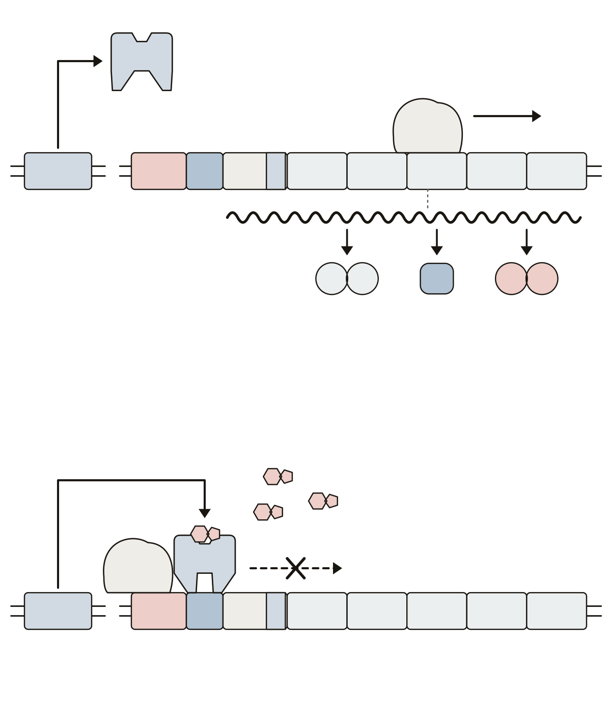 Trp Operon DiagramtrpR, promoter, operator, leader, attenuator and the genes trpE, trpD, trpC, trpB and trpA, with the repressor, tryptophan as corepressor, RNA polymerase, the mRNA and the enzymes.
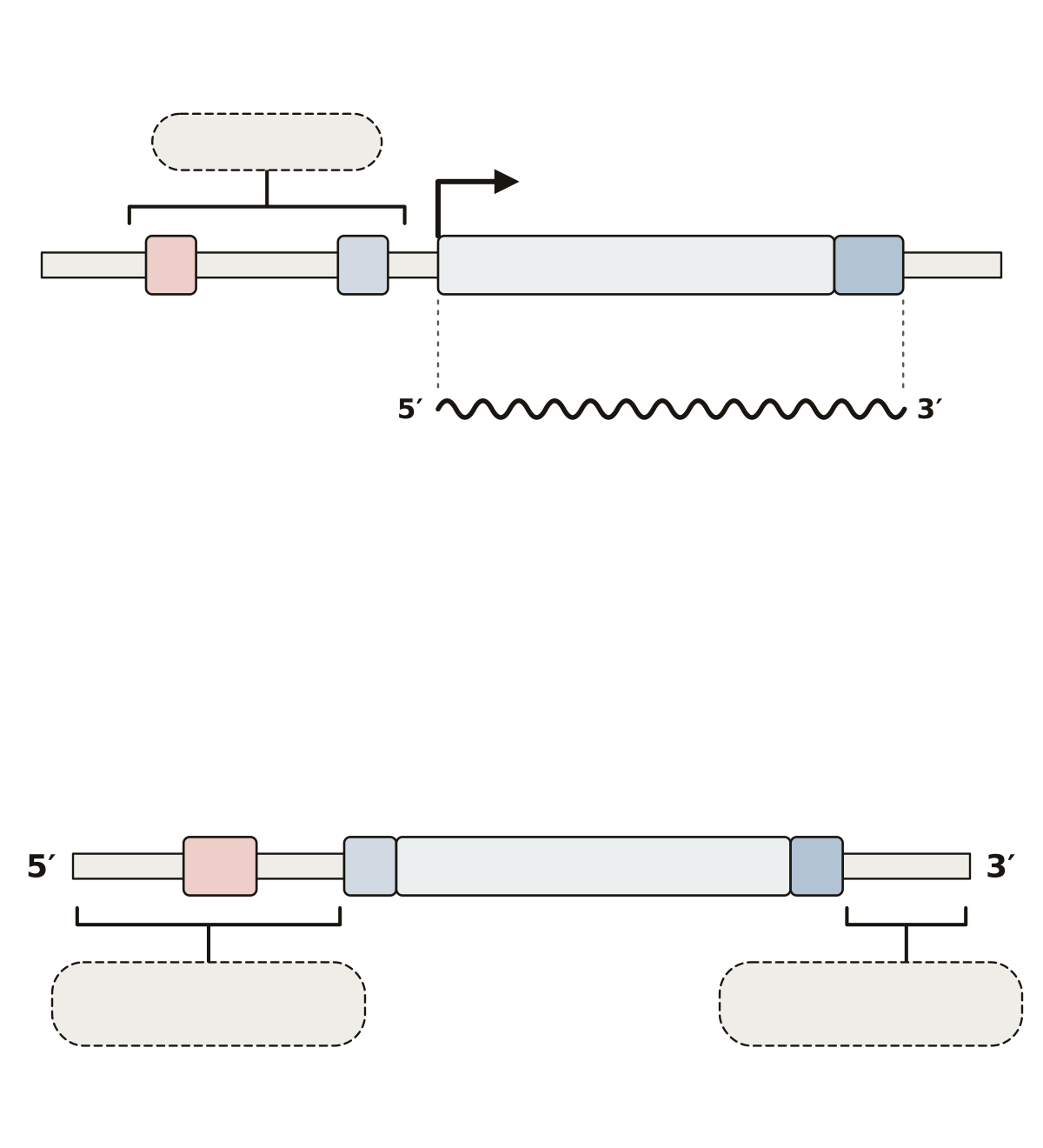 Bacterial Gene Structure DiagramPromoter, −35 and −10 sequences, transcription start site and terminator on the DNA; ribosome-binding site, start codon, coding sequence, stop codon and untranslated regions on the mRNA.
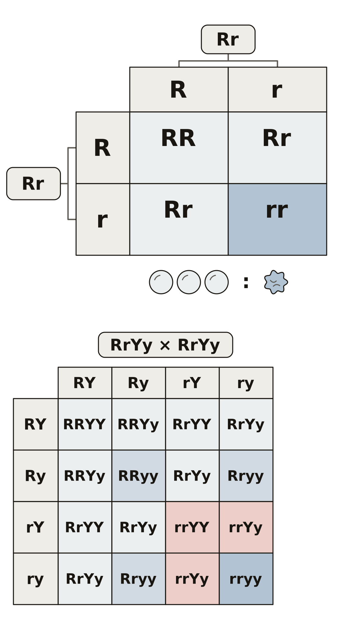 Punnett Square LabeledParent genotypes, dominant and recessive alleles, the three genotypes and two phenotypes of a four-box square, then gametes and four classes in sixteen boxes.
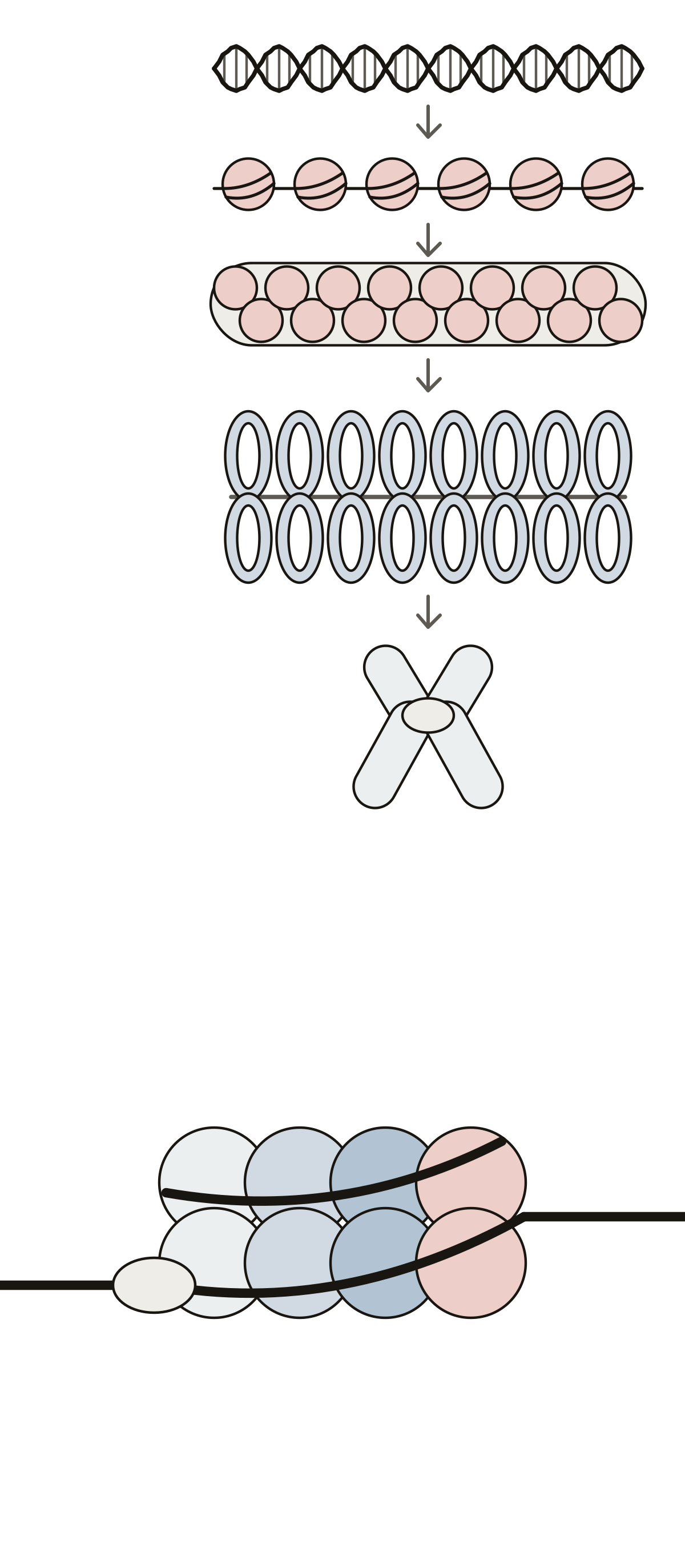 DNA Packaging DiagramDouble helix, beads on a string, the 30-nanometer fiber, chromatin loops and the metaphase chromosome, then the histone core, linker DNA and histone H1.
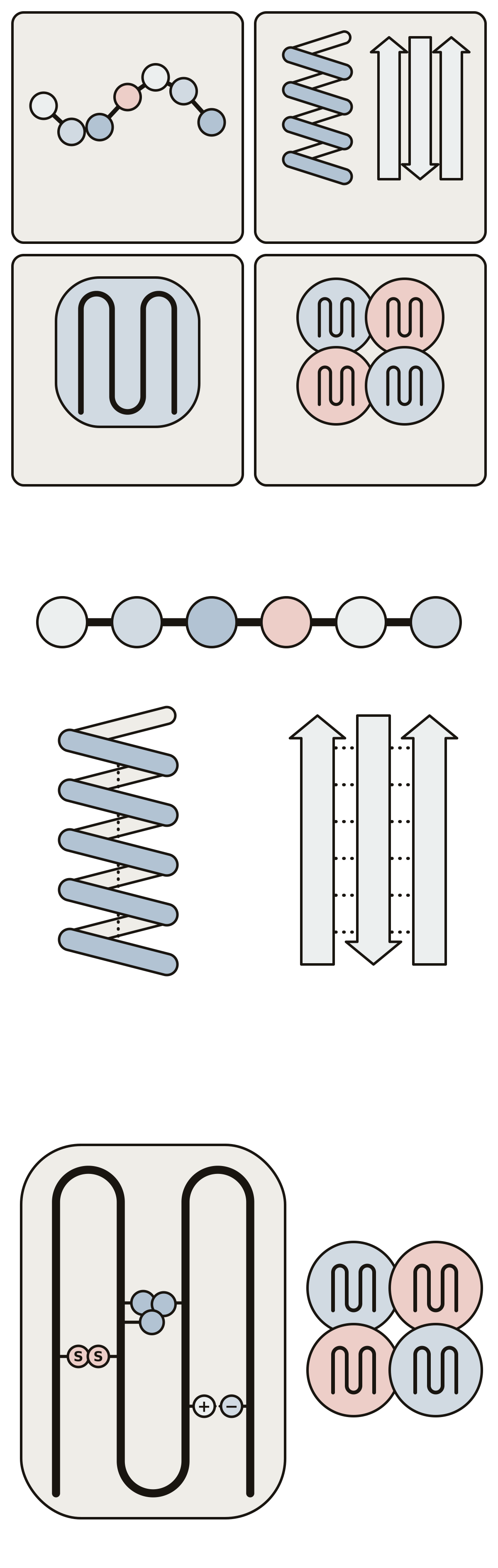 Protein Structure DiagramPrimary, secondary, tertiary and quaternary structure, then the peptide bond, alpha helix and beta sheet, and the bonds that hold a folded chain.
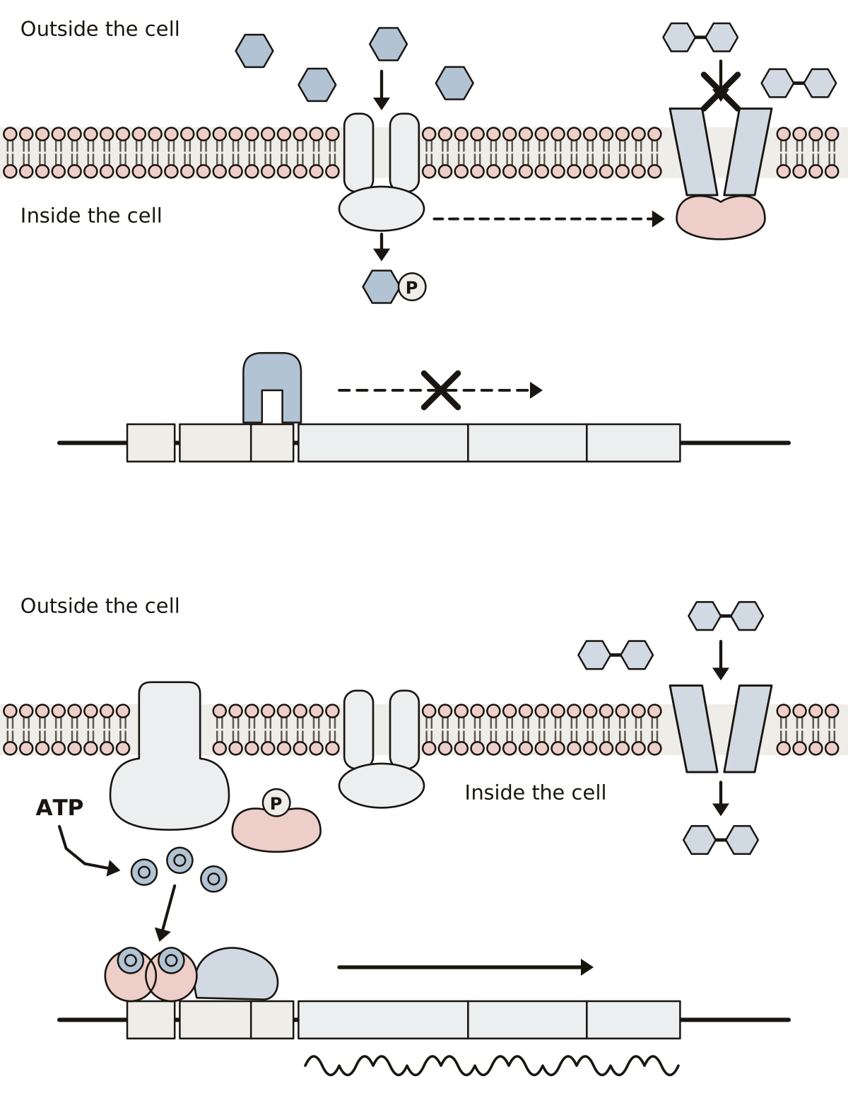 Inducer Exclusion DiagramGlucose, its PTS transporter, EIIA on lactose permease and the lactose left outside; then EIIA with its phosphate, adenylate cyclase, cyclic AMP and CAP on the DNA.
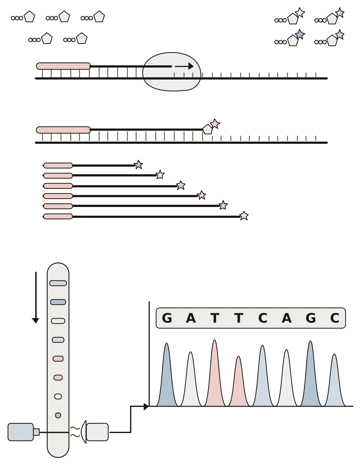 Sanger Sequencing DiagramTemplate strand, primer, DNA polymerase, normal and dideoxy nucleotides and the fragments they give; then the capillary, laser, detector, a chromatogram peak and the sequence.
- Lac Operon Quiz DiagramA quiz from the first question: find lacI, the promoter, the operator, the three genes, the repressor and RNA polymerase, then allolactose, the mRNA and the three proteins.
How a cell divides 14
The cell cycle as a wheel, the stages of mitosis and meiosis in order, and the chromosome they move. Then stem cells, and a bacterium dividing in two.
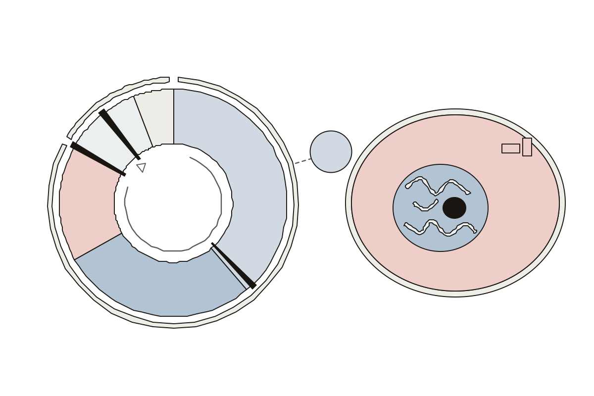 Interphase Cell Diagram and the Cell CycleThe cell cycle as one wheel, then a cell in interphase: 15 parts to name. Cell membrane · Cytokinesis · Cytoplasm
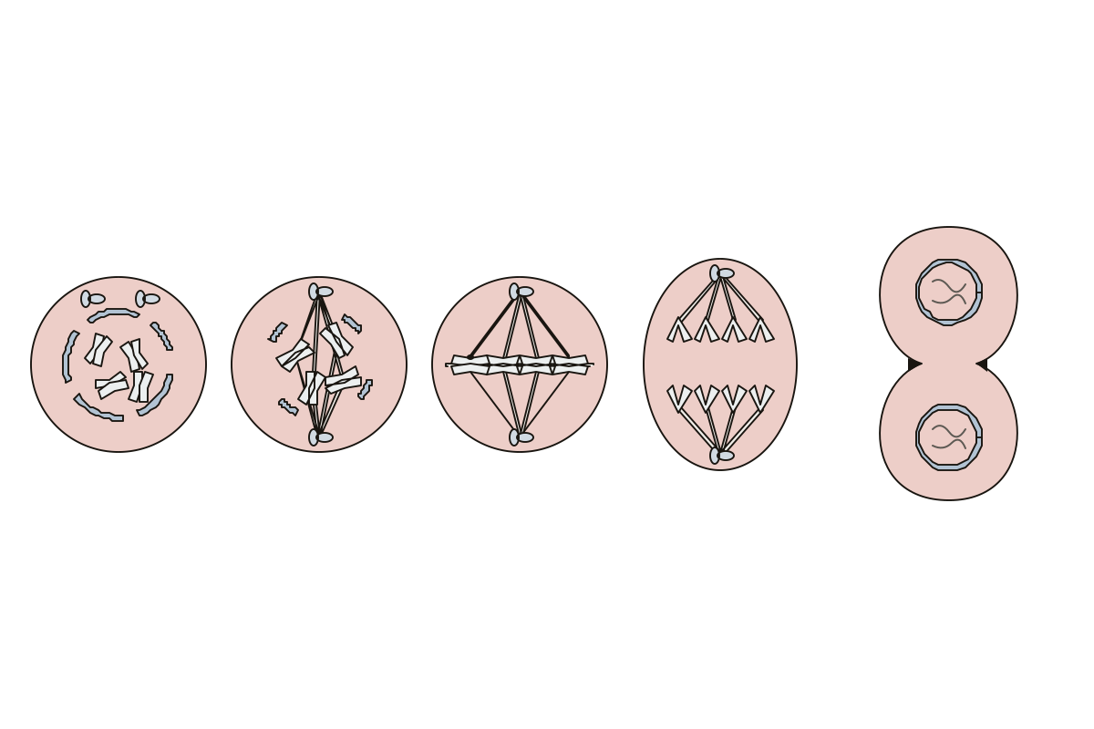 Mitosis Labeled DiagramFive cells, one per stage of mitosis: 13 parts to name, labeled or blank. Telophase · Nuclear envelope · Prophase
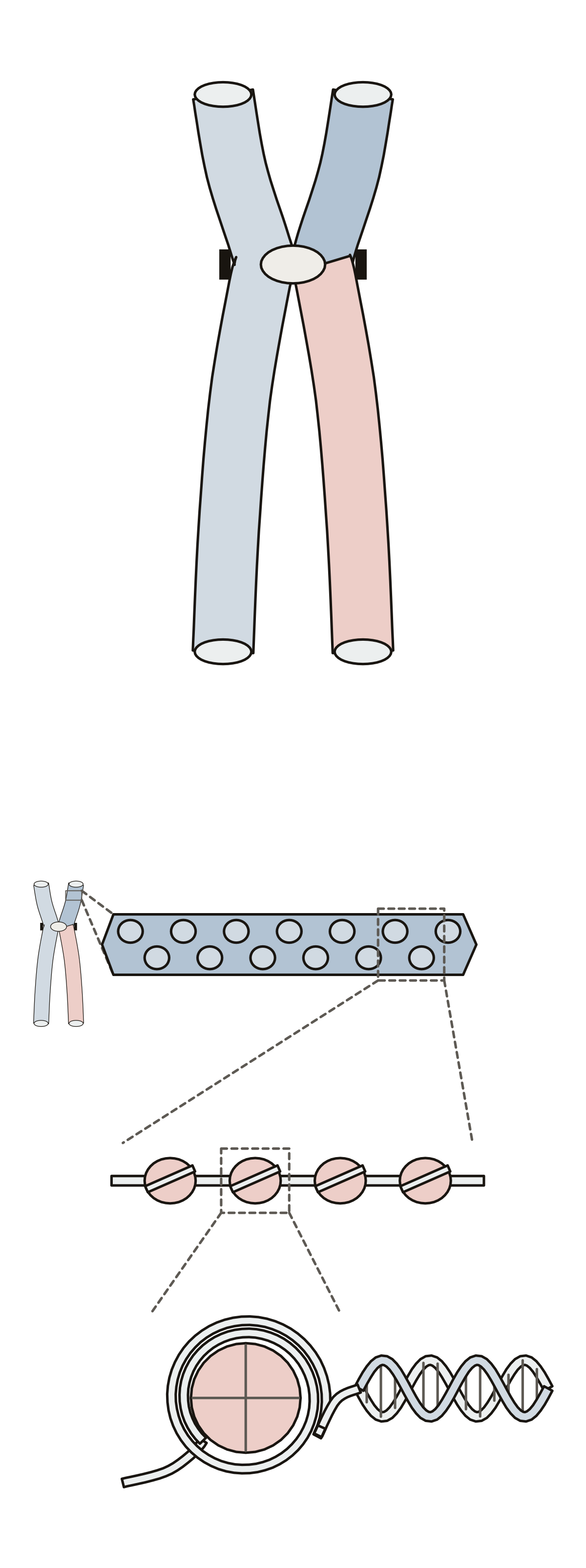 Chromosome Diagram LabeledOne chromosome and its packed DNA: 11 parts to name, labeled or blank. Telomere · Sister chromatid · Chromatin
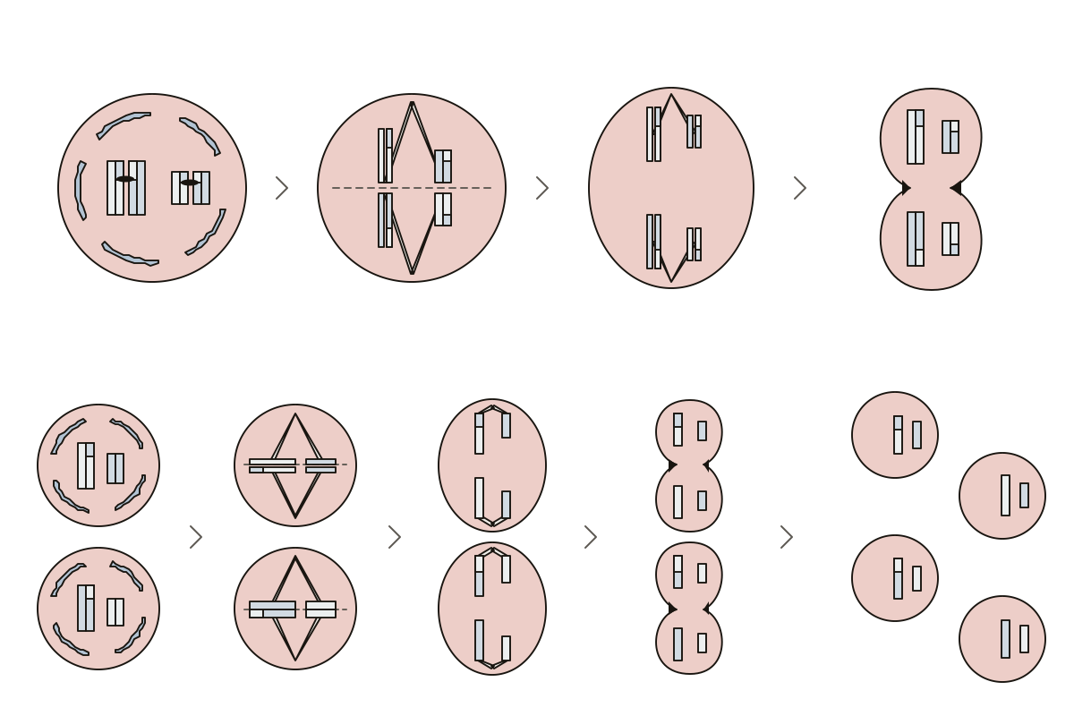 Meiosis Stages Diagram LabeledEight stages and the chromosomes that make them: 14 parts to name, labeled or blank. Homologous chromosomes · Telophase II · Chiasma
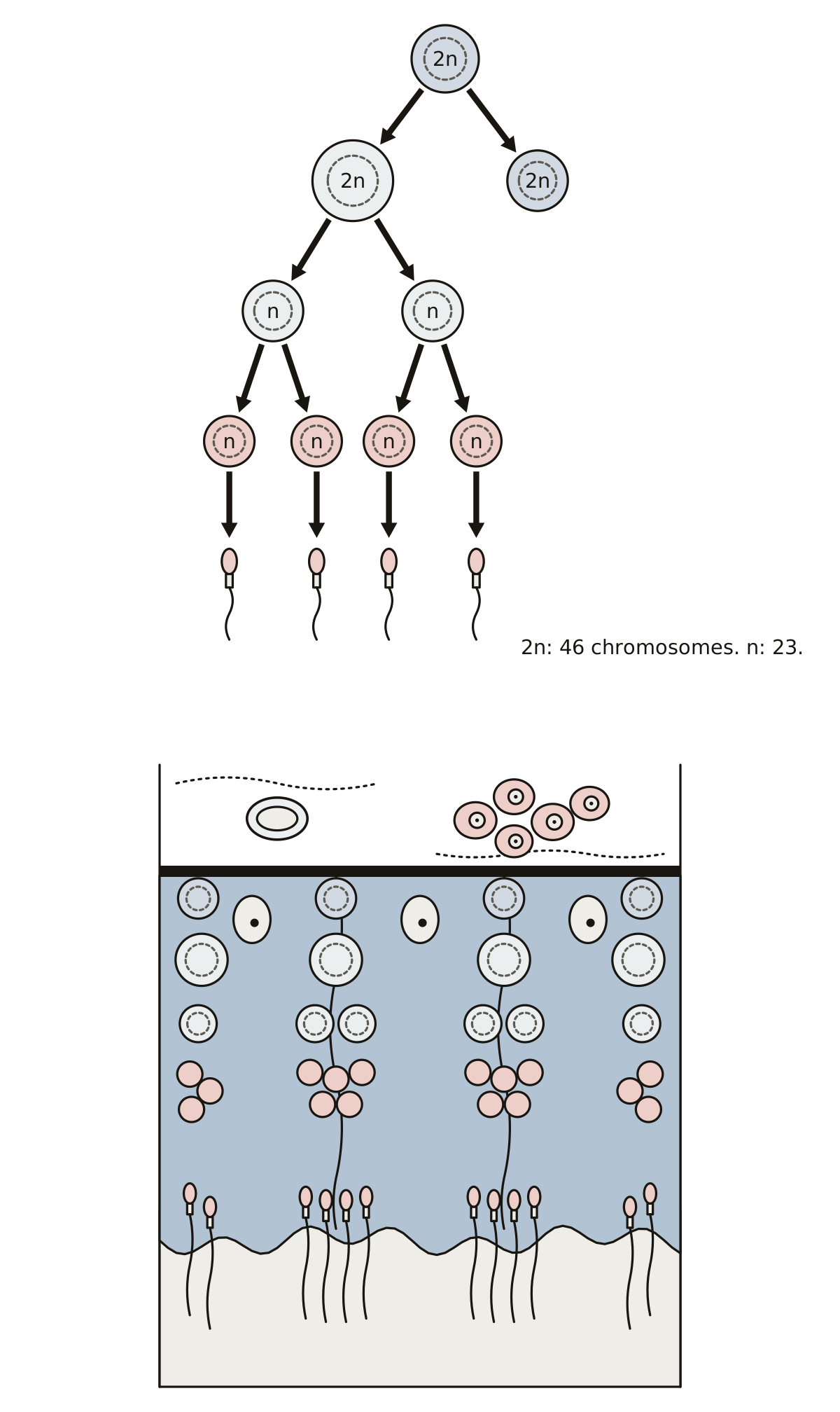 Spermatogenesis Diagram LabeledFrom one spermatogonium to four sperm: the cells, the divisions between them, and the wall of the seminiferous tubule.
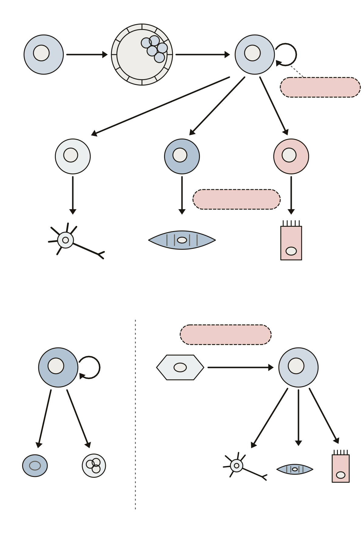 Stem Cell DiagramTotipotent, pluripotent and multipotent cells, the blastocyst and inner cell mass, the three germ layers, self-renewal, differentiation and induced pluripotent stem cells.
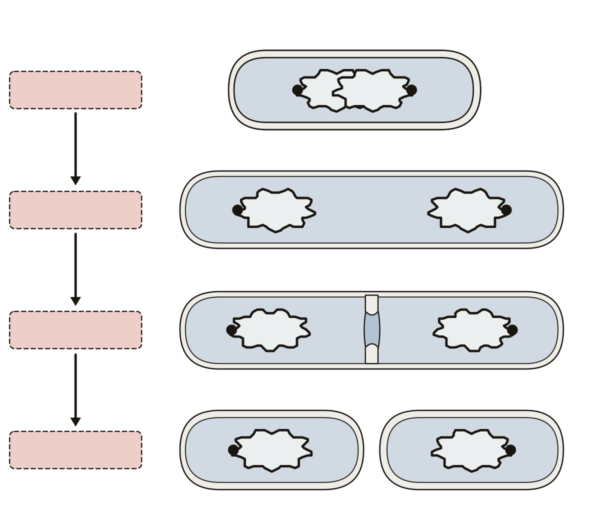 Bacterial Cell Division DiagramDNA replication, cell growth, septum formation and cell separation, with the circular chromosome, the FtsZ ring, the septum and the daughter cells.
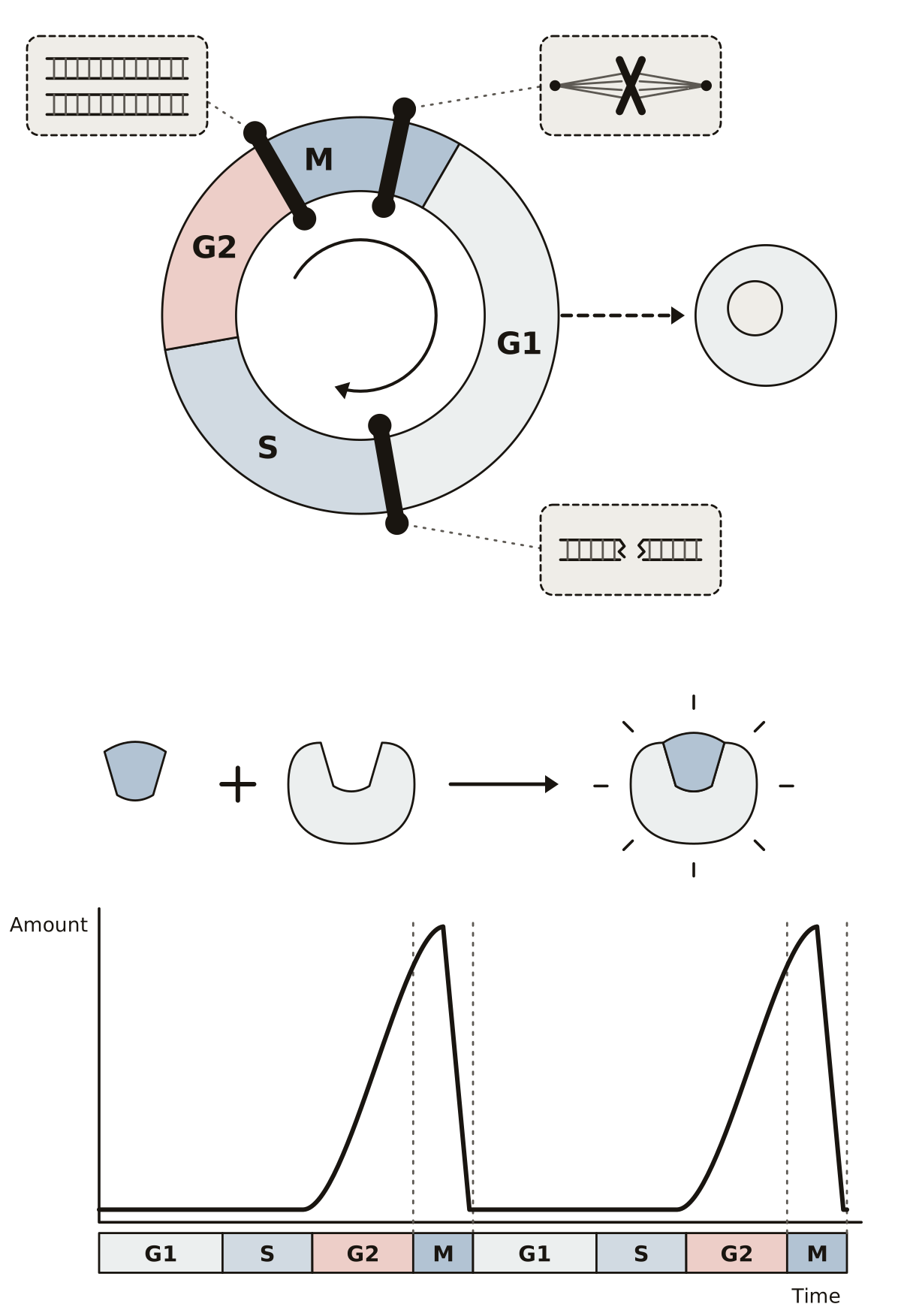 Cell Checkpoint DiagramThe G1, G2 and spindle checkpoints as gates on the wheel, what each one checks, the way out to G0, and the rise and fall of cyclin with its Cdk.
- Blank Cell Cycle Diagram With CheckpointsThe cell cycle wheel and the cyclin curve with nothing named, to print and fill in: three checkpoints, what each checks, G0, cyclin, Cdk and MPF, with an answer key.
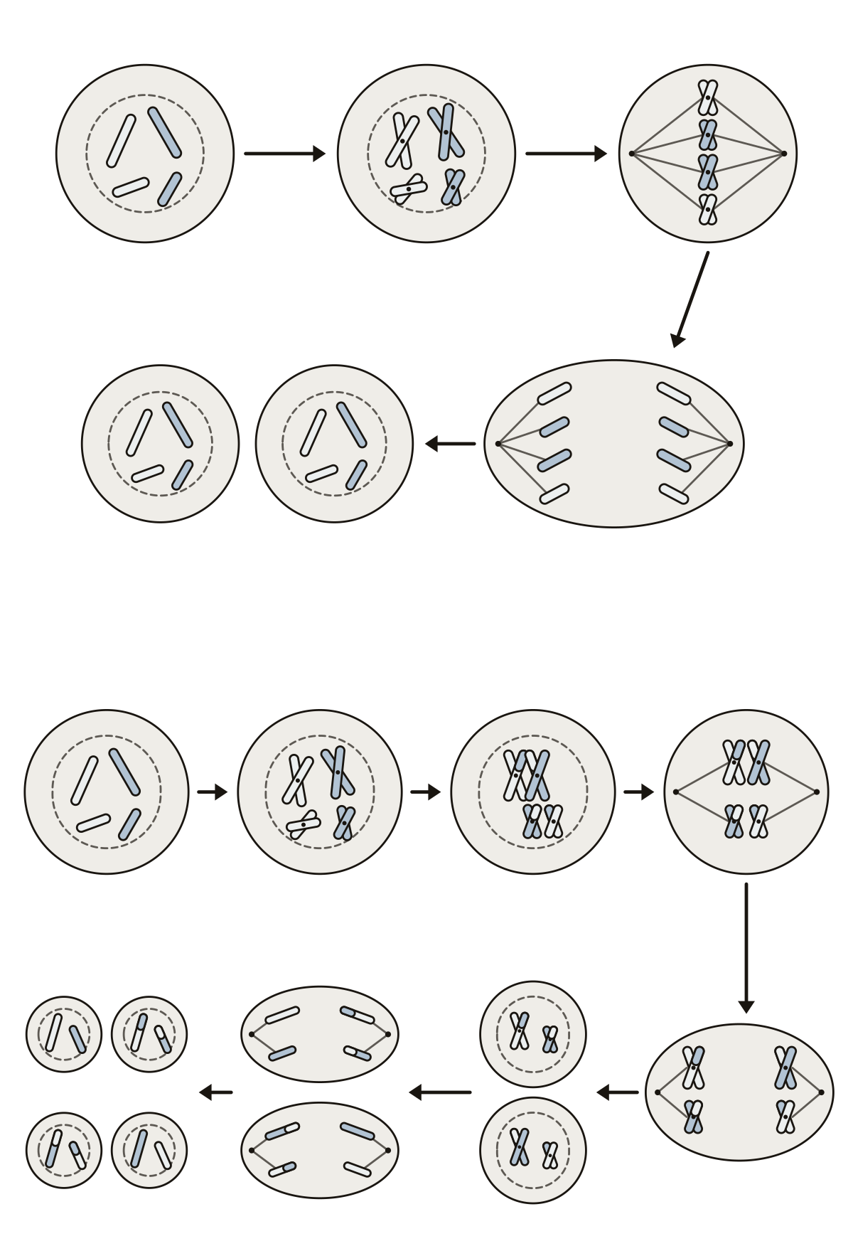 Cell Division Mitosis and Meiosis DiagramThe same parent cell taken through mitosis in five steps and meiosis in eight: DNA copied, chromosomes or pairs lined up, what separates, and the cells at the end.
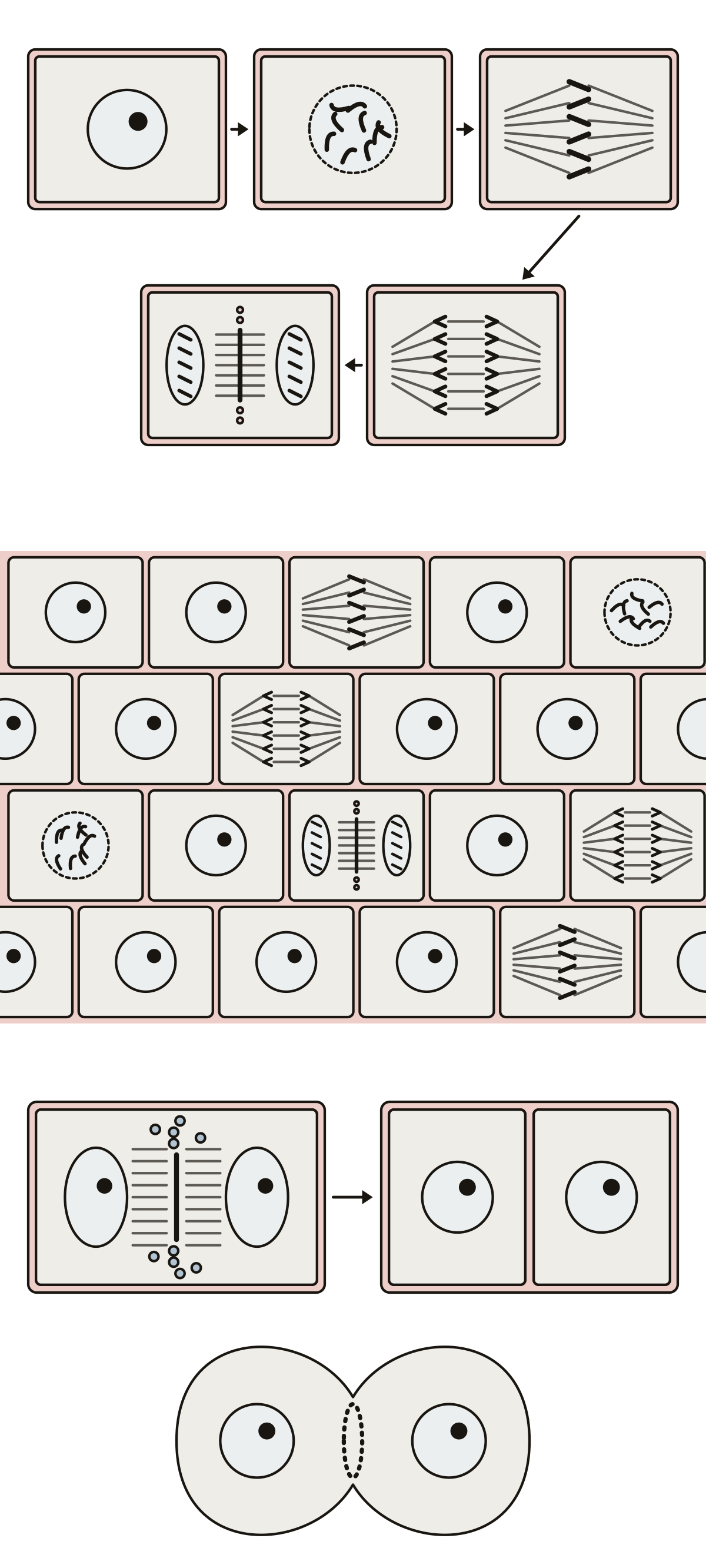 Labeled Plant Cells in MitosisInterphase, prophase, metaphase, anaphase and telophase in box-shaped cells, ten root tip cells to sort by stage, and the cell plate beside an animal cell's cleavage furrow.
- Plant Cell Mitosis Unlabeled DiagramPlant cells in mitosis with nothing written on them, to print and fill in: five stages in order, a root tip field to sort by stage, cytokinesis, and an answer key.
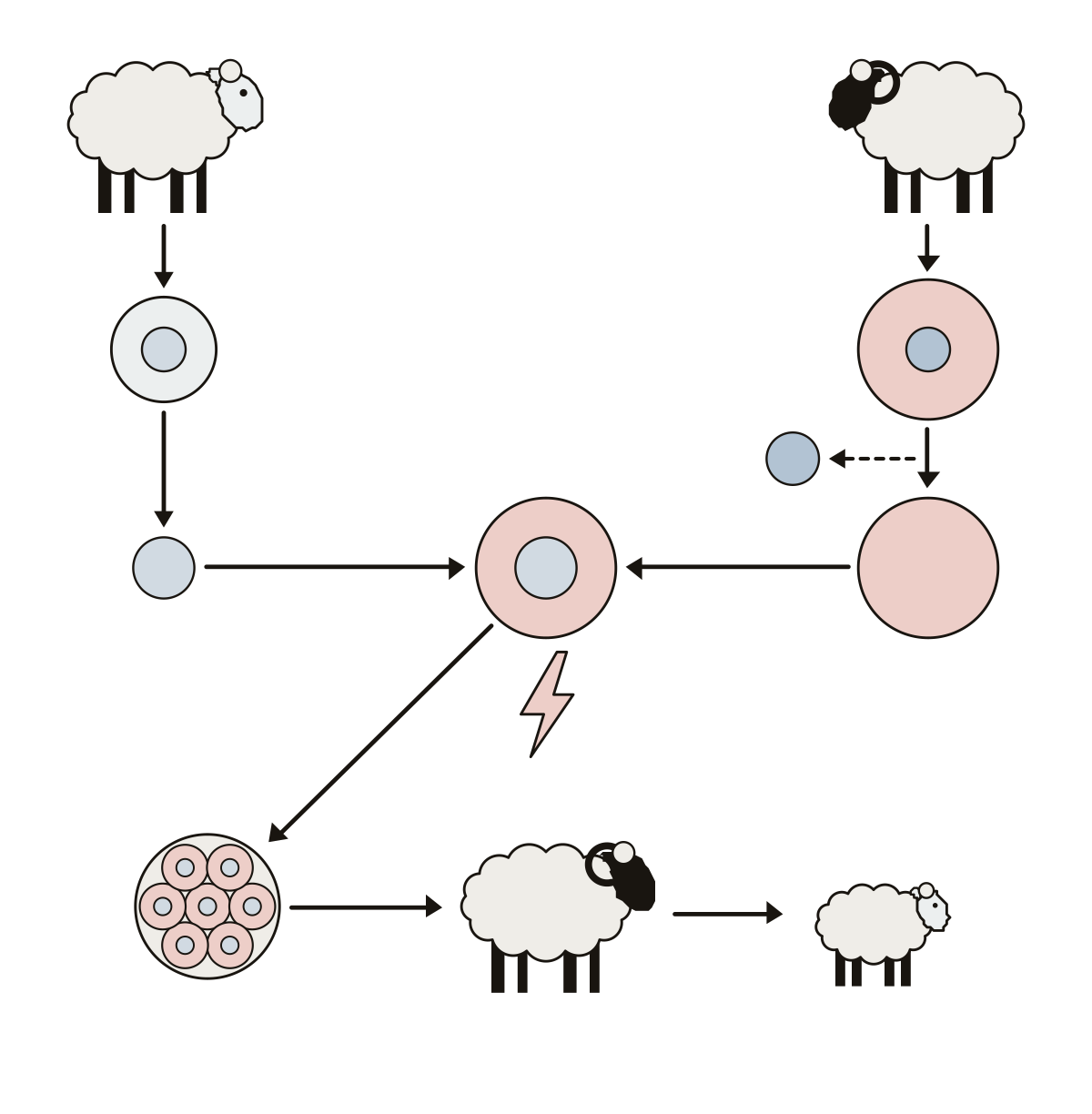 Somatic Cell Nuclear Transfer DiagramThe somatic cell and its nucleus, the egg cell and the enucleated egg, the electric current that fuses them, the early embryo, the surrogate mother and the clone.
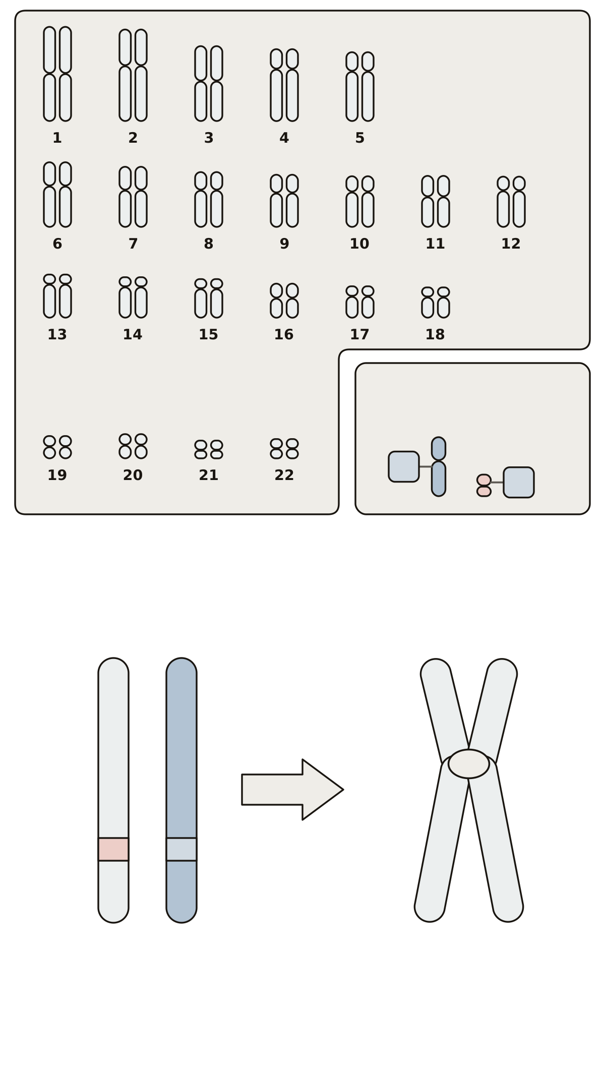 Human Chromosomes LabeledAutosomes, sex chromosomes, X and Y on a karyotype, then a homologous pair, its alleles, sister chromatids and the centromere.
Energy, cycles and enzymes 18
The pathways drawn as rings and chains, from the light-dependent reactions to the electron transport chain, the molecules that carry energy between them, eight sugars drawn as rings, and how an enzyme works.
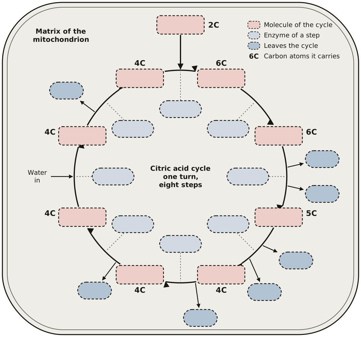 Citric Acid Cycle QuizThe Krebs cycle as a ring of 24 numbered blanks: 9 molecules, 8 enzymes and the 7 points where CO₂, NADH, FADH₂ and GTP or ATP leave. Prints blank and labeled.
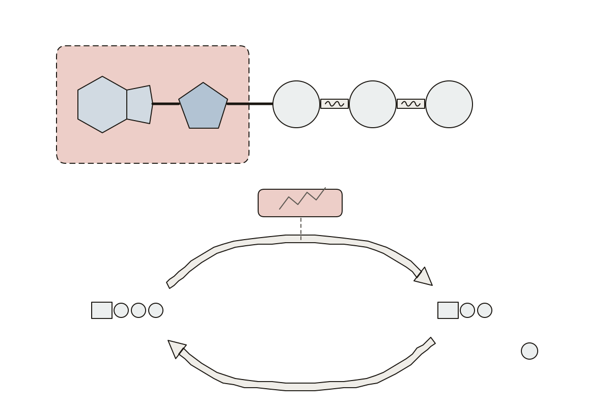 ATP Molecule Diagram LabeledThe ATP molecule, then the cycle that spends and remakes it: 13 parts to name. Regeneration · Gamma phosphate · ATP
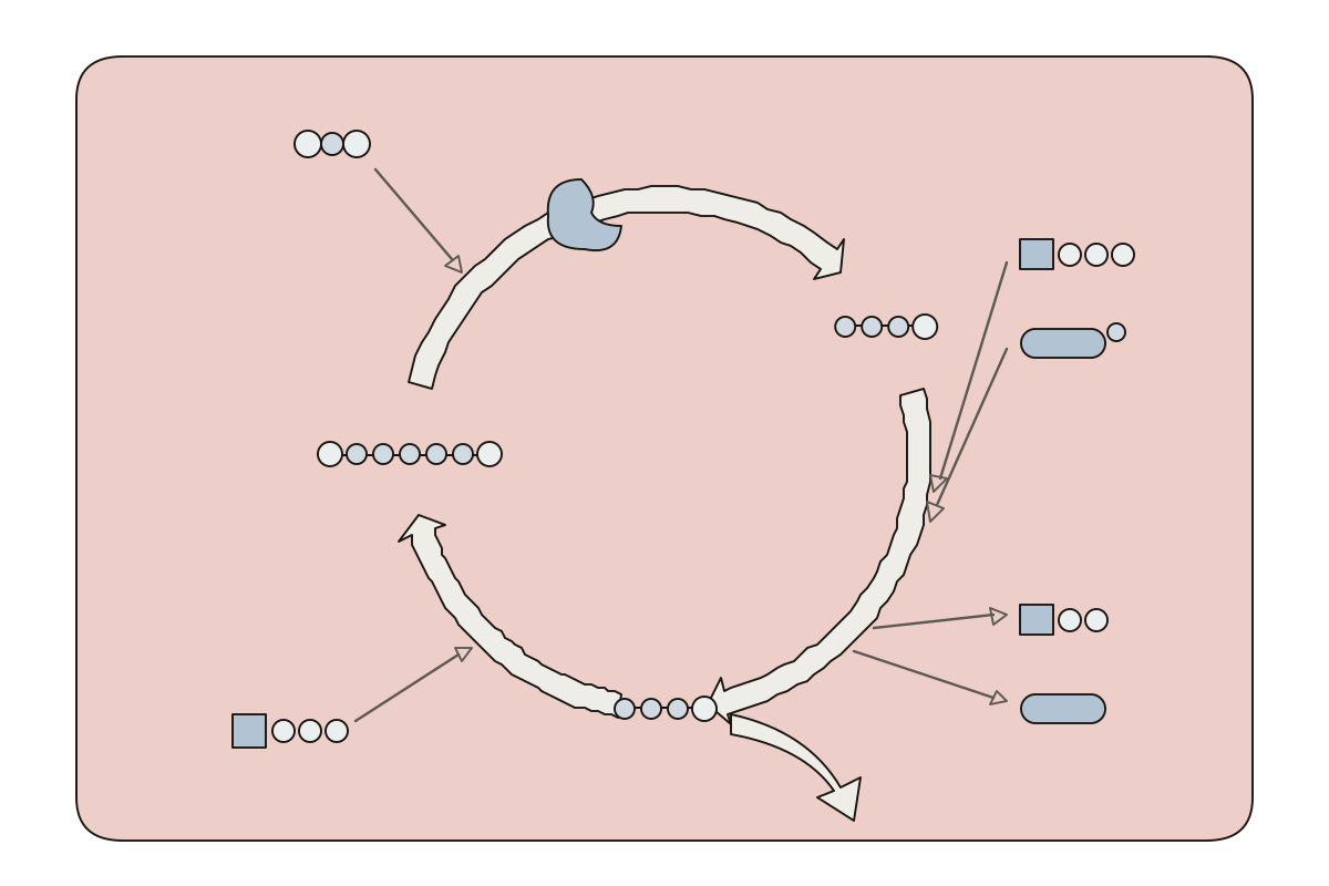 Calvin Cycle Diagram LabeledThe light-independent reactions as one circle of three stages: 14 parts to name. 3-PGA · NADP+ · Regeneration
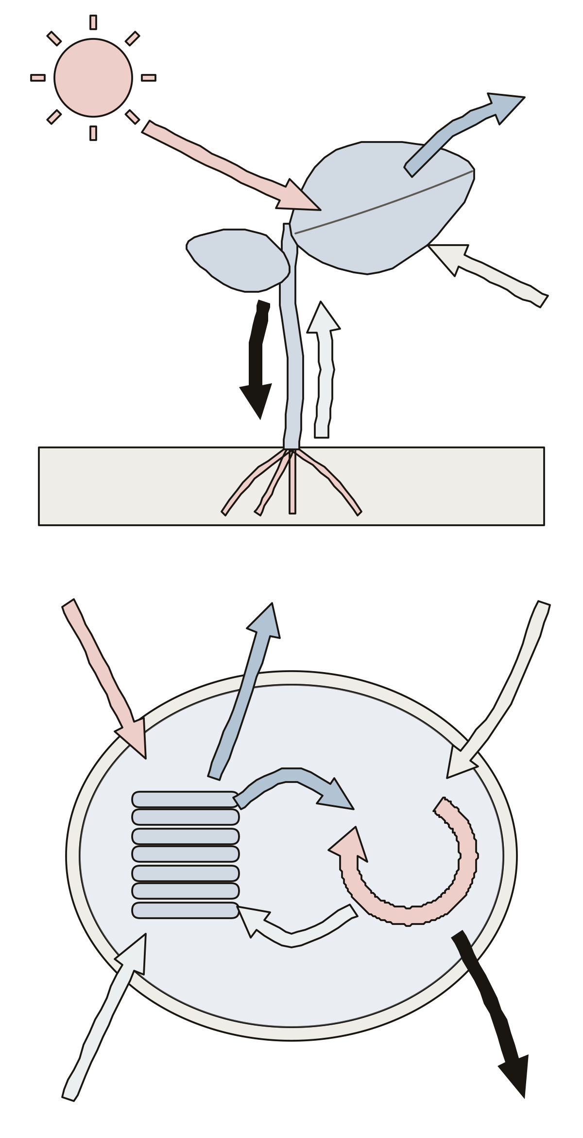 Photosynthesis Diagram LabeledA plant in sunlight and one chloroplast: 11 numbered parts, from sunlight, water, carbon dioxide, oxygen and glucose to the light reactions and Calvin cycle. Carbon dioxide · Glucose · Water
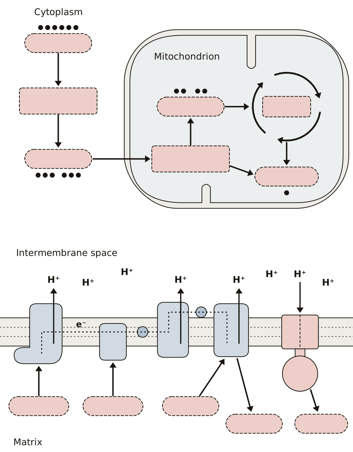 Cellular Respiration DiagramGlucose, glycolysis, pyruvate, acetyl CoA, the citric acid cycle, NADH, FADH₂, the electron transport chain, ATP synthase, oxygen, water and ATP.
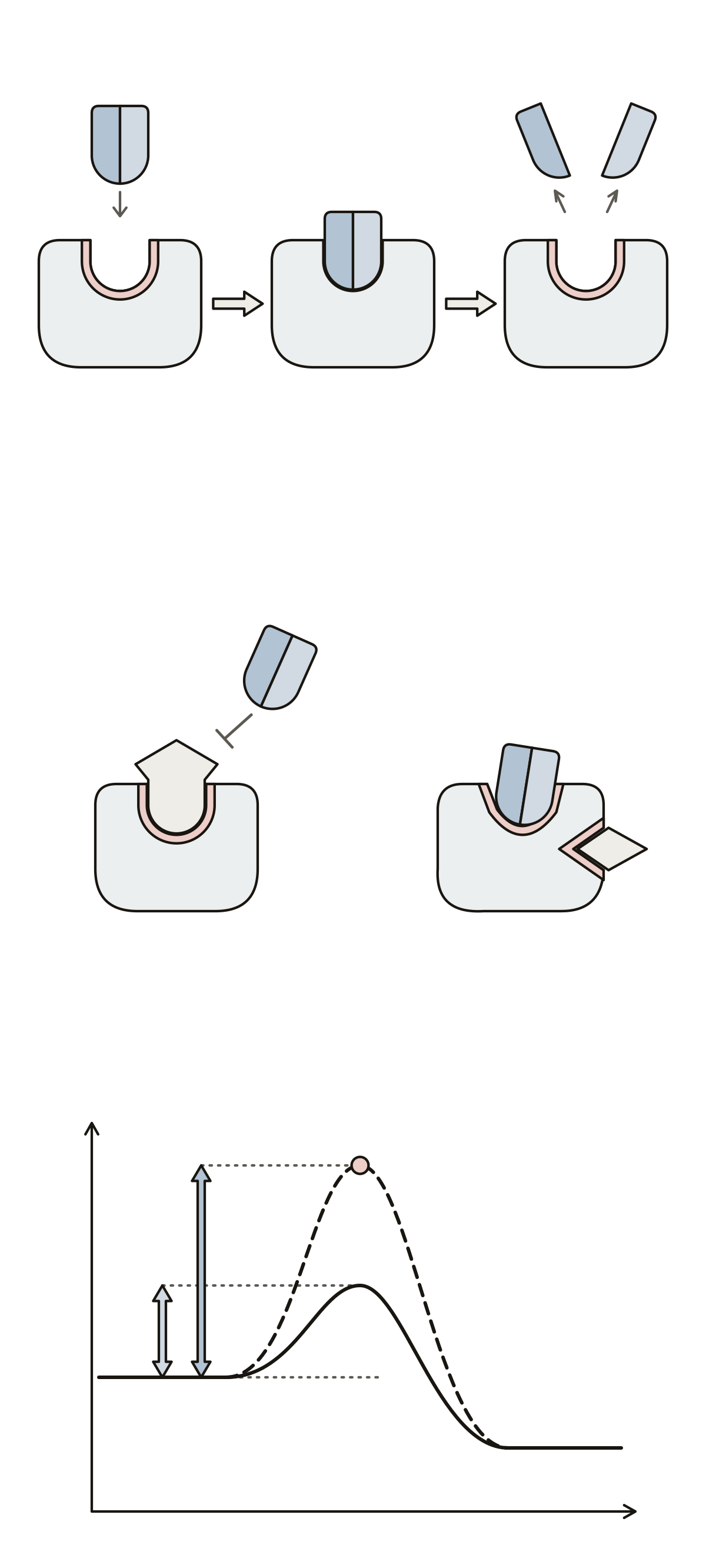 Label the EnzymeAn enzyme with its active site, substrate and products, a competitive and a noncompetitive inhibitor, and the activation energy graph.
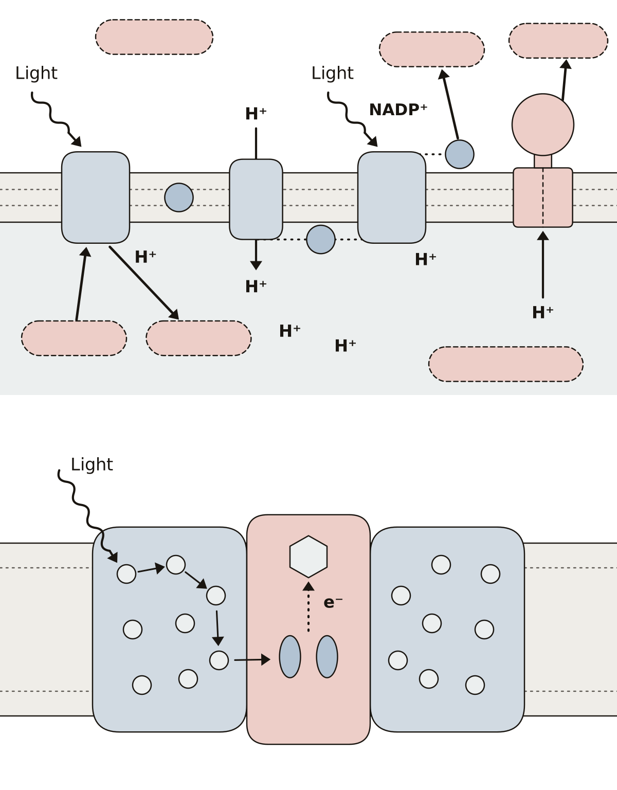 Light-Dependent Reactions DiagramPhotosystem II and I, plastoquinone, the cytochrome complex, plastocyanin, ferredoxin, ATP synthase, the lumen and stroma, and the inside of one photosystem.
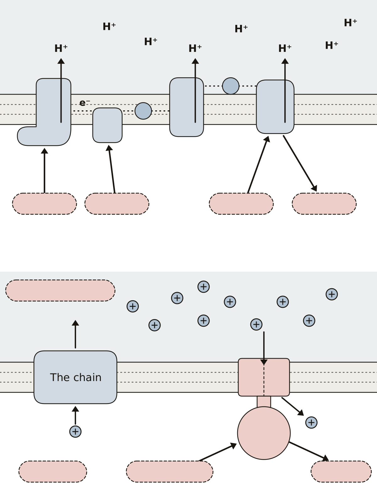 Electron Transport Chain DiagramComplexes I to IV, ubiquinone, cytochrome c, NADH, FADH₂, oxygen, water, the hydrogen ion gradient, ATP synthase and ATP.
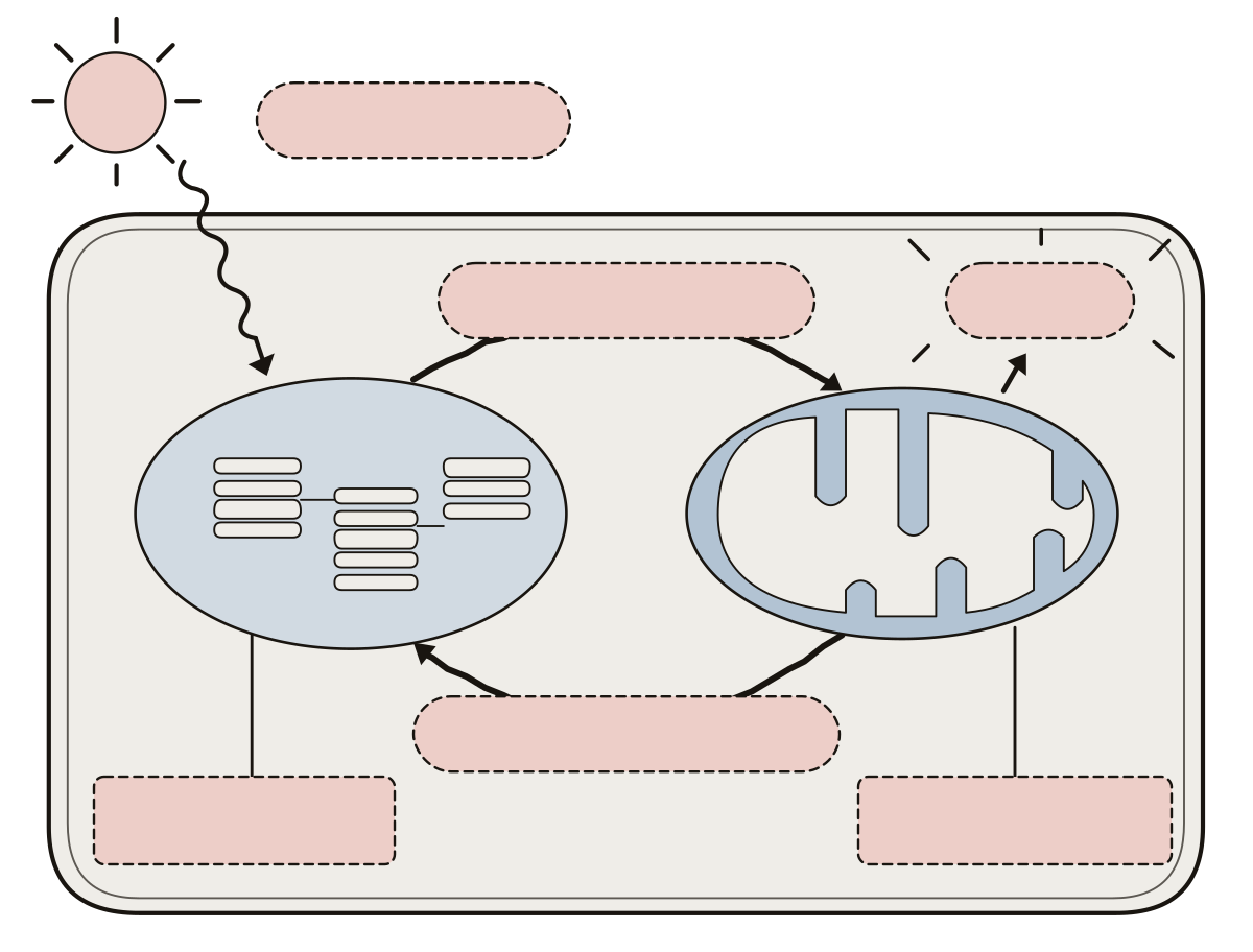 Cell Respiration and Photosynthesis DiagramChloroplast, mitochondrion, light energy, photosynthesis, cellular respiration, ATP, and the two pairs of molecules that pass between them.
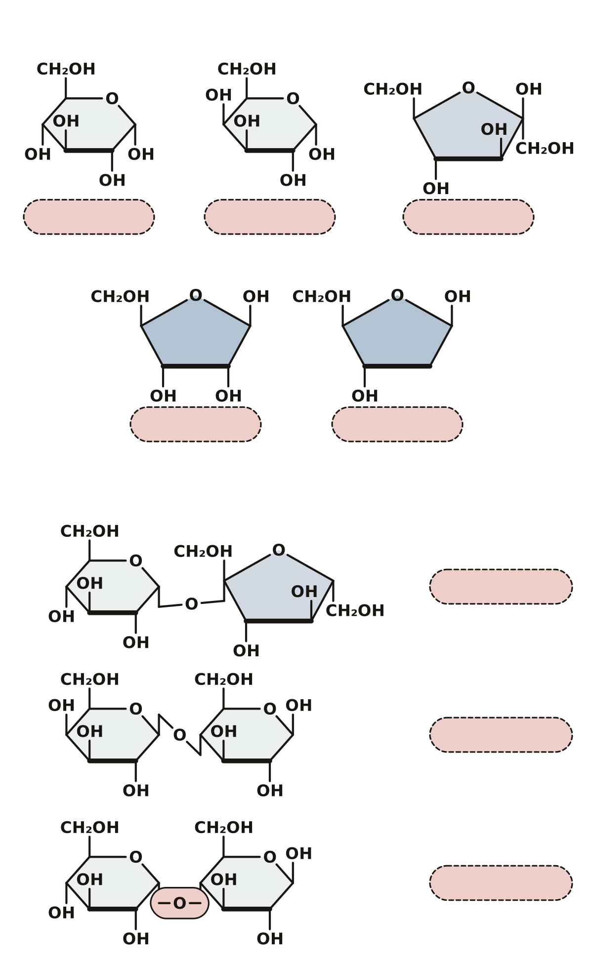 Monosaccharides and Disaccharides DiagramGlucose, galactose, fructose, ribose and deoxyribose, then sucrose, lactose and maltose and the glycosidic bond that joins two rings.
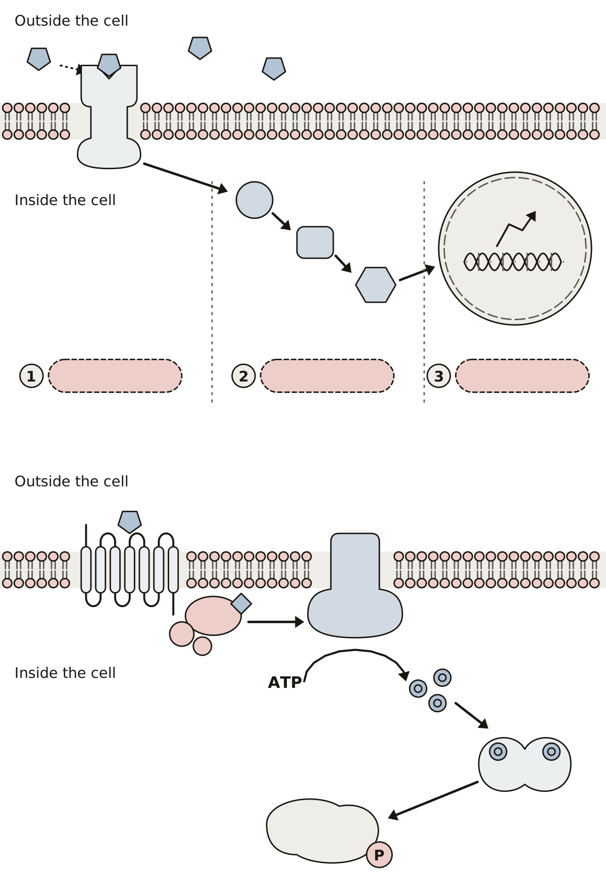 Cell Signaling Diagram LabeledSignaling molecule, receptor, cascade and response on one chart; then a G protein-coupled receptor, its G protein, adenylyl cyclase, cyclic AMP and protein kinase A.
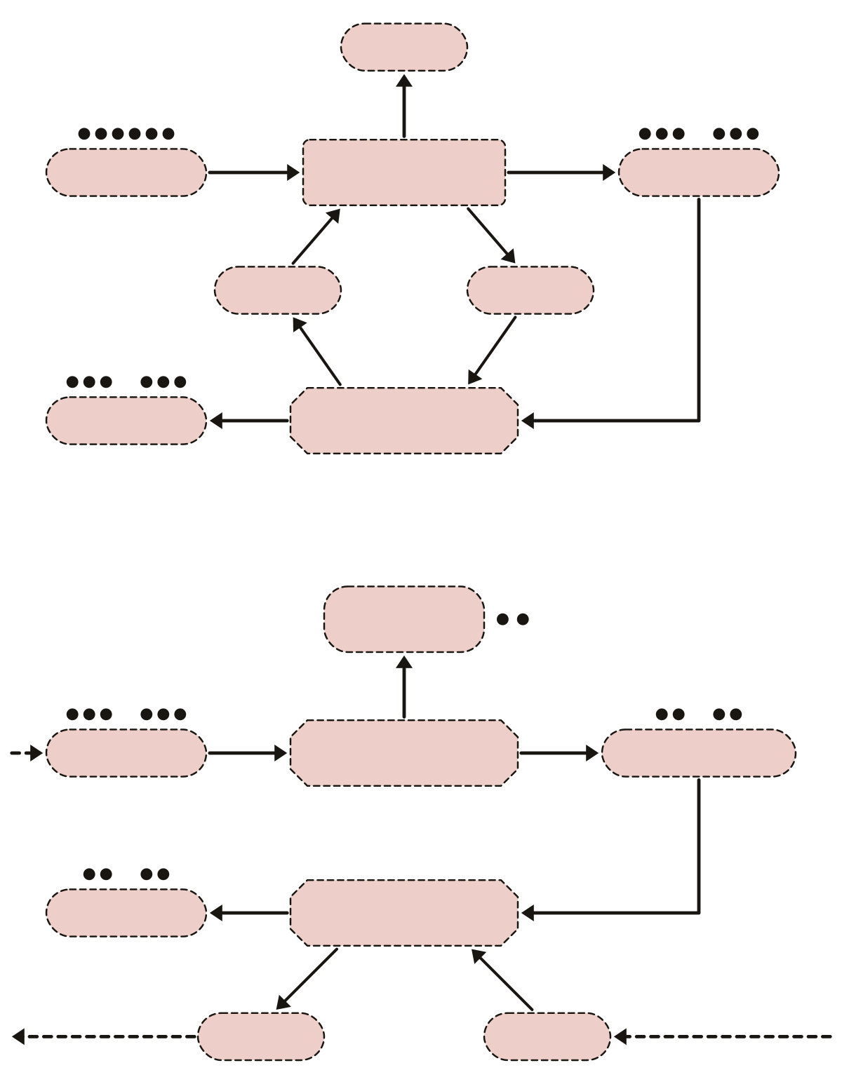 Fermentation DiagramGlucose, glycolysis, ATP, pyruvate, NADH and NAD⁺, then lactate dehydrogenase and lactate; pyruvate decarboxylase, carbon dioxide, acetaldehyde, alcohol dehydrogenase and ethanol.
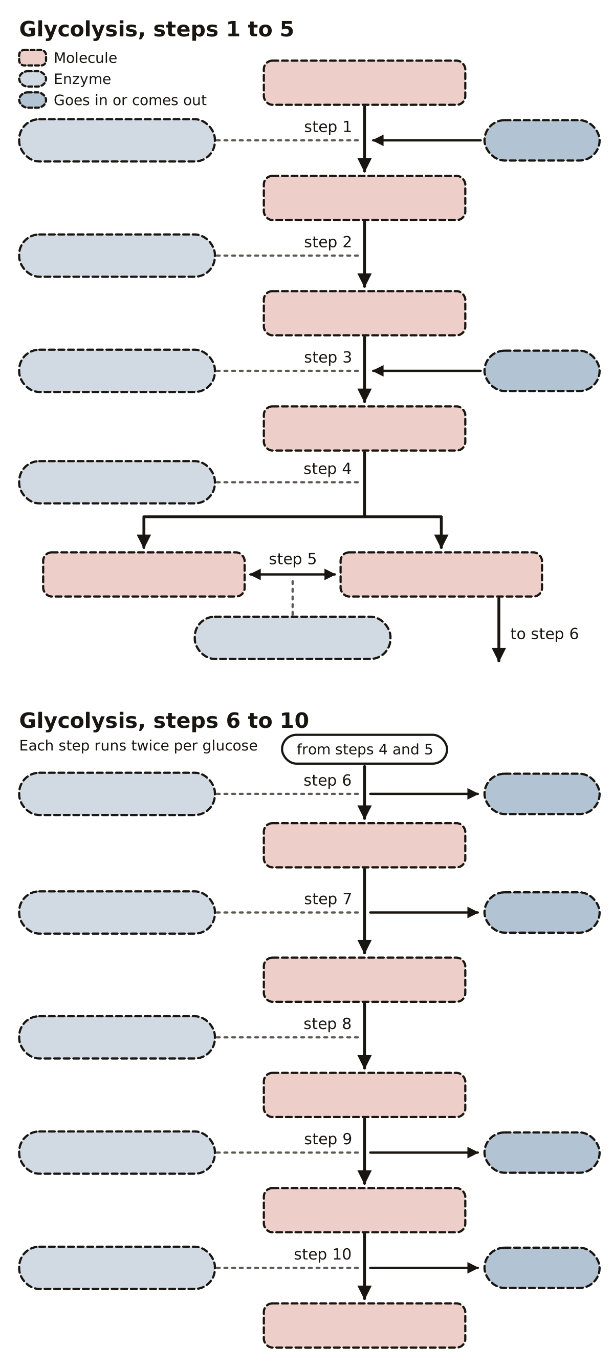 Glycolysis Labeling QuizGlycolysis as a chart of numbered blanks: the molecules from glucose to pyruvate, the enzyme of each step, and where ATP, NADH and water go in or come out.
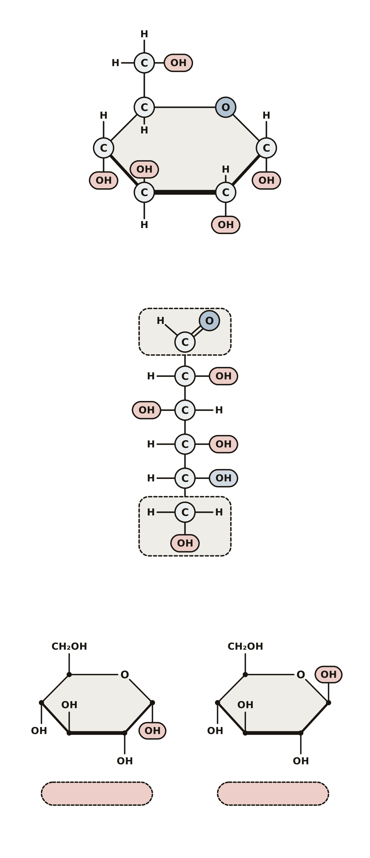 Glucose Molecule LabeledCarbons 1 to 6, the ring oxygen and the hydroxyl groups on the ring; the aldehyde and CH₂OH groups on the chain; alpha-glucose beside beta-glucose.
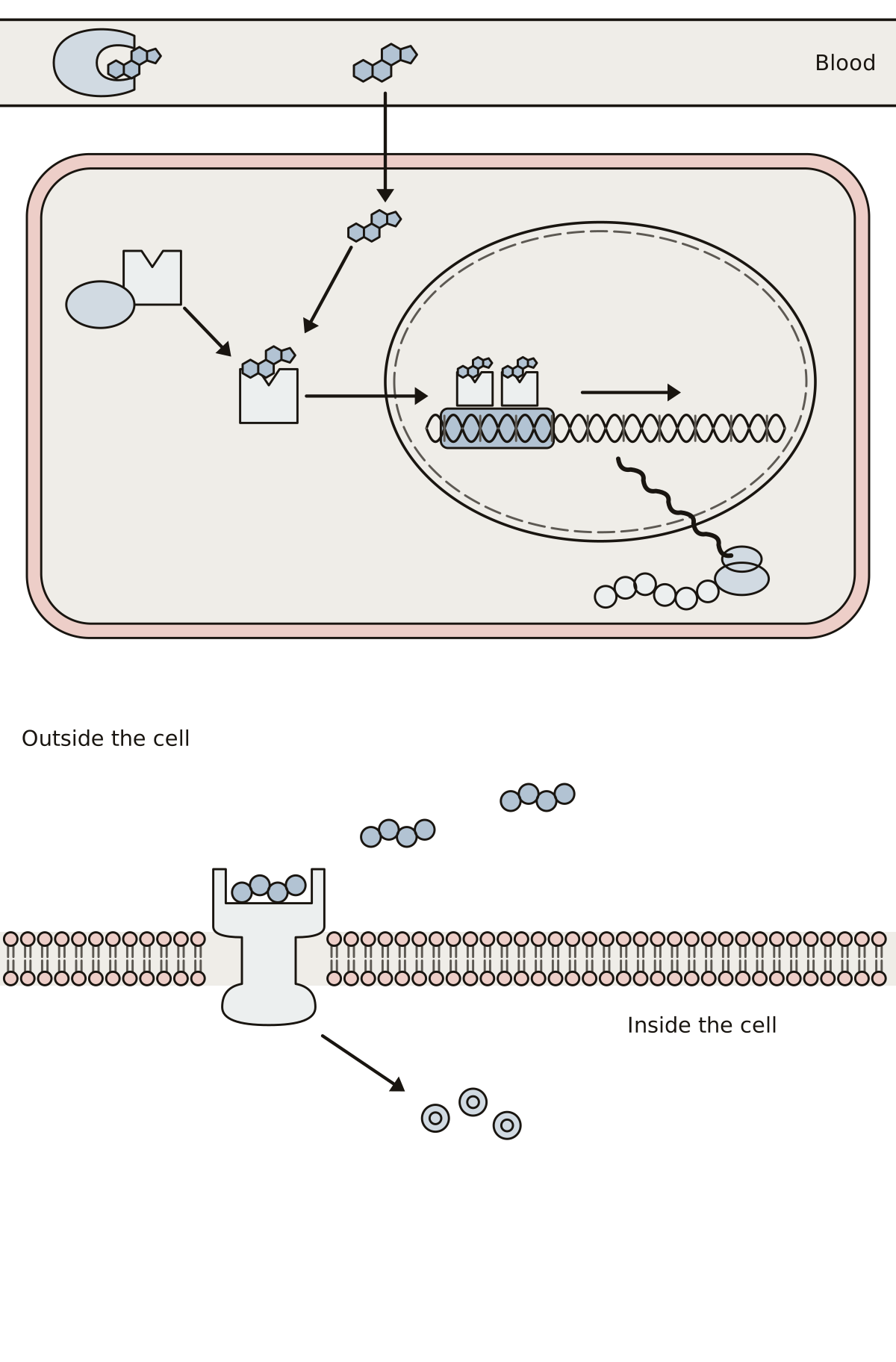 Steroid Hormone Action DiagramSteroid hormone, carrier protein, plasma membrane, receptor, heat shock protein, the complex on its response element, mRNA and new protein; then a peptide hormone, its membrane receptor and a second messenger.
- Photosynthesis and Cellular Respiration Diagram QuizA quiz from the first question on one plant cell: find the chloroplast, the mitochondrion, the two processes, light energy, ATP and the two pairs of molecules the organelles trade.
- Cell Communication Diagram QuizA quiz from the first question: find the signaling molecule, receptor, cascade and the three stages on one chart, then the G protein, adenylyl cyclase, cyclic AMP and protein kinase A on the next.
- Glucose Molecule Diagram QuizA quiz from the first question: find carbons 1 to 6, the ring oxygen and a hydroxyl group on the ring, then the aldehyde and CH₂OH groups, alpha-glucose and beta-glucose.
The set covers the figures a molecular and cell biology course asks you to label: DNA and how it is copied and read, how a cell divides, and the cycles that move energy through a cell. Each page is one drawing with a number on every part.
More about these tools
A number lights up and you choose or type what belongs there, or you get a name and tap its place. Study shows every answer with a line on what it does, and missed ones come back when the round ends.
Use a page before a unit quiz to find the names you do not know, or print the blank copy and its key for a class. The first page is the citric acid cycle: nine molecules, eight enzymes and seven points where something leaves. Names and steps were checked against an open college biology textbook on the date each page shows.
Keep exploring
- Animal body-part diagrams (94)
- Blank US maps (61)
- Blank maps (159)
- Bone labeling quizzes (49)
- Capitals quizzes (11)
- Car and engine part diagrams (54)
- Cell and organelle diagrams (63)
- Clothing, shoes and jewelry (45)
- Computer, phone and camera diagrams (43)
- Anatomy concept maps (1)
- Country map quizzes (28)
- Animal anatomy diagrams (100)
- Science Quizzes
- All quizzes