Cell Diagrams to Label: Plant, Animal, Bacteria and Organelles
One drawing per page, a numbered ring on every part, and a blank copy to print when you want to work on paper.
Every page here is one drawing of a cell or of a single organelle, with a numbered ring on each part. In Find it you get a name and tap its ring.
63 pages
Whole cells 6
Plant, animal and bacterial cells, alone and side by side, each organelle's job, and the cell as a factory or city.
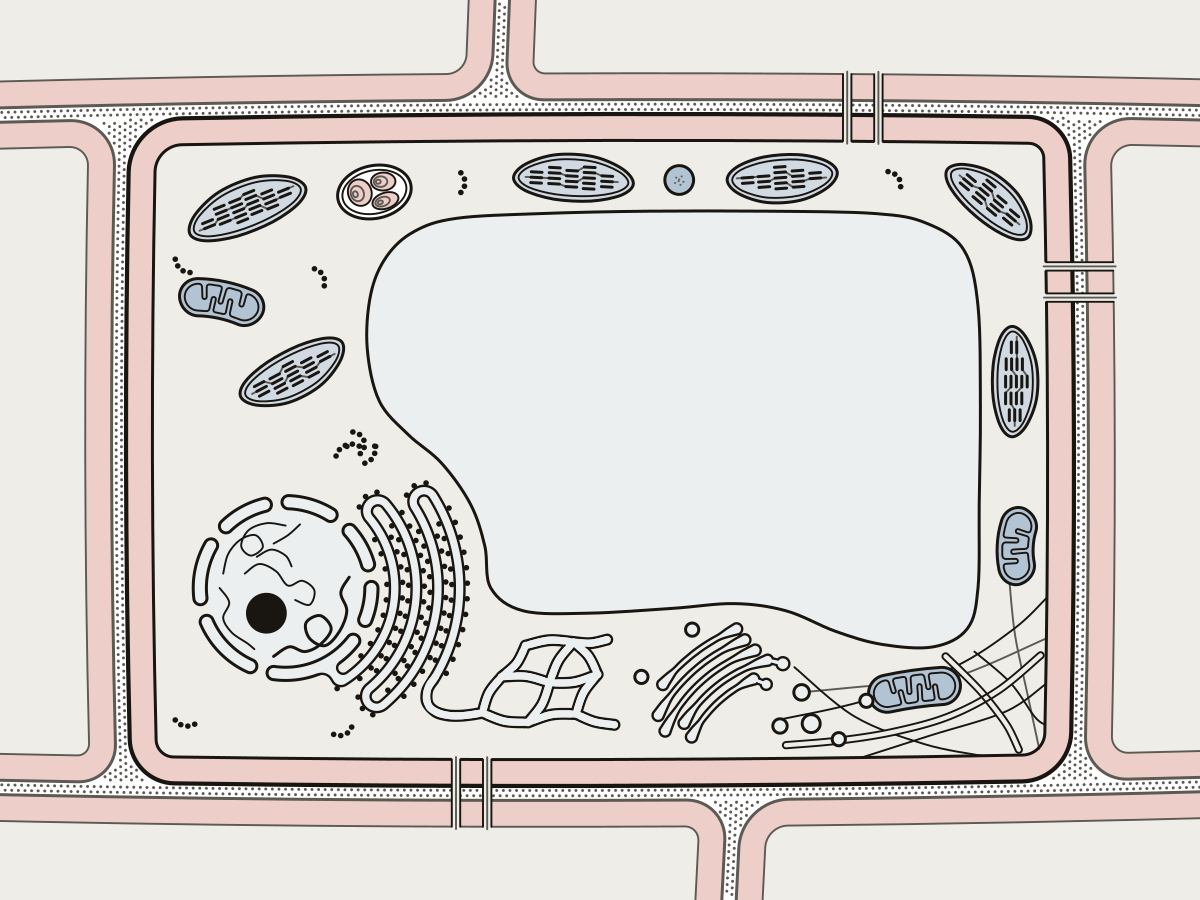 Plant Cell DiagramA cut-open plant cell with 22 parts to find or name, a simple set of 8 for younger grades, and blank and labeled versions to print.
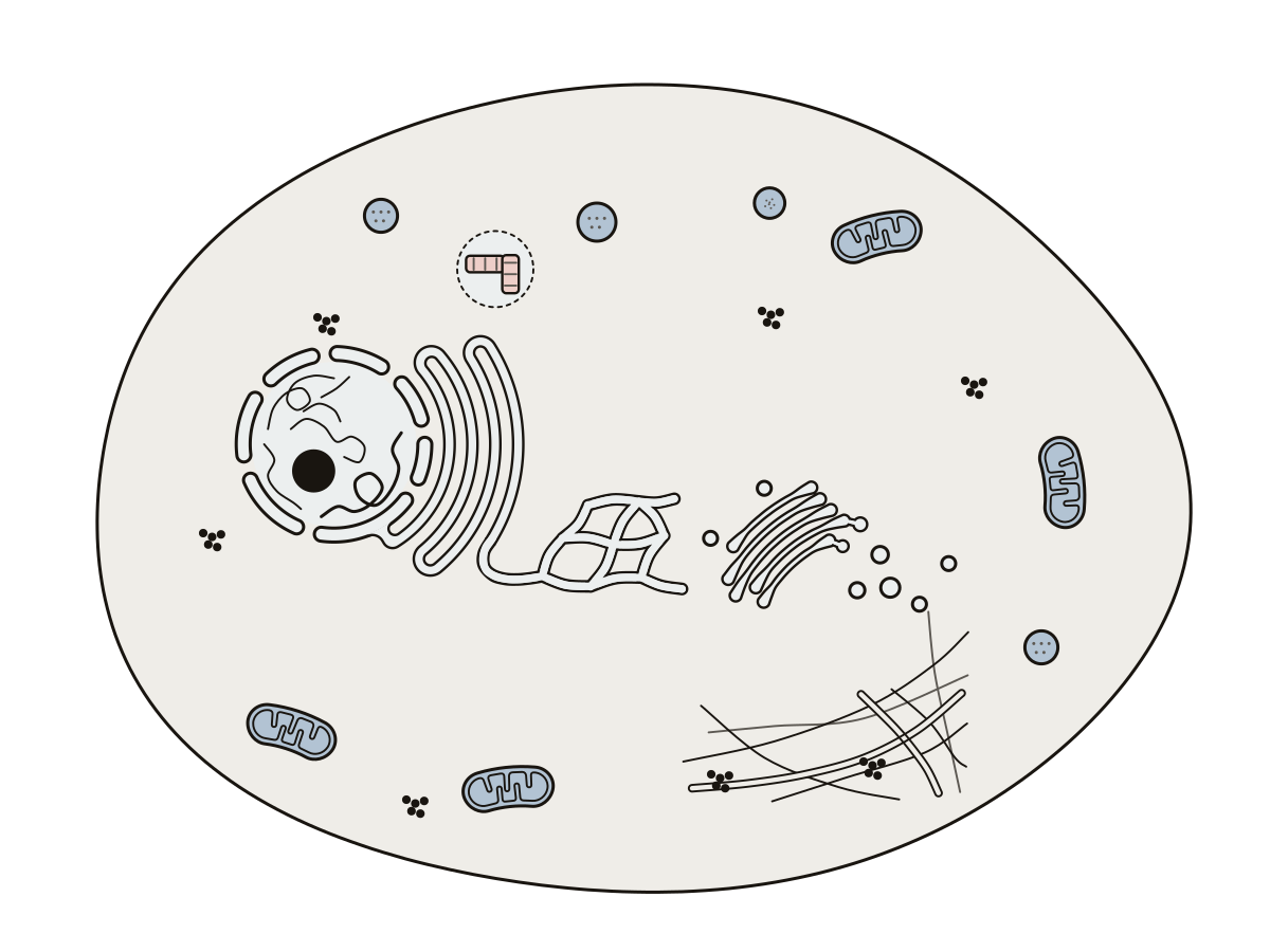 Parts of an Animal CellOne animal cell cut open: 17 parts to find or name, from the nucleus and rough ER to lysosomes and centrioles, with blank and labeled prints.
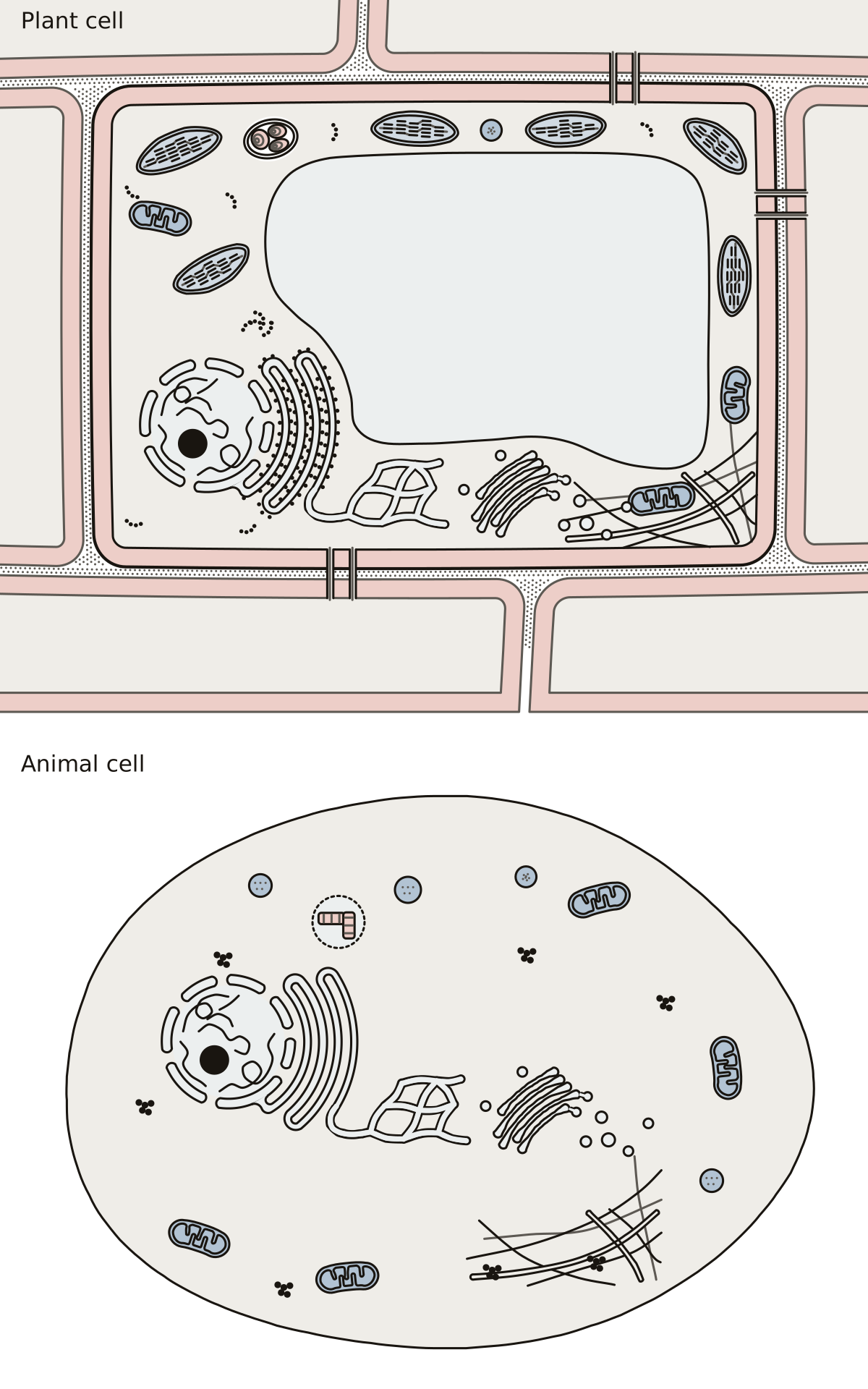 Plant vs Animal Cell DiagramA plant cell and an animal cell on one sheet: wall, chloroplasts and central vacuole above, centrosome and lysosomes below, and the organelles both share.
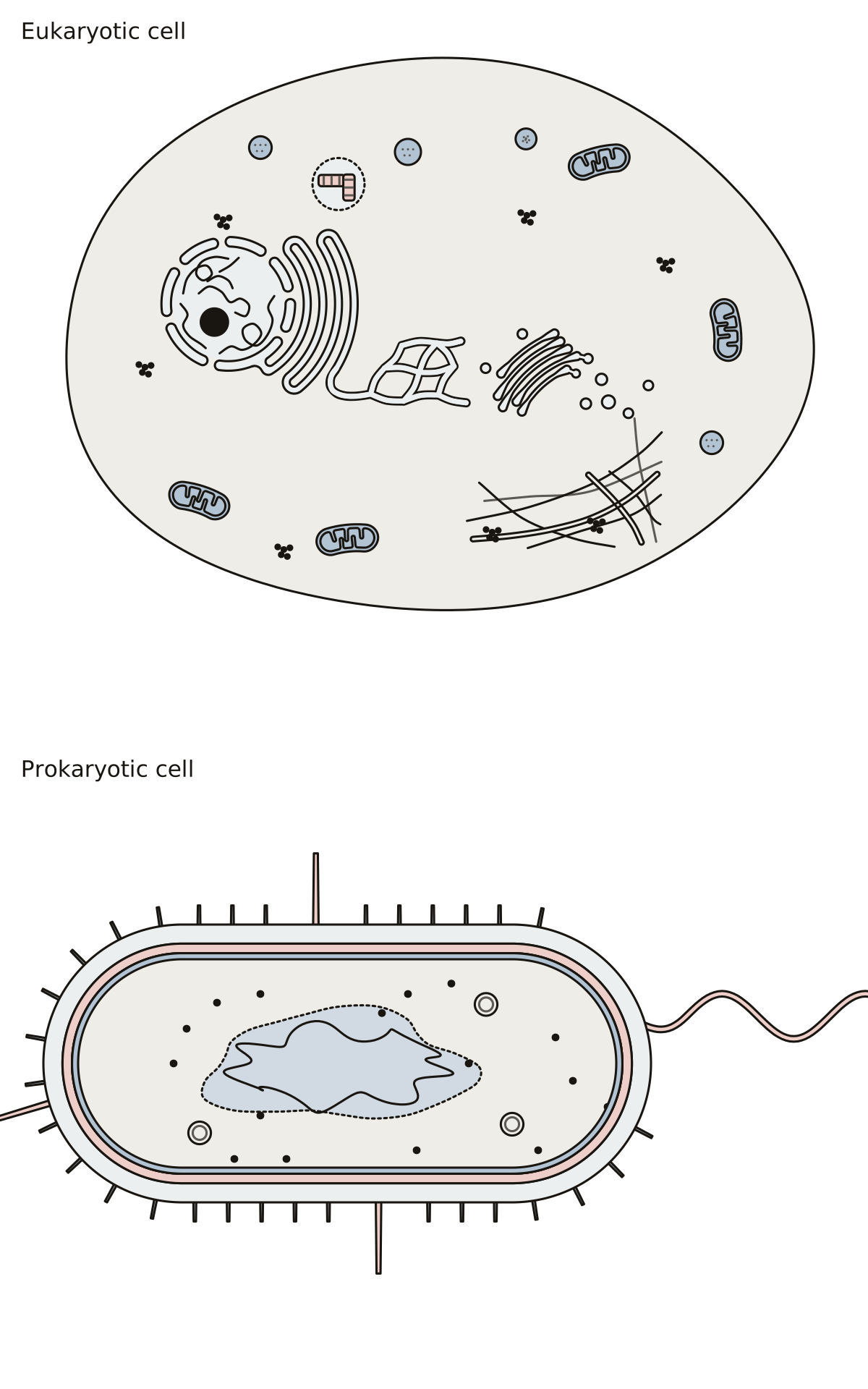 Eukaryotic Cell Diagram Labeled, Next to a Prokaryotic CellA eukaryotic cell and a prokaryotic cell on one sheet: nucleus and organelles above, nucleoid, wall and capsule below, with the parts both cells share.
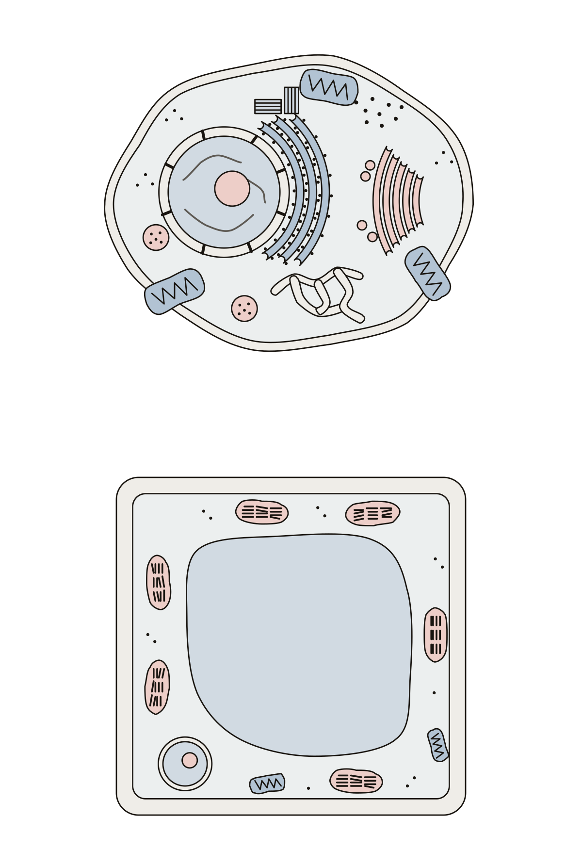 Parts of a Cell DiagramName the parts of an animal cell and a plant cell, then match each part to the job it does.
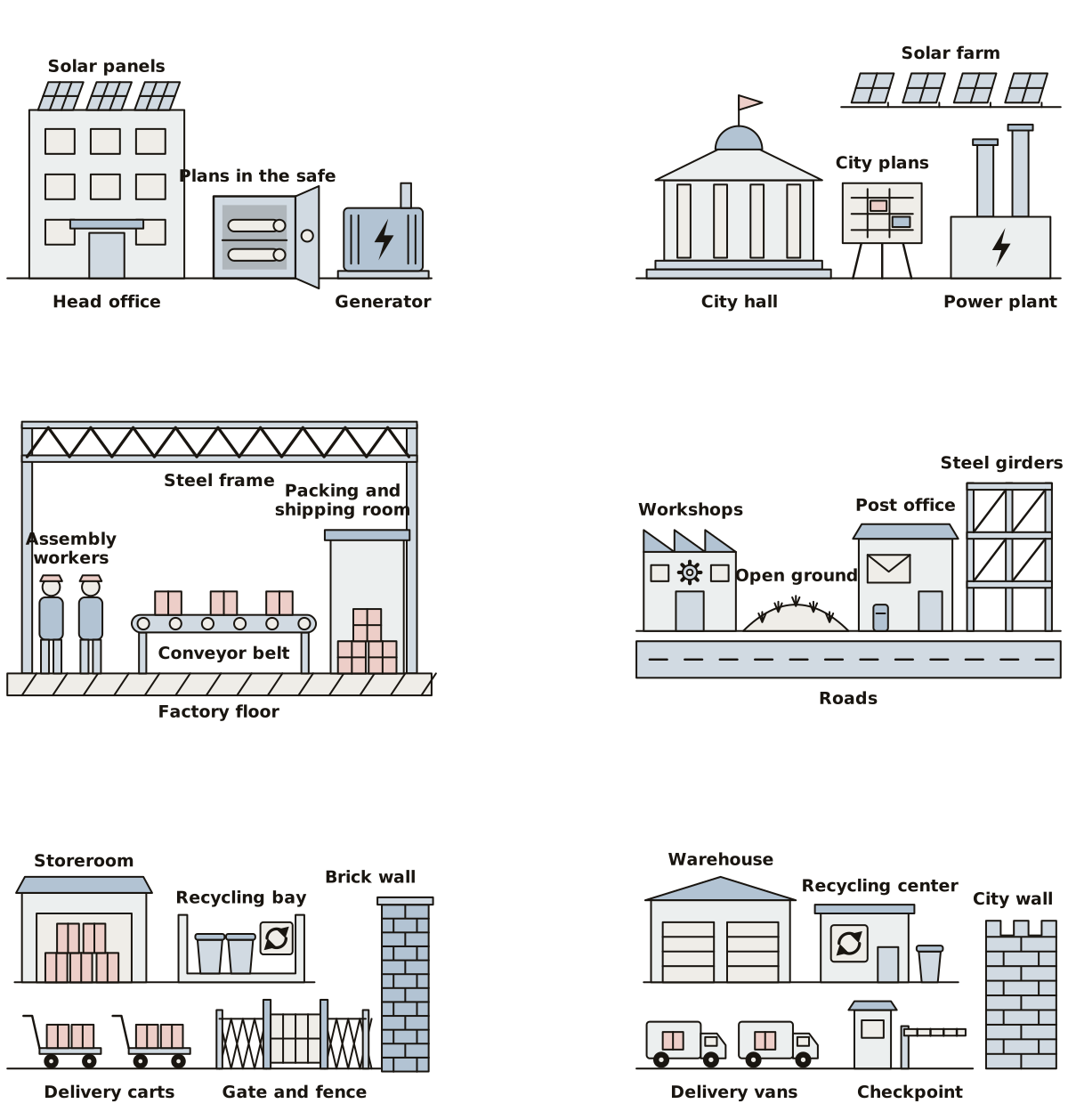 Cell Factory Diagram and Cell City DiagramA cell drawn as a factory and as a city, fourteen places each, to match to the nucleus, mitochondria, Golgi apparatus and the rest.
Organelles, close up 21
One organelle per drawing, enlarged until its membranes, folds and inner spaces are big enough to tap, plus how substances cross the cell membrane and what a cell does in a hypertonic, an isotonic and a hypotonic solution. The plant cell wall, cell junctions, cilia and flagella, and a triglyceride are drawn the same way.
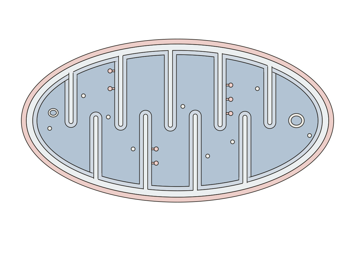 Mitochondria Diagram LabeledA mitochondrion cut lengthwise: 8 numbered parts, from the outer membrane and the folded cristae to the matrix, its DNA and ATP synthase.
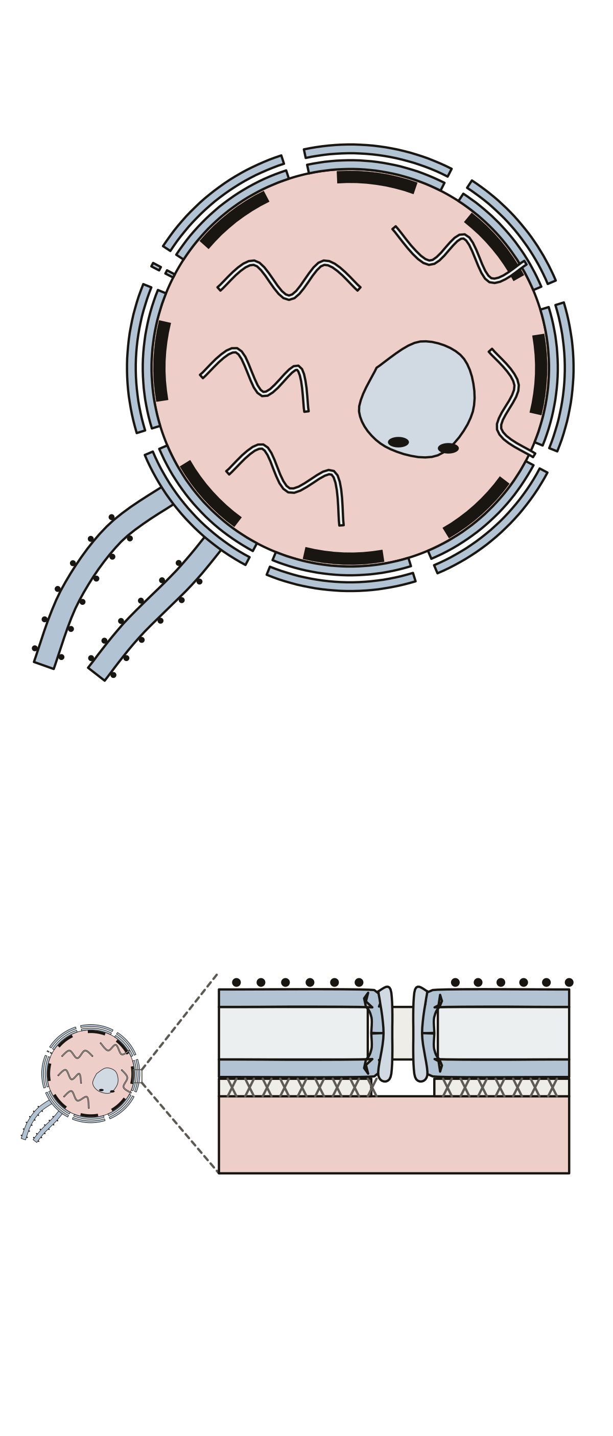 Nucleus Diagram LabeledThe nucleus in section and its envelope close up: 11 parts to name. Inner nuclear membrane · Outer nuclear membrane · Heterochromatin
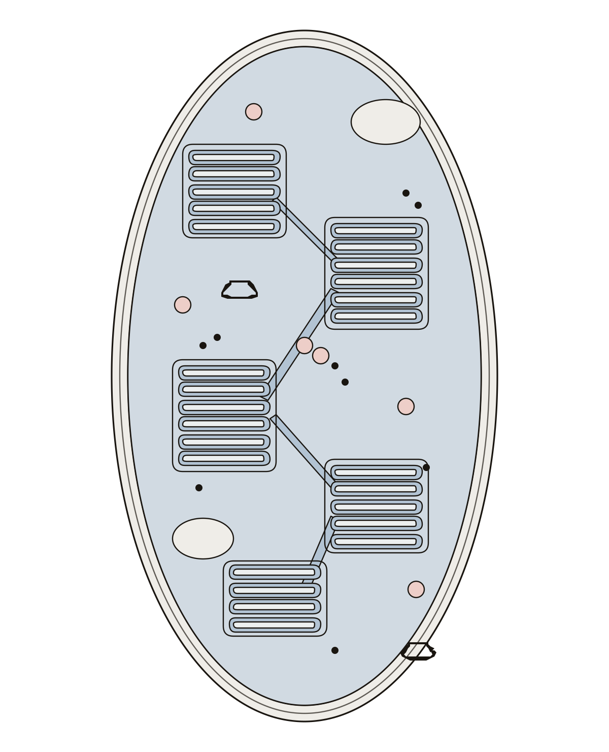 Chloroplast Labeled DiagramA chloroplast cut open: envelope, stroma, thylakoids, thylakoid lumen, grana, stroma lamellae, starch granules, plastoglobuli, DNA and ribosomes. Starch · DNA · Ribosomes
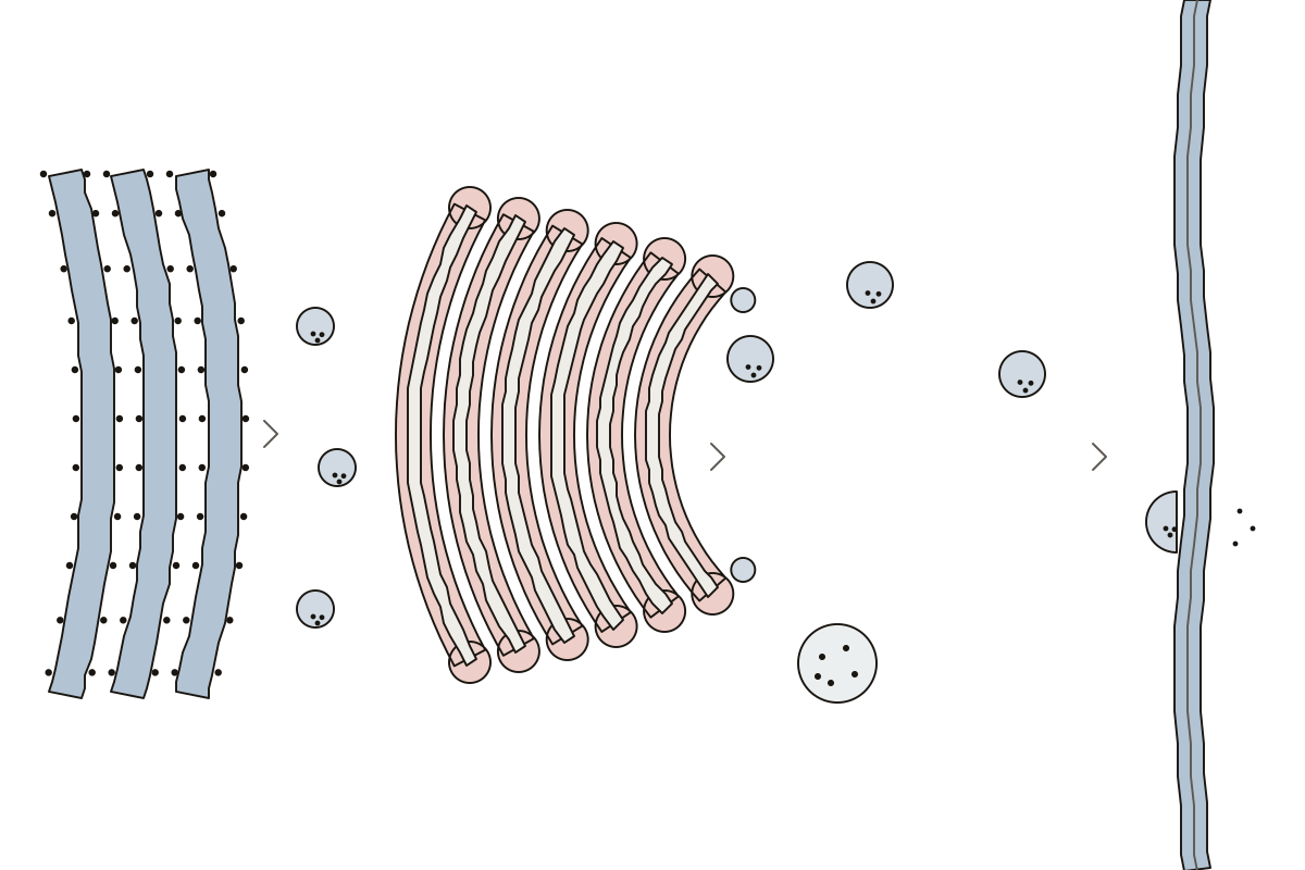 Golgi Apparatus Diagram LabeledThe Golgi stack and the route through it: 9 parts to name, labeled or blank. Trans face · Transport vesicle · Cisternae
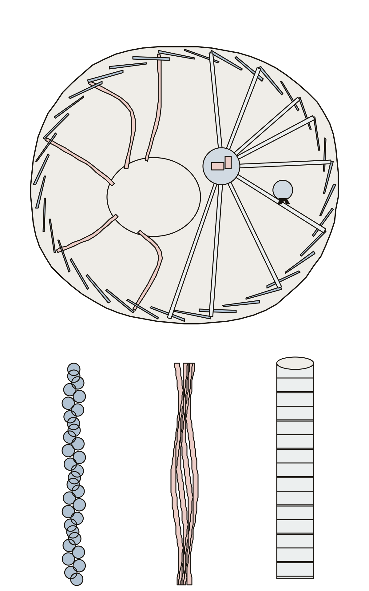 Cytoskeleton Diagram LabeledThe cytoskeleton: microfilaments, intermediate filaments, microtubules, the centrosome and its centrioles, and a motor protein. Centrosome · Motor protein · Centrioles
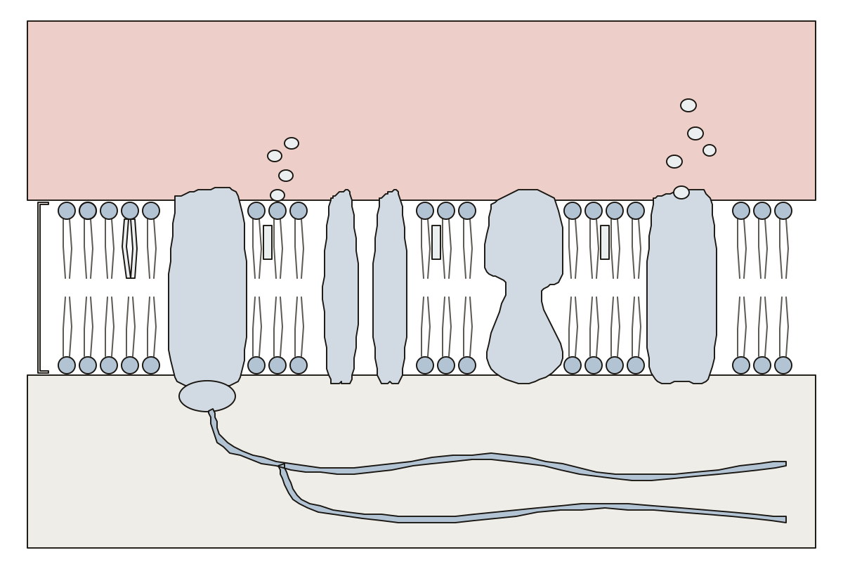 Cell Membrane Labeled DiagramA strip of the fluid mosaic, outside above and cytoplasm below: 13 parts to name. Extracellular fluid · Carrier protein · Cholesterol
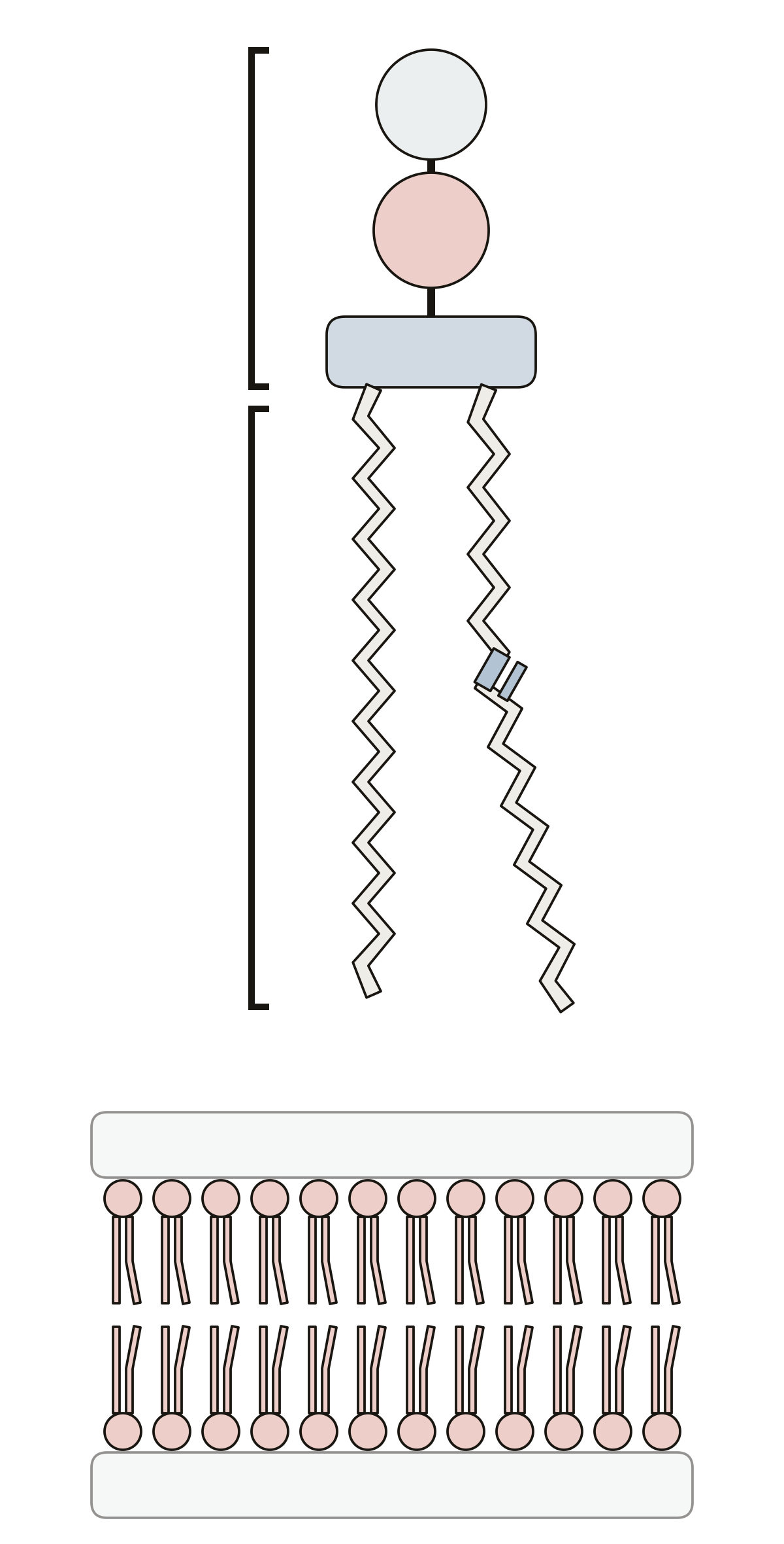 Parts of a PhospholipidOne phospholipid and a piece of membrane: 10 numbered parts, from the hydrophilic head and hydrophobic tails to glycerol, a double bond and the bilayer. Double bond · Phosphate group · Choline
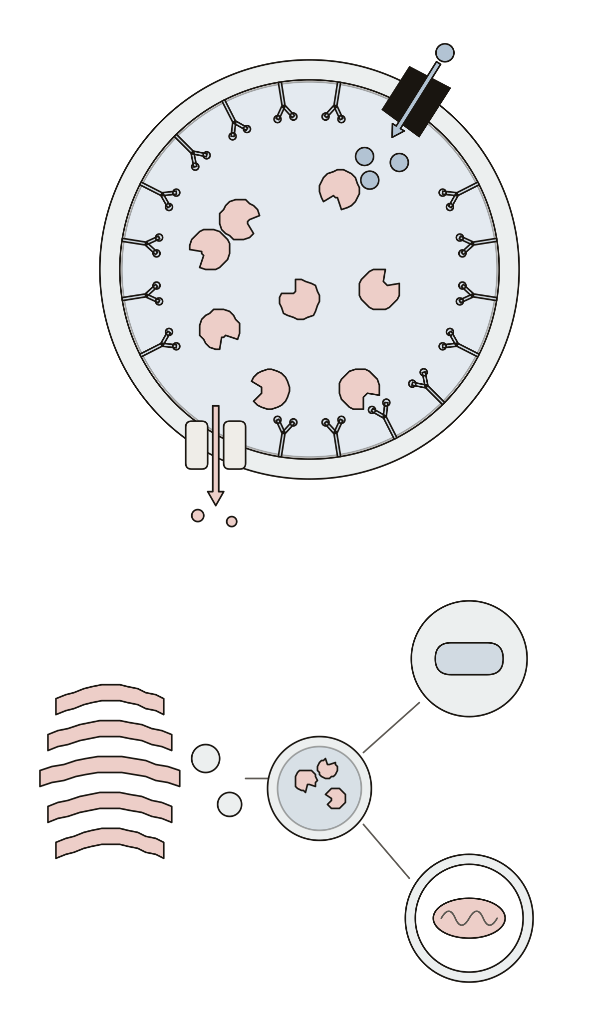 Lysosome Diagram LabeledA lysosome cut open: 10 numbered parts, from the membrane, glycocalyx and acidic lumen to the enzymes, proton pump, phagosome and autophagosome. Lumen · Phagosome · Autophagosome
- Lysosome Without LabelsThe lysosome drawing with nothing written on it, to print and fill in: one lysosome cut open and the three structures it works with, plus the answer key. Golgi apparatus · Transport protein · Phagosome
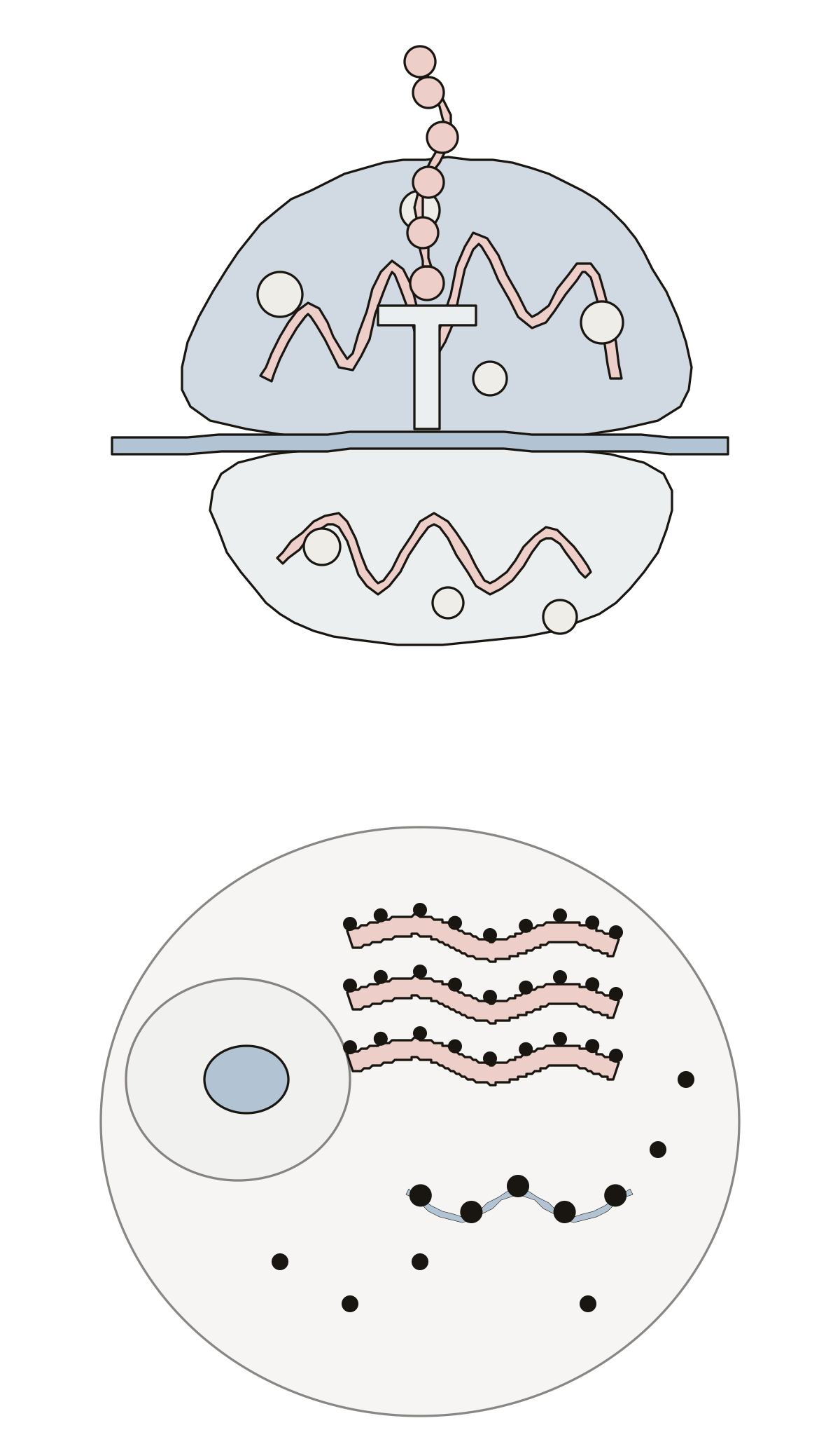 Ribosome Diagram LabeledA ribosome close up and in a cell: 12 numbered parts, from the large and small subunits, rRNA and tRNA to free and bound ribosomes and a polysome. tRNA · Rough ER · Small subunit
- Unlabeled Diagram of RibosomeThe ribosome drawing with no names on it, to print and label: one ribosome cut open while it builds a protein, a cell with its ribosomes, and the answer key. mRNA · tRNA · Polypeptide chain
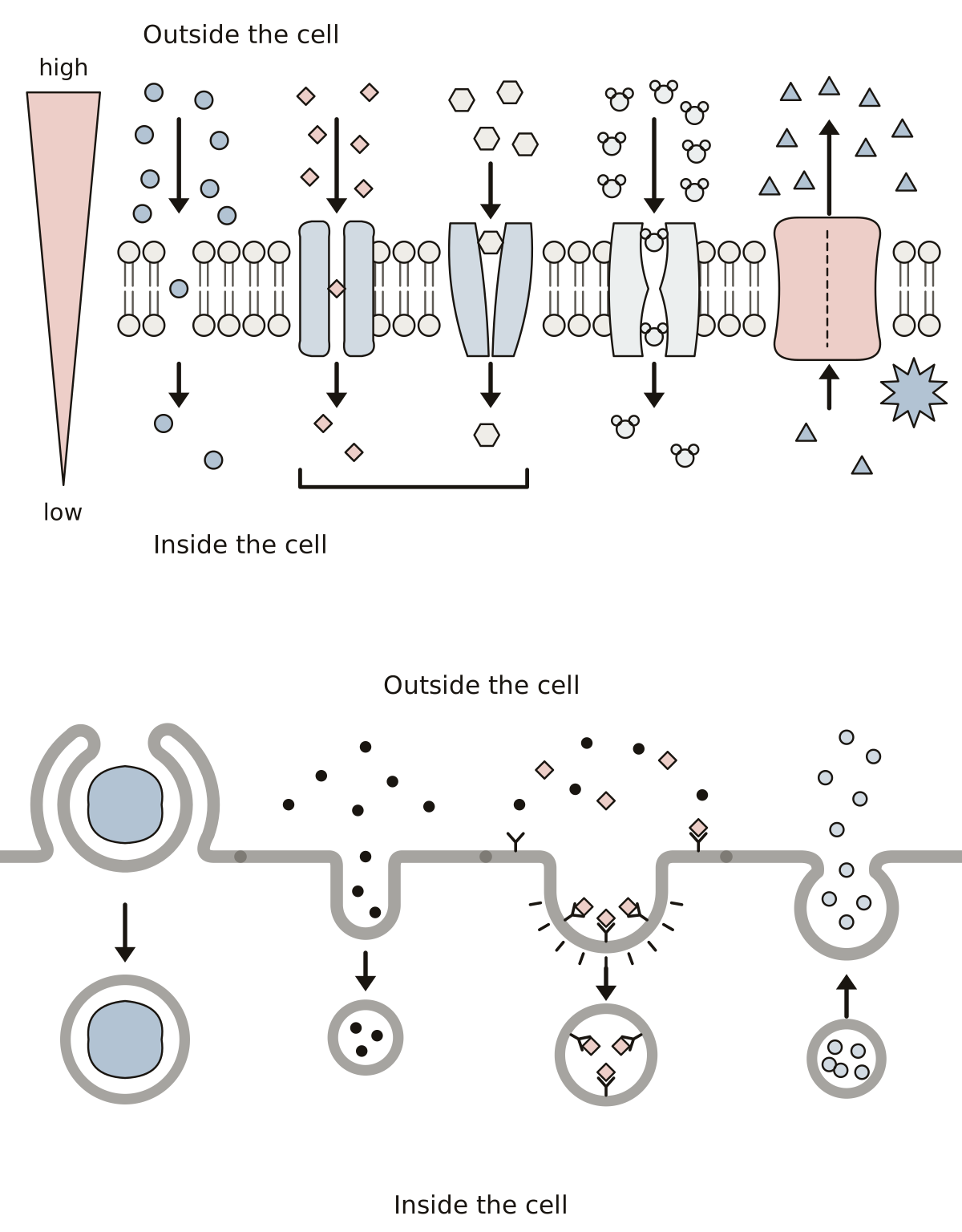 Cell Transport DiagramSimple and facilitated diffusion, osmosis, active transport with ATP, phagocytosis, pinocytosis, receptor-mediated endocytosis and exocytosis.
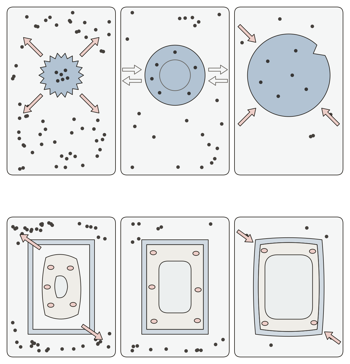 Hypertonic Hypotonic Isotonic DiagramCells in three solutions: hypertonic, isotonic and hypotonic solution, crenated, normal and swollen red blood cell, water moving out and in, plasmolyzed, flaccid and turgid plant cell, cell wall, central vacuole.
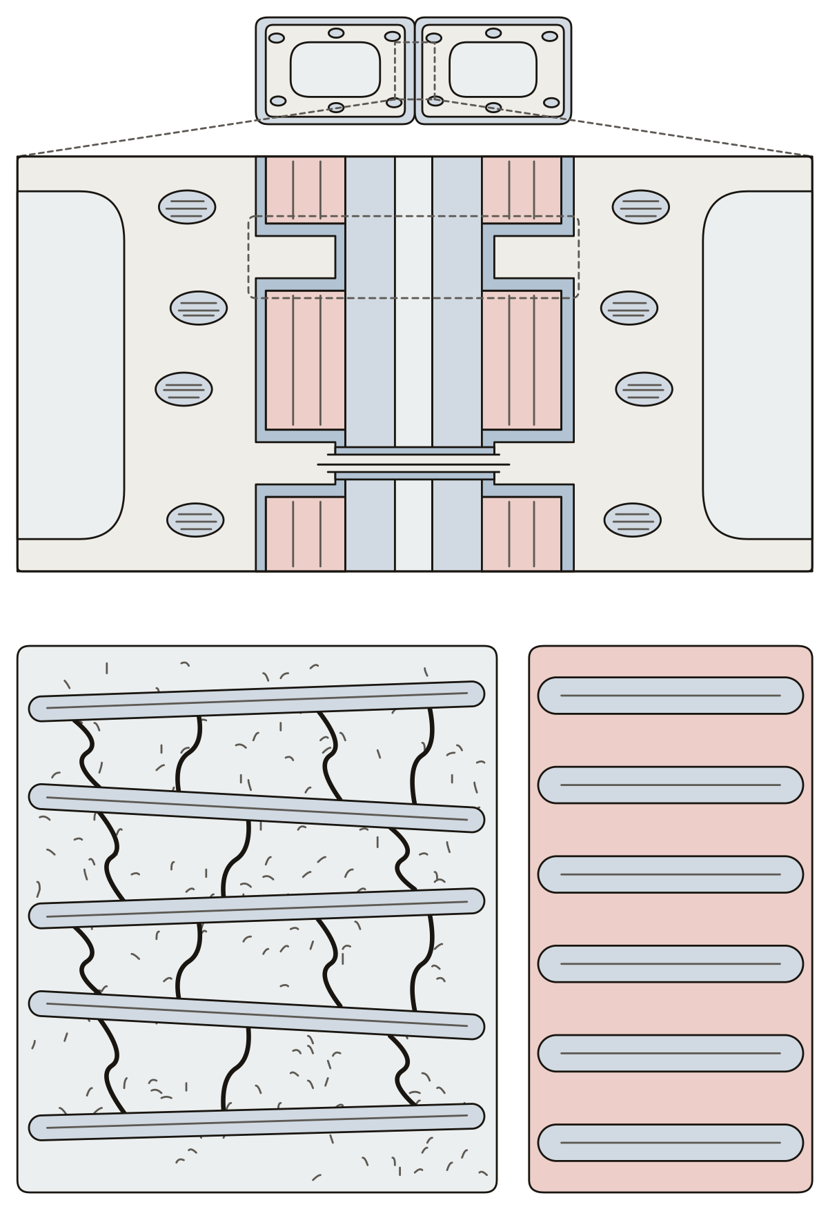 Diagram of Plant Cell WallThe wall between two plant cells: middle lamella, primary and secondary cell wall, plasma membrane, pit, plasmodesma, then cellulose microfibrils, hemicellulose, pectin and lignin.
- Cell Wall in Plant Cell Diagram With No LabelsThe plant cell wall drawing with nothing written on it, to print and label: the layers between two cells, a pit, a plasmodesma and the four materials of the wall, with an answer key.
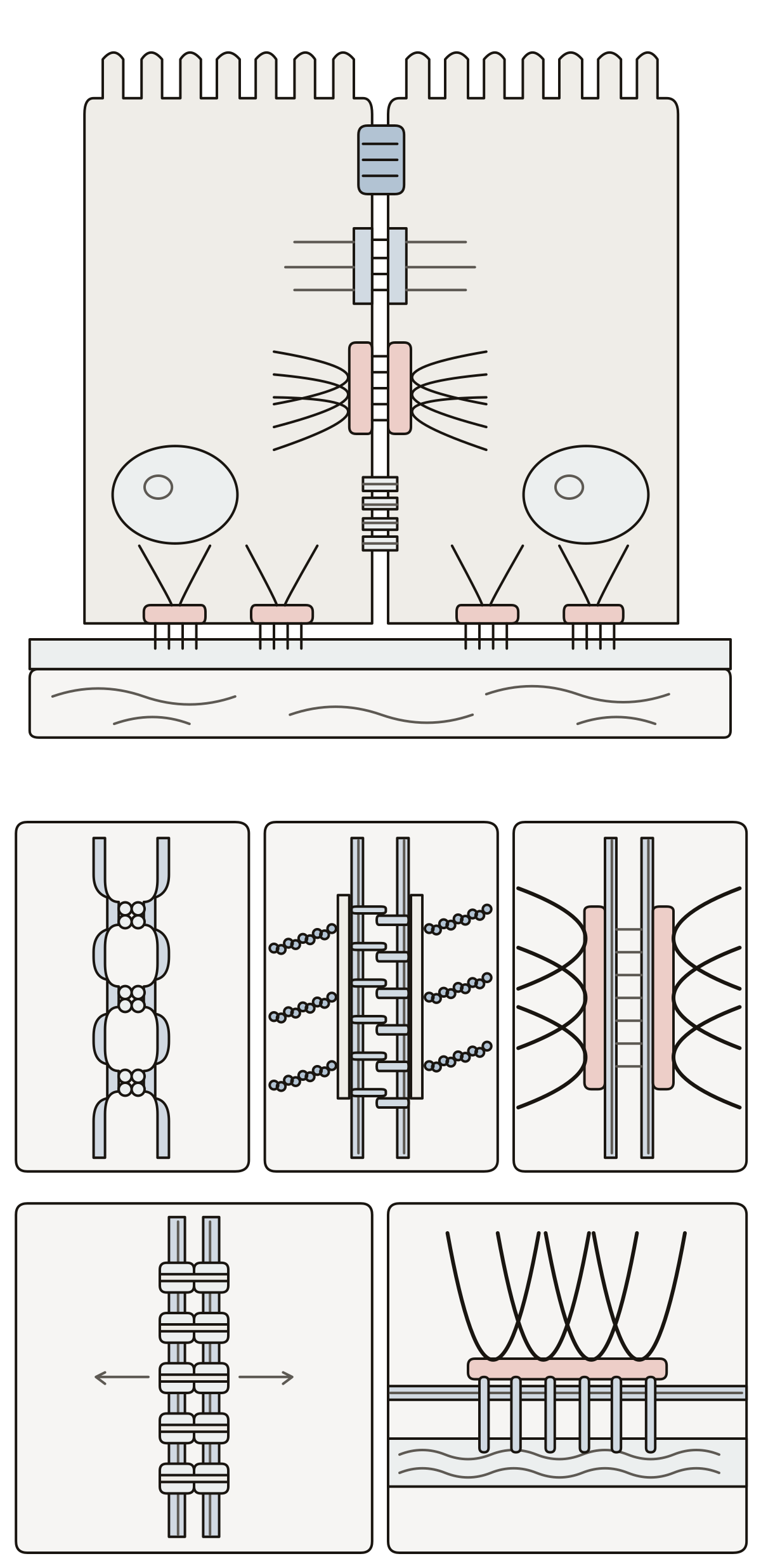 Cell Junctions DiagramJunctions between animal cells: tight junction, adherens junction, desmosome, gap junction, hemidesmosome, claudins, cadherins, actin filaments, plaque, intermediate filaments, connexon, integrins.
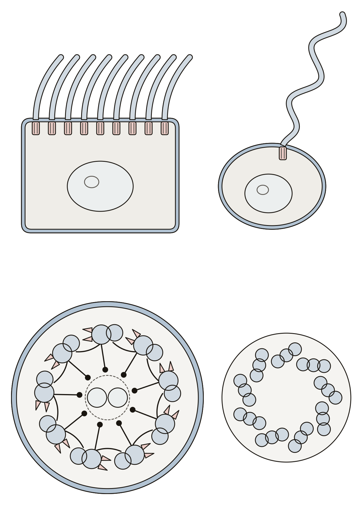 Animal Cell Diagram With Cilia and FlagellaCilia and flagella of animal cells: cilia, flagellum, basal body, plasma membrane, outer doublet, central pair, dynein arms, radial spoke, nexin link, microtubule triplet.
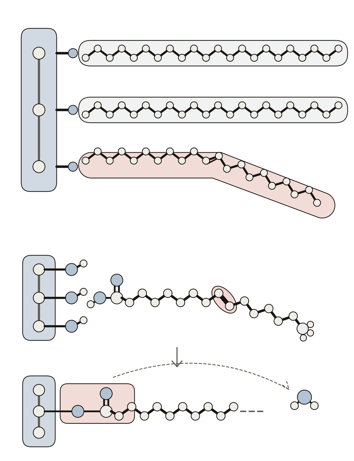 Parts of a TriglycerideA fat molecule: glycerol, saturated fatty acid, unsaturated fatty acid, hydroxyl group, carboxyl group, hydrocarbon chain, double bond, omega end, ester bond, water molecule.
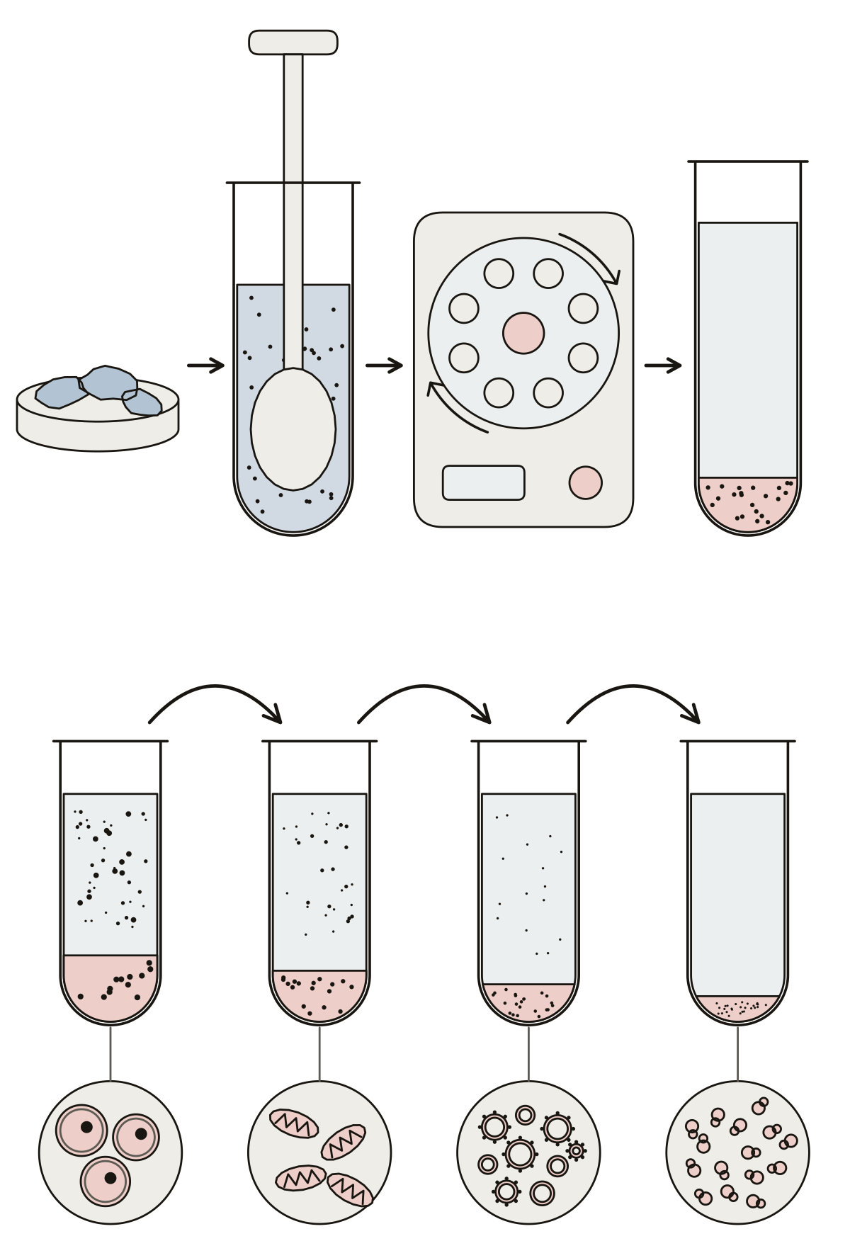 Cell Fractionation DiagramTissue, homogenizer, homogenate, centrifuge, supernatant and pellet, then the pellets in order: nuclei, mitochondria, microsomes, ribosomes, with the cytosol left at the end.
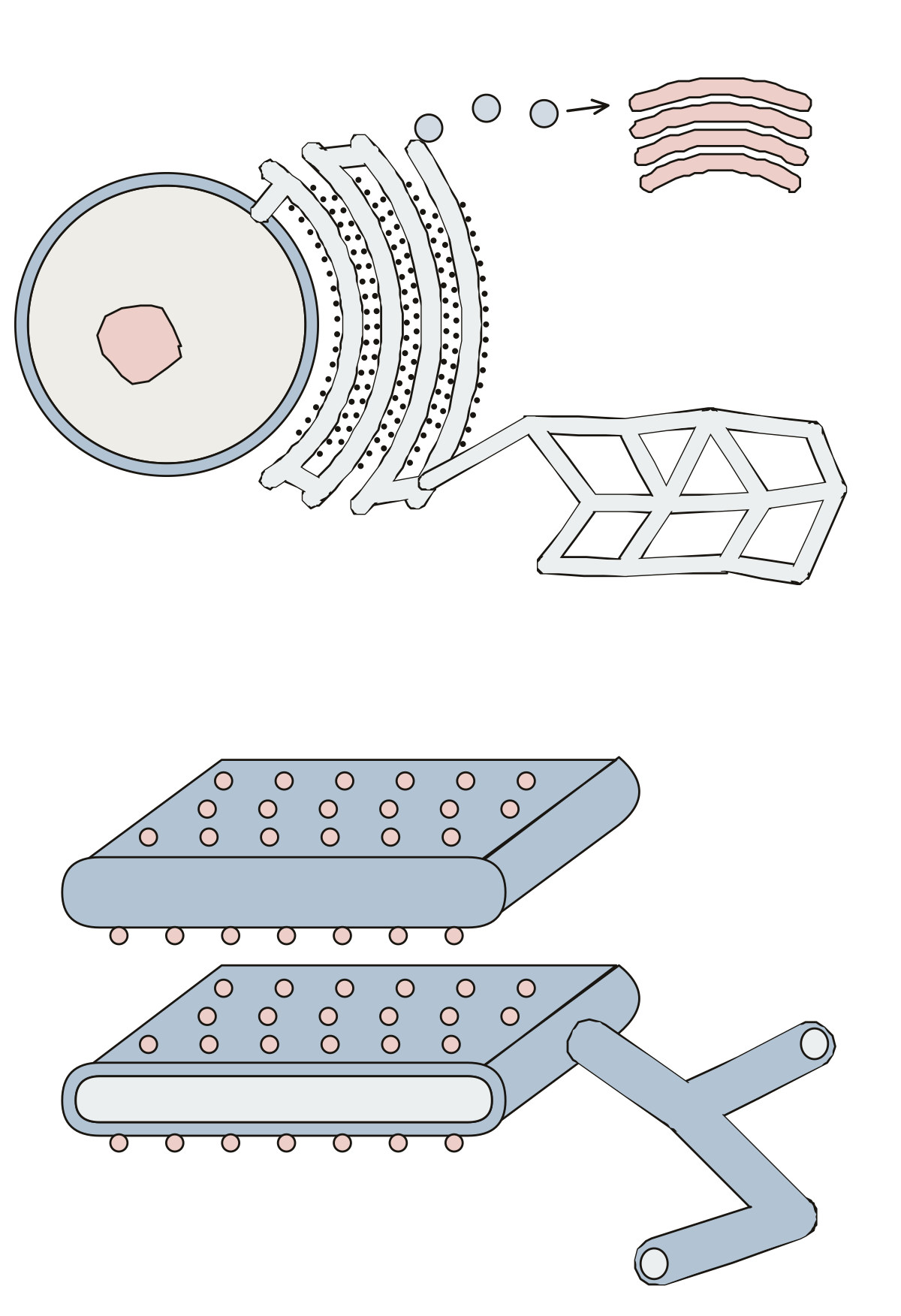 ER Cell Diagram: Endoplasmic ReticulumRough ER, smooth ER, the nuclear envelope, a transport vesicle and the Golgi apparatus, then a cut sac with its ribosomes, cisterna, lumen, membrane and tubules.
- Unlabeled Diagram of Smooth Endoplasmic ReticulumThe endoplasmic reticulum with no names on it, to print and label: rough ER, smooth ER, nuclear envelope, vesicle and Golgi above, then a cut sac with its ribosomes, lumen, membrane and tubule.
Specialized cells and microscope slides 10
Cells shaped for one job, a fat cell among them, and the onion, cheek and pondweed cells that show up under a school microscope.
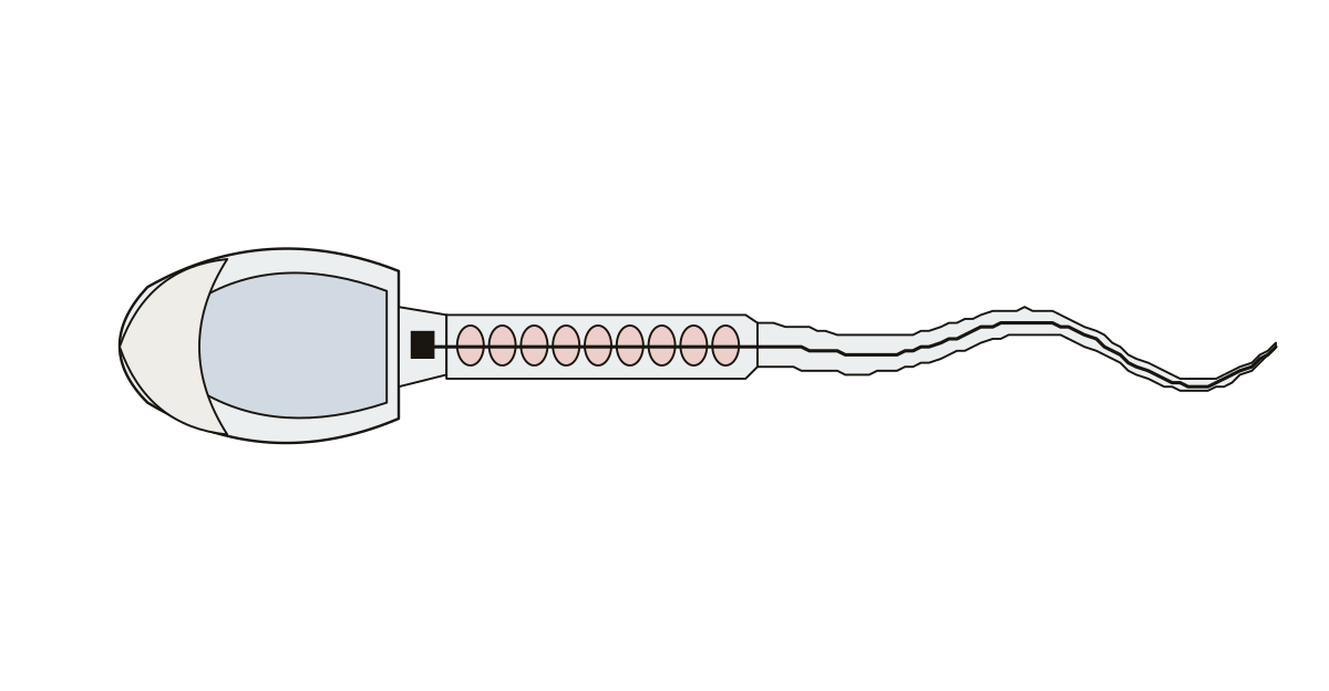 Sperm Cell DiagramHead, acrosome, nucleus, neck, centriole, mid-piece, mitochondria, tail and flagellum.
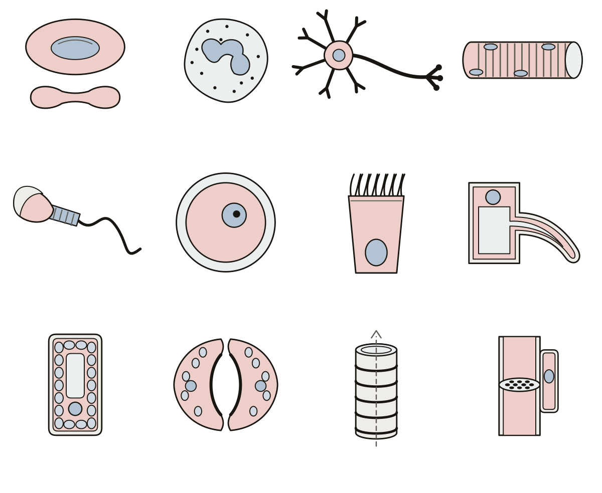 Specialized Cells WorksheetName twelve specialized animal and plant cells and match each to the job it is adapted for. Egg cell · Red blood cell · Muscle cell
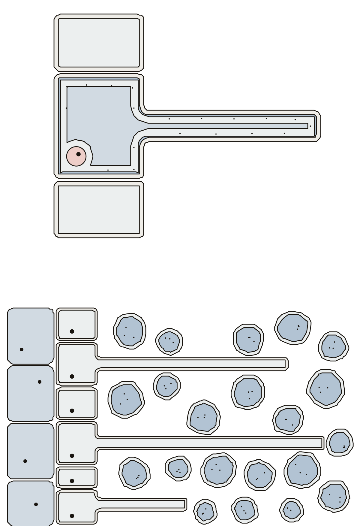 Root Hair Cell DiagramA root hair cell with its hair, wall, membrane, cytoplasm, nucleus and vacuole, and a second view of root hairs among soil particles.
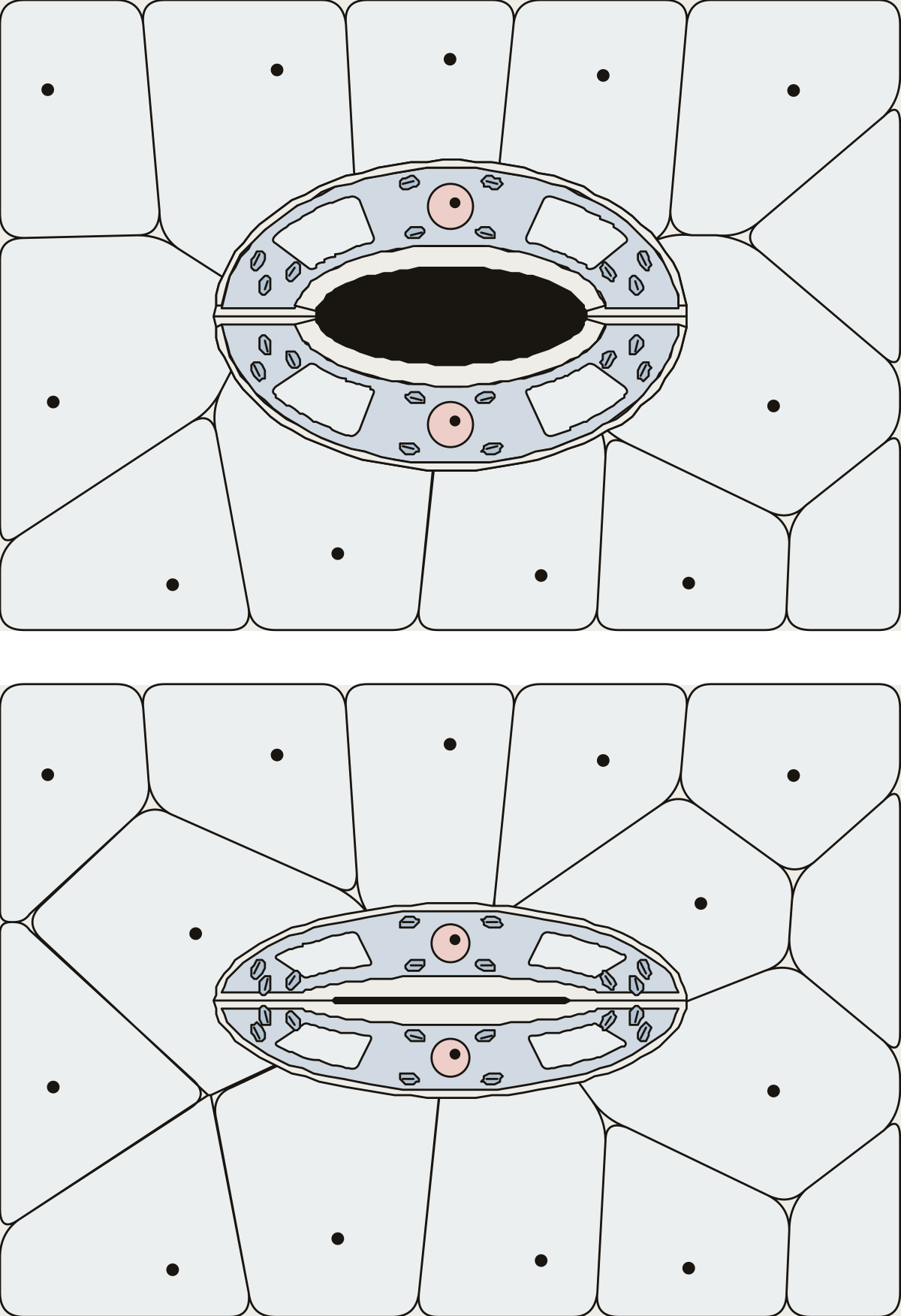 Guard Cell DiagramA stoma open and closed: guard cells, pore, thick inner wall, thin outer wall, chloroplasts, nucleus, vacuole and the epidermal cells round it.
- Stomata and Guard Cells Without LabelsA stoma open and closed with nothing written on it, to print and label: guard cells, pore, thick and thin wall, chloroplast, nucleus, vacuole and epidermal cells, with an answer key.
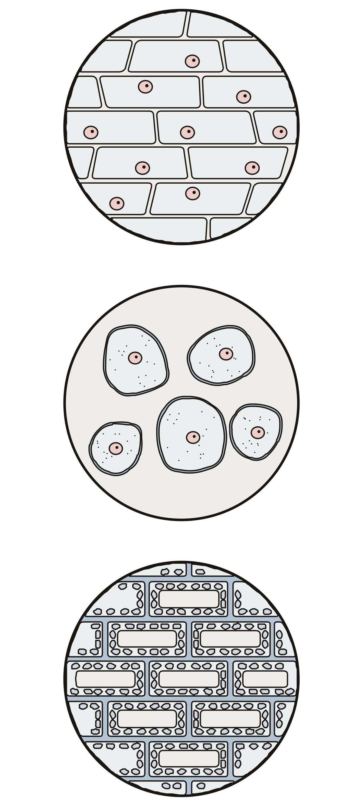 Onion Cell DiagramOnion skin, cheek cells and an Elodea leaf as the school microscope shows them, with the parts you can see on each slide.
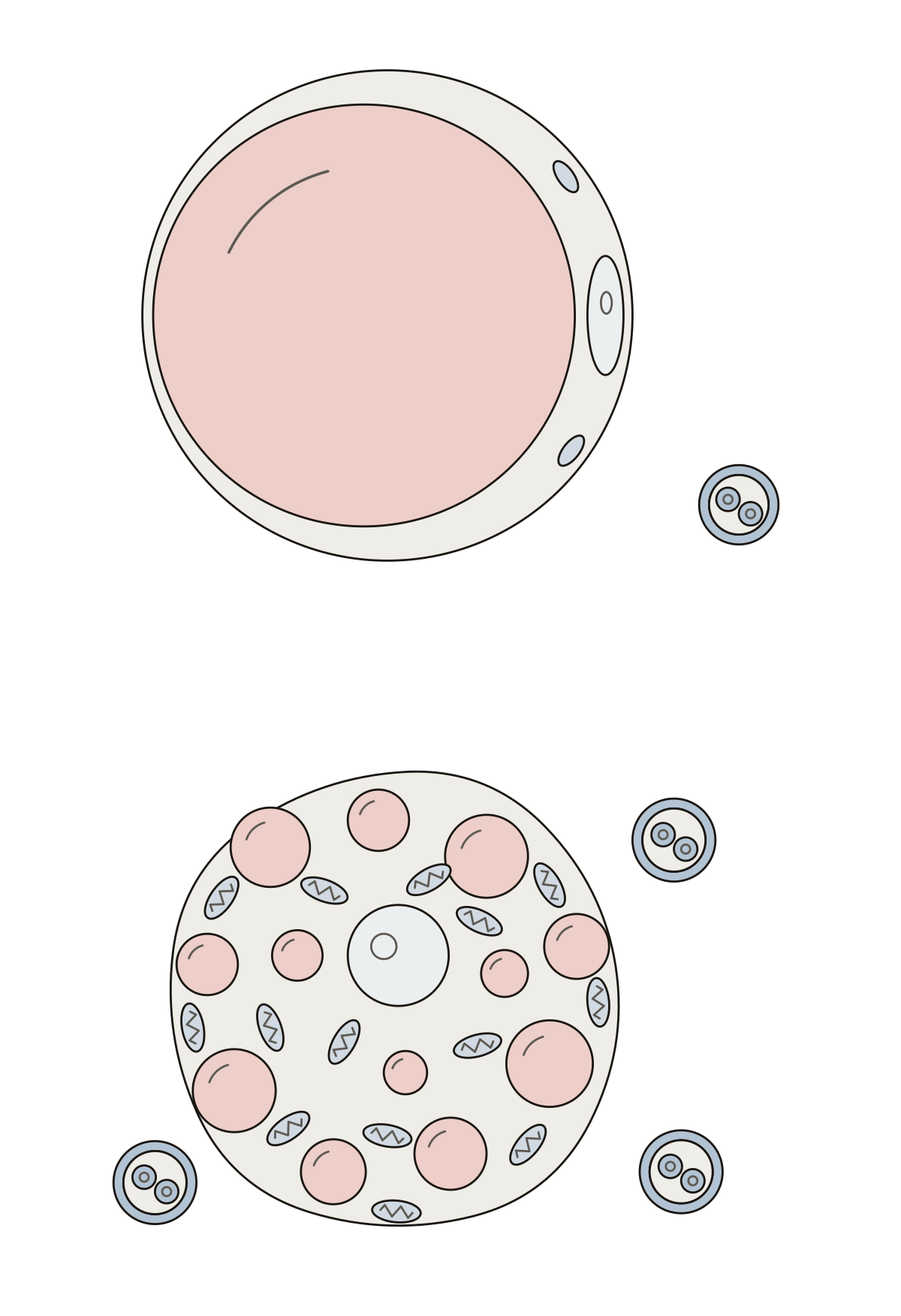 Adipocyte Cell DiagramWhite and brown fat cells: lipid droplet, rim of cytoplasm, flattened nucleus, many small lipid droplets, round nucleus, mitochondria, capillary.
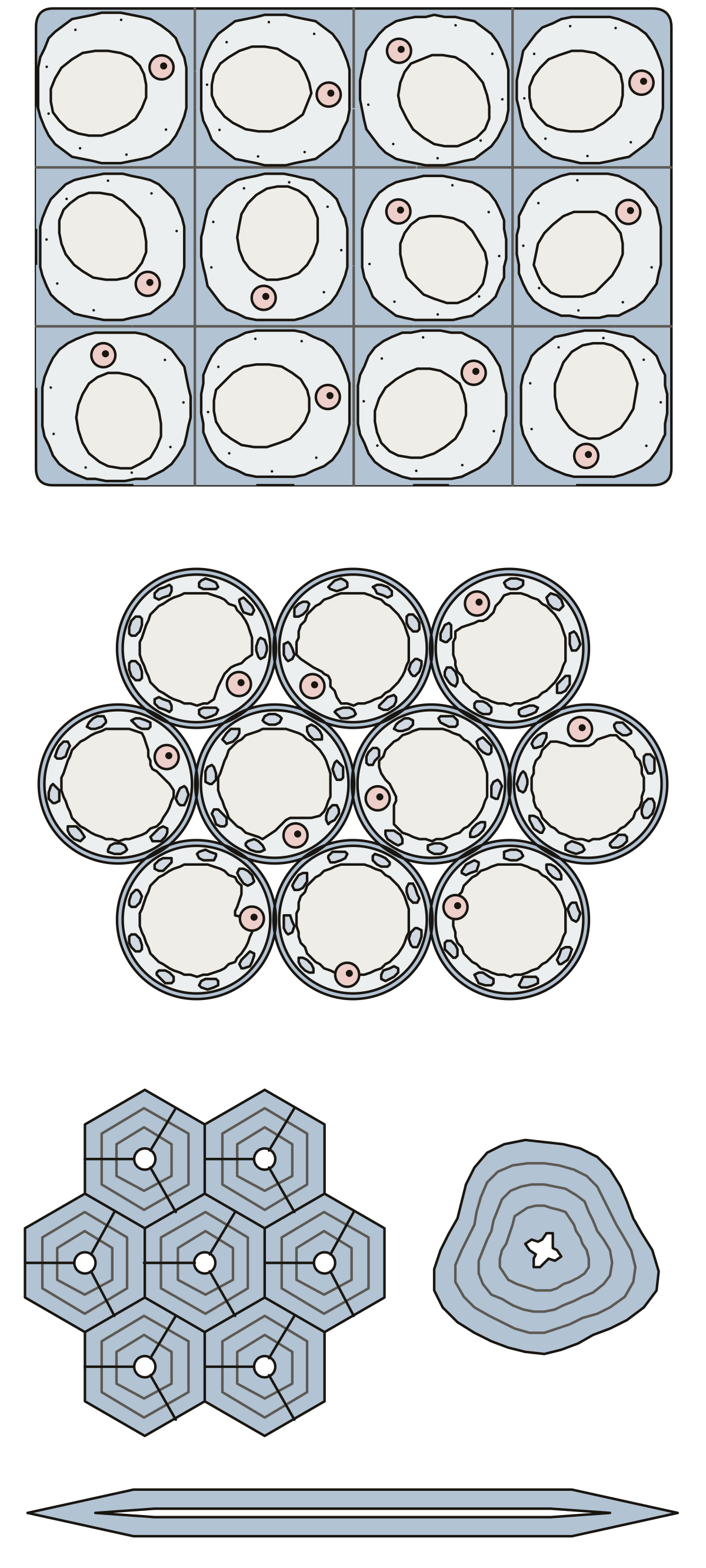 Collenchyma Cells DiagramCollenchyma with its thickened corners, parenchyma with thin walls, vacuoles and air spaces, and sclerenchyma with lignified walls, pits, fibers and a stone cell.
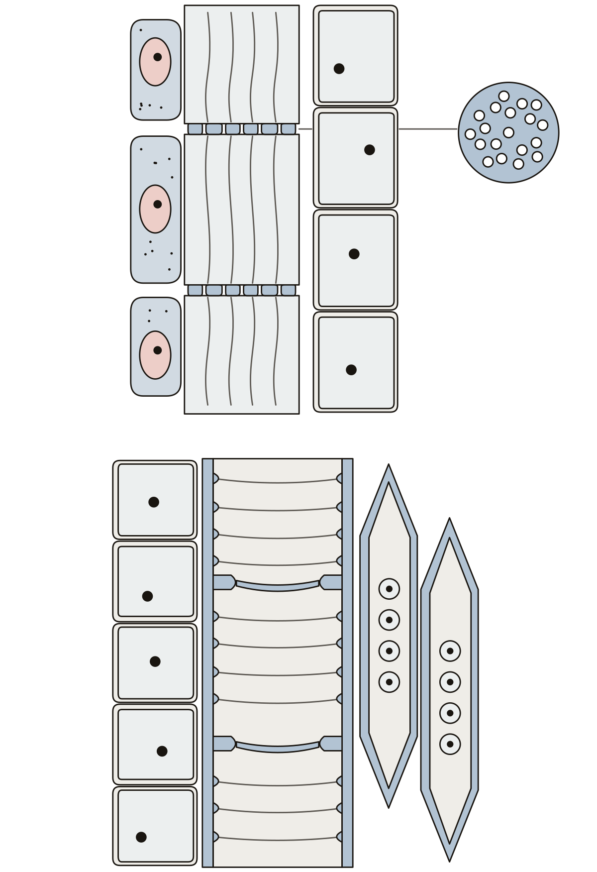 Xylem and Phloem DiagramCompanion cell, sieve tube element, sieve plate and phloem parenchyma, then vessel element, tracheid, pit, perforation plate, lignified wall and xylem parenchyma.
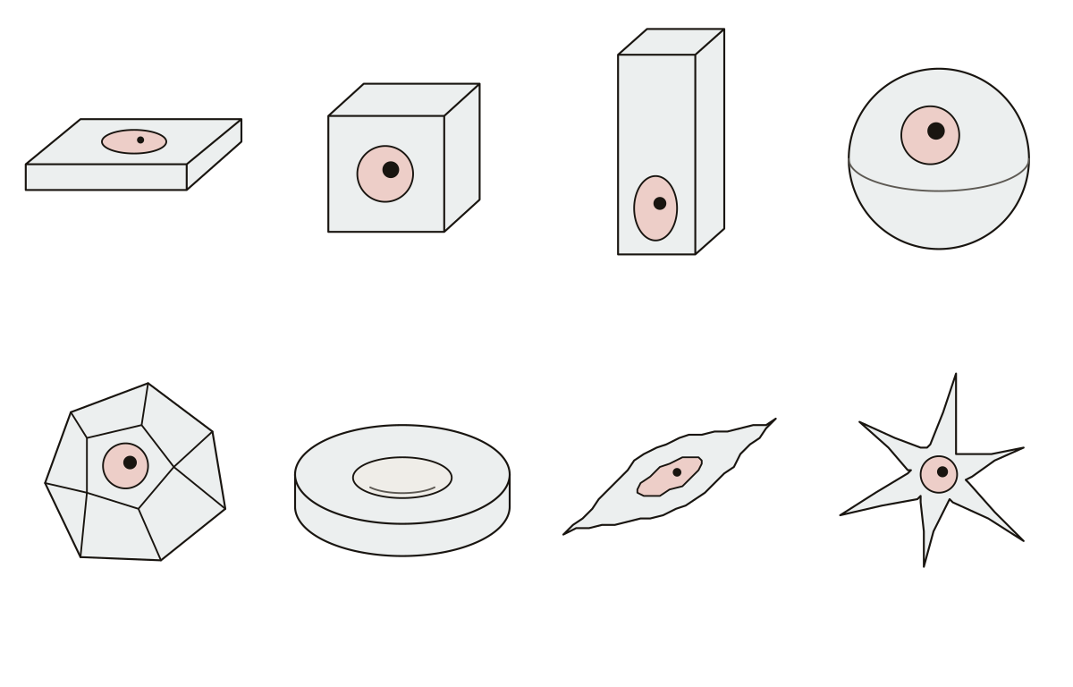 Cell Shape DiagramSquamous, cuboidal, columnar, spherical, polyhedral, discoid, fusiform and stellate cells, each drawn as a small solid, with a cell of that shape.
Single-celled organisms and viruses 26
Paramecium, amoeba, yeast and a green alga are each a whole organism in one cell, and the shapes of bacteria and three bacterial cell walls sit beside them. A virus is not a cell at all, but it is drawn and labeled the same way, so the two virus diagrams sit here.
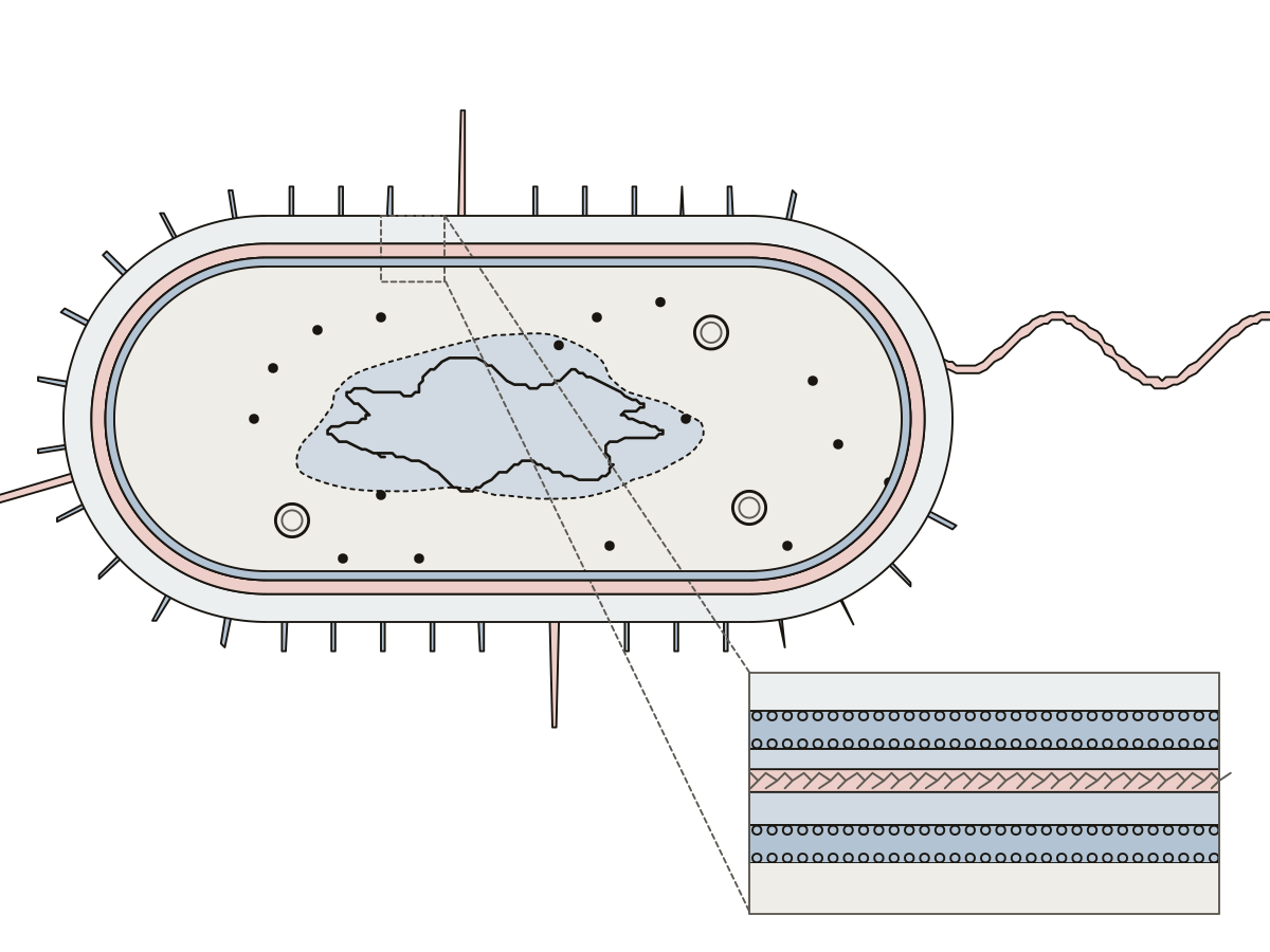 Bacteria Cell DiagramA rod-shaped bacterium cut open and its gram-negative envelope enlarged: 13 numbered parts, from capsule and flagellum to peptidoglycan and periplasm.
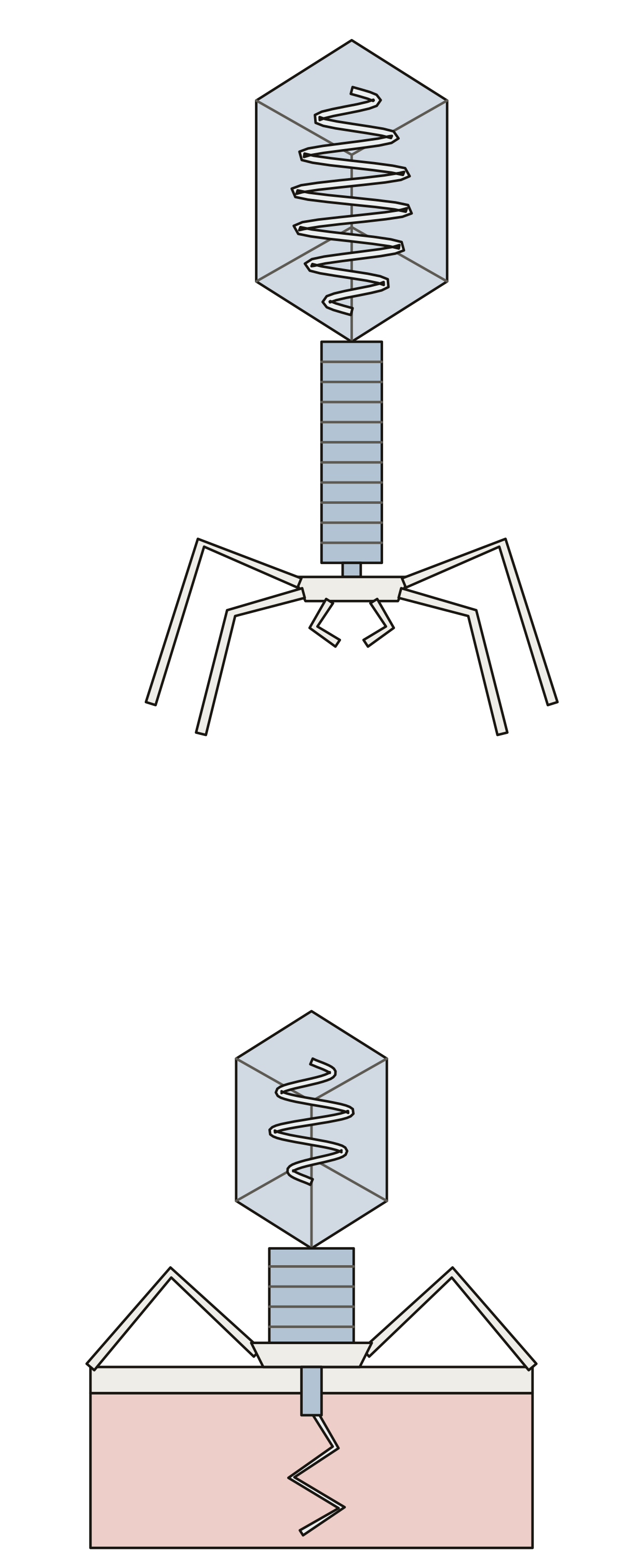 Parts of a BacteriophageA T4 phage: head, DNA, tail sheath, baseplate, long and short tail fibres, and the contracted sheath and tail tube during infection. Head · Outer membrane · DNA
- Bacteriophage Unlabeled DiagramThe bacteriophage drawing with nothing written on it, to print and label: a T4 phage free and landed on a bacterium, nine parts and the answer key. Short tail fibres · Head · Contracted tail sheath
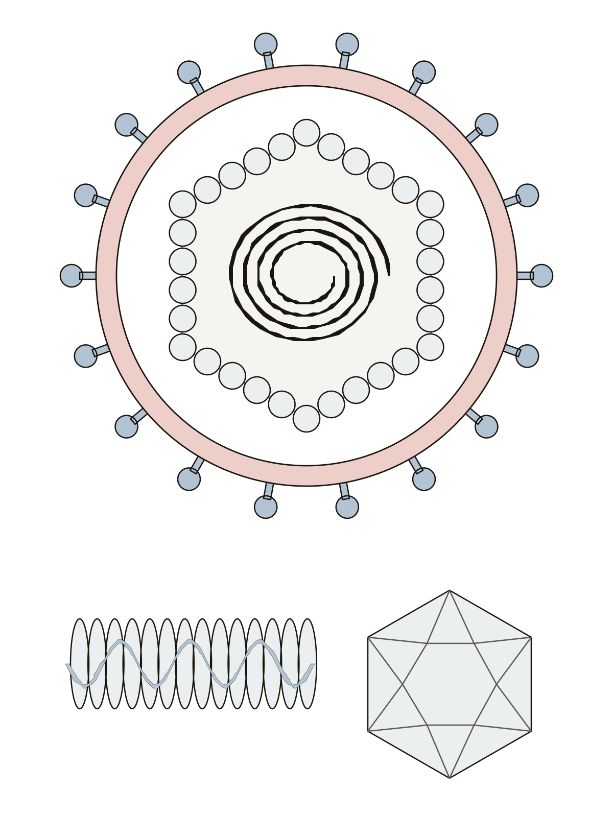 Virus Diagram LabeledA virus cut in half: 7 numbered parts, from the genetic material, capsid and capsomeres to the envelope, spike proteins and the two capsid shapes. Genetic material · Icosahedral virus · Spike proteins
- Virus Diagram UnlabeledThe virus drawing with nothing written on it, to print and fill in: an enveloped virus cut in half, a rod and a many-sided ball, with the answer key. Genetic material · Capsid · Spike proteins
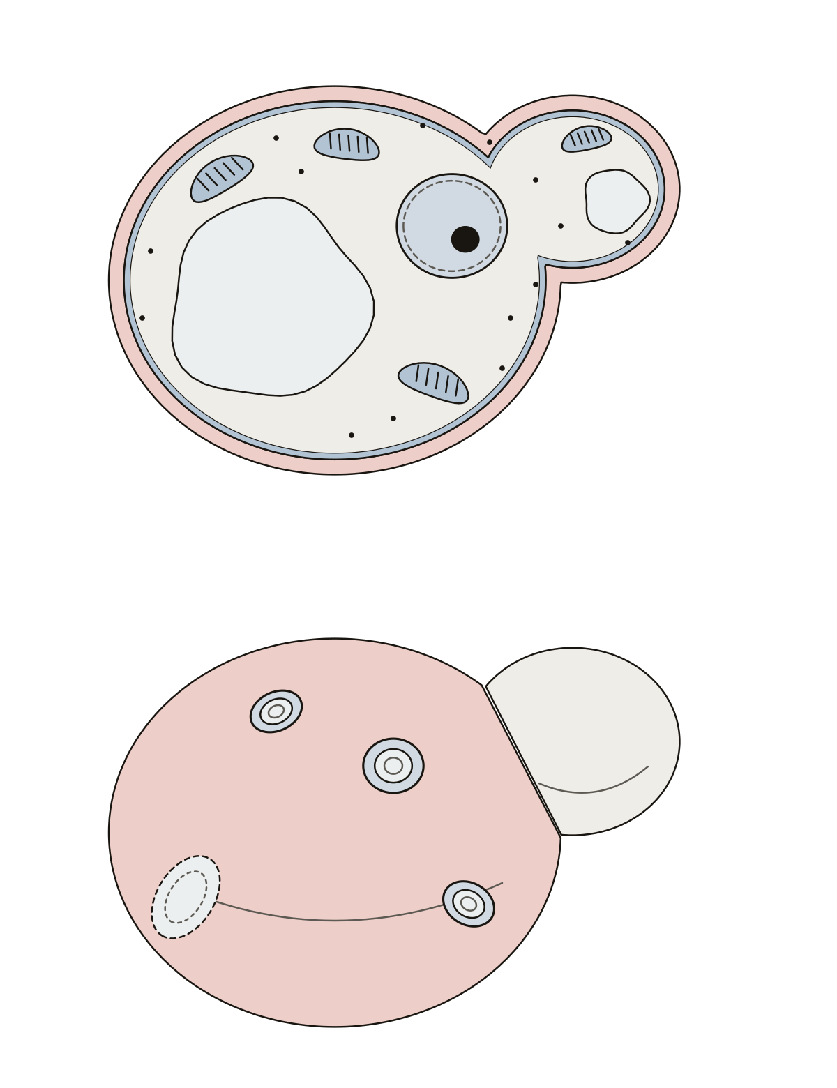 Yeast Cell DiagramA budding yeast cell cut open (wall, membrane, nucleus, vacuole, mitochondria) and from outside, with the bud, the bud scars and the birth scar.
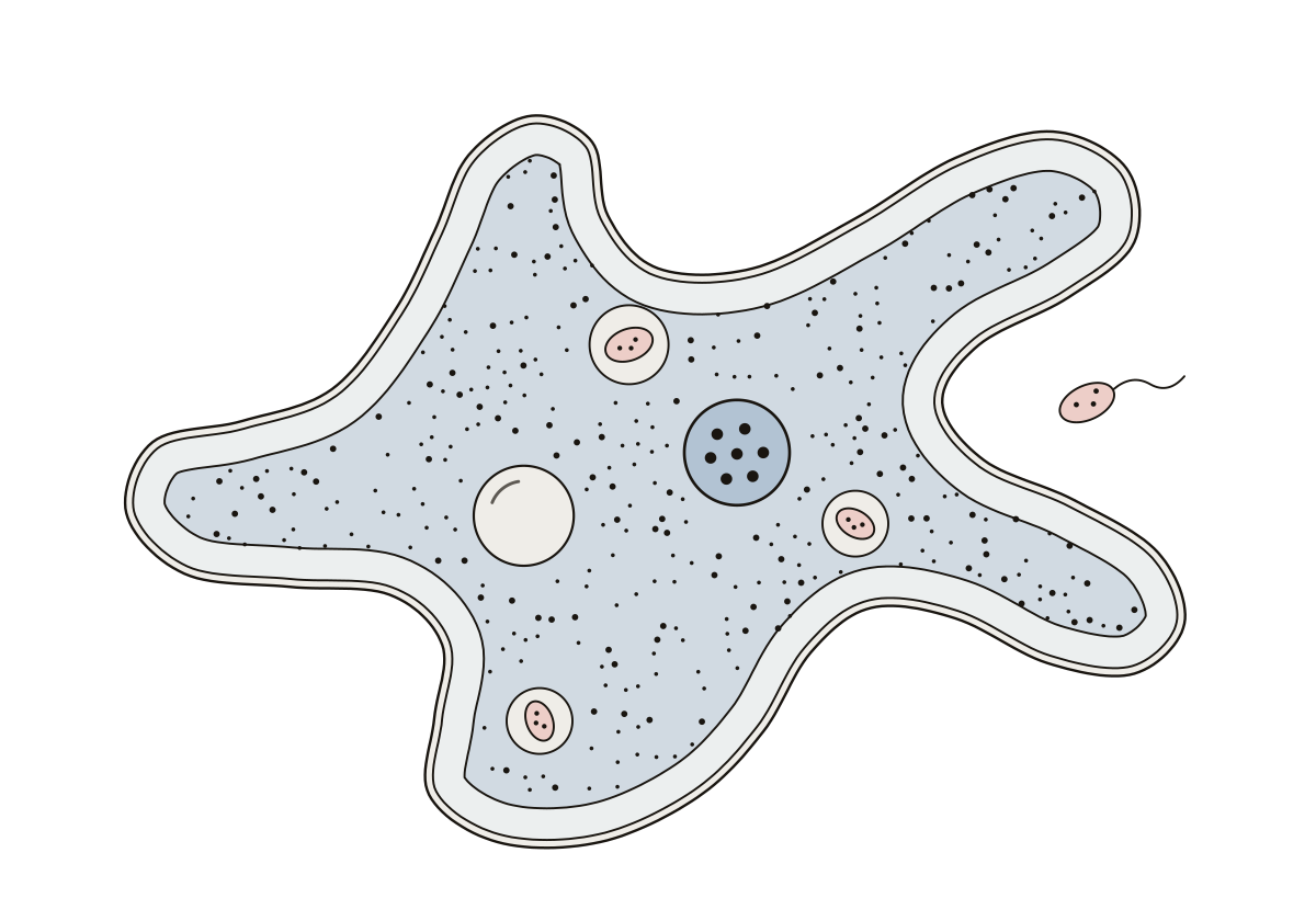 Amoeba Diagram LabeledAn amoeba with its pseudopods, plasma membrane, ectoplasm, endoplasm, nucleus, food vacuoles and contractile vacuole.
- Unlabeled Diagram of the Amoeba WorksheetThe amoeba drawing with nothing written on it, as a worksheet to print: seven parts to name and the answer key, with a word bank on the page.
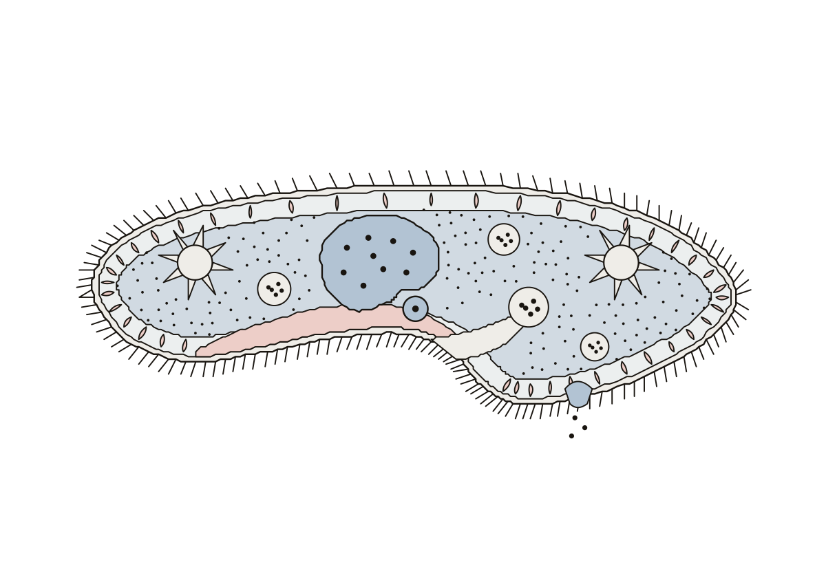 Paramecium Diagram LabeledA paramecium with its cilia, pellicle, trichocysts, oral groove, gullet, food vacuoles, two nuclei, contractile vacuoles and anal pore.
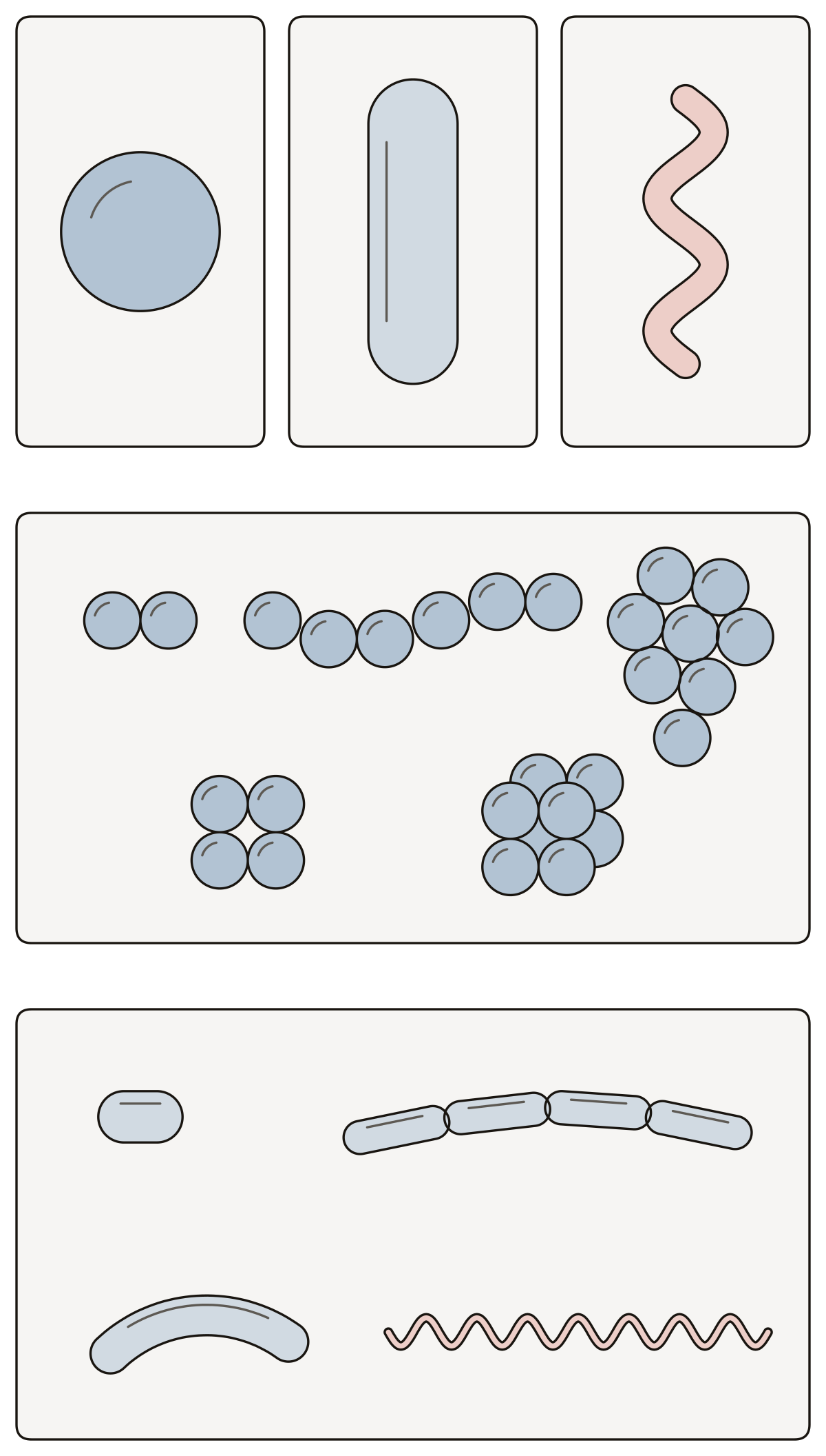 Shapes of BacteriaShapes and arrangements of bacteria: coccus, bacillus, spirillum, diplococcus, streptococcus, staphylococcus, tetrad, sarcina, coccobacillus, streptobacillus, vibrio, spirochete.
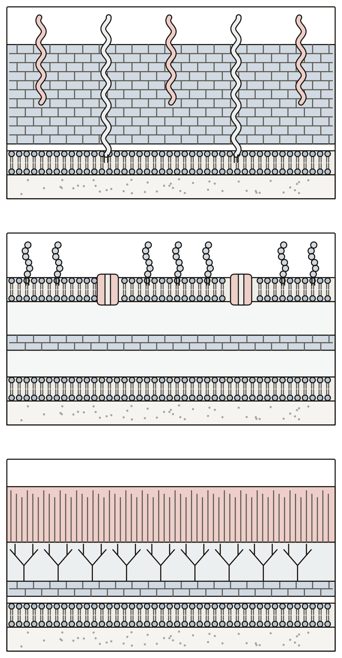 Bacterial Cell Wall Structure DiagramThree bacterial walls in section: peptidoglycan, teichoic and lipoteichoic acid, outer membrane, lipopolysaccharide, porin, periplasm, arabinogalactan, mycolic acids, plasma membrane.
- Blank Bacterial Cell Wall DiagramThree bacterial cell walls in section with nothing written on them, to print and label: Gram-positive, Gram-negative and acid-fast, fourteen names in all, with an answer key.
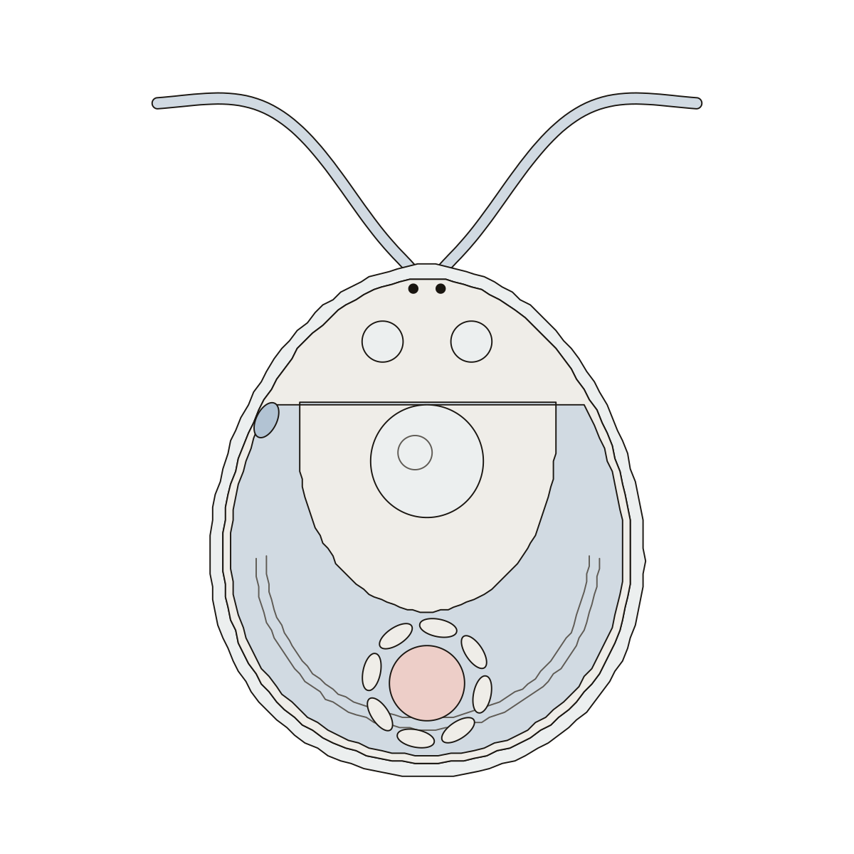 Algae Cell DiagramA single-celled green alga, Chlamydomonas: flagella, cell wall, cup-shaped chloroplast, nucleus, eyespot, contractile vacuoles, pyrenoid, starch sheath.
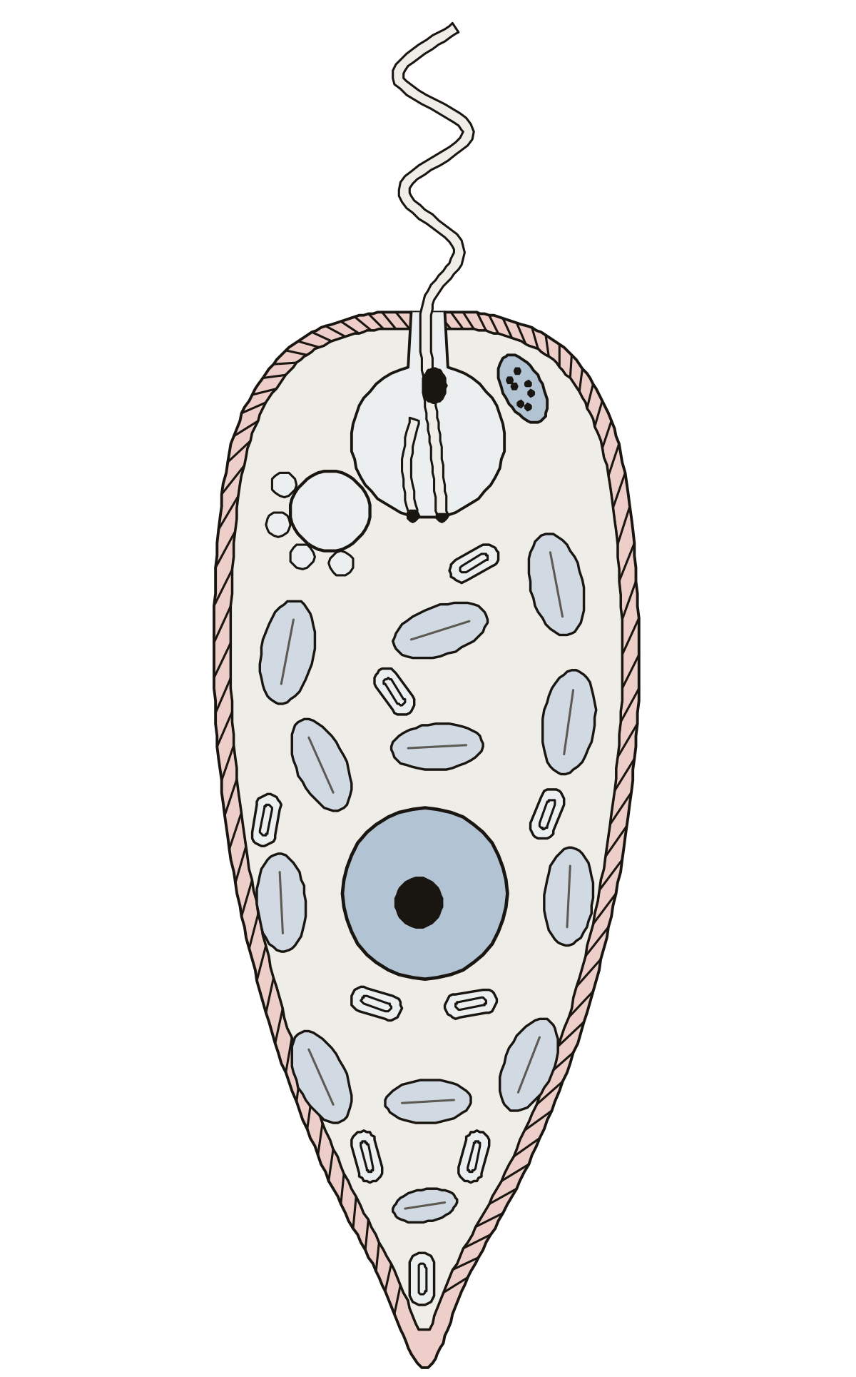 Euglena Cell DiagramA Euglena cut open: flagellum, reservoir, eyespot, photoreceptor, contractile vacuole, pellicle, chloroplasts, nucleus and paramylon granules.
- Fill in the Blank Diagram of a Euglena CellThe euglena drawing with nothing written on it, to print and fill in: nine blanks, a word bank on the page and the answer key.
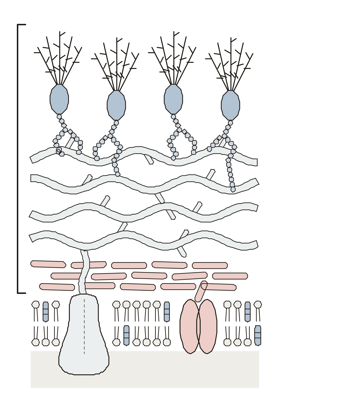 Fungal Cell Wall DiagramA slice of fungal cell wall: plasma membrane with ergosterol, glucan synthase and chitin synthase, then chitin, beta-1,3-glucan, beta-1,6-glucan and mannoproteins.
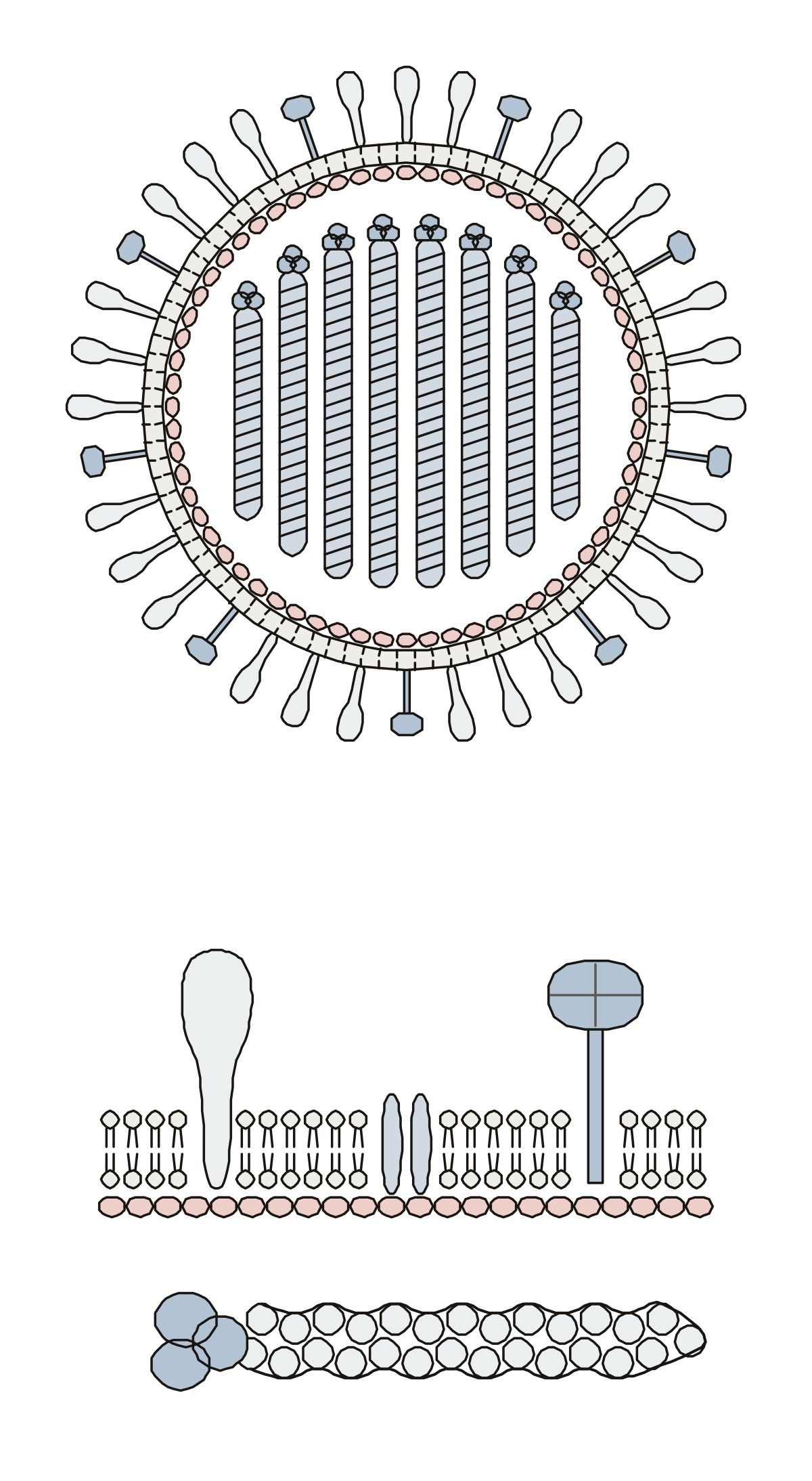 Label the Parts of This Enveloped Influenza VirusAn influenza A virus in two views: hemagglutinin and neuraminidase spikes, envelope and RNA segments, then the M2 ion channel, matrix protein, nucleoprotein and polymerase complex.
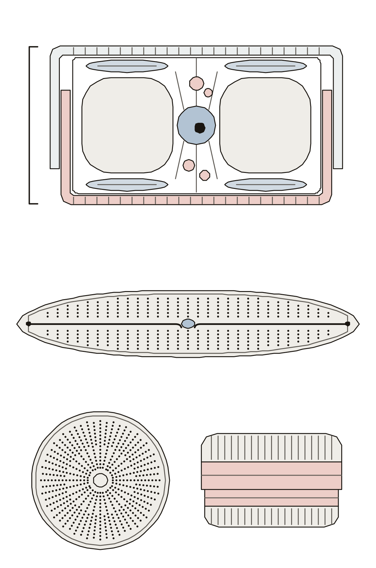 Diatom Cell DiagramA diatom in two views: frustule, epitheca, hypotheca, chloroplasts, nucleus, vacuole and oil droplets, then the raphe, striae, central nodule and girdle band of the shell.
- Label the Diatom CellA diatom as a worksheet: seven numbers on the cell cut open, four on its empty shells, a line for each name, and a key.
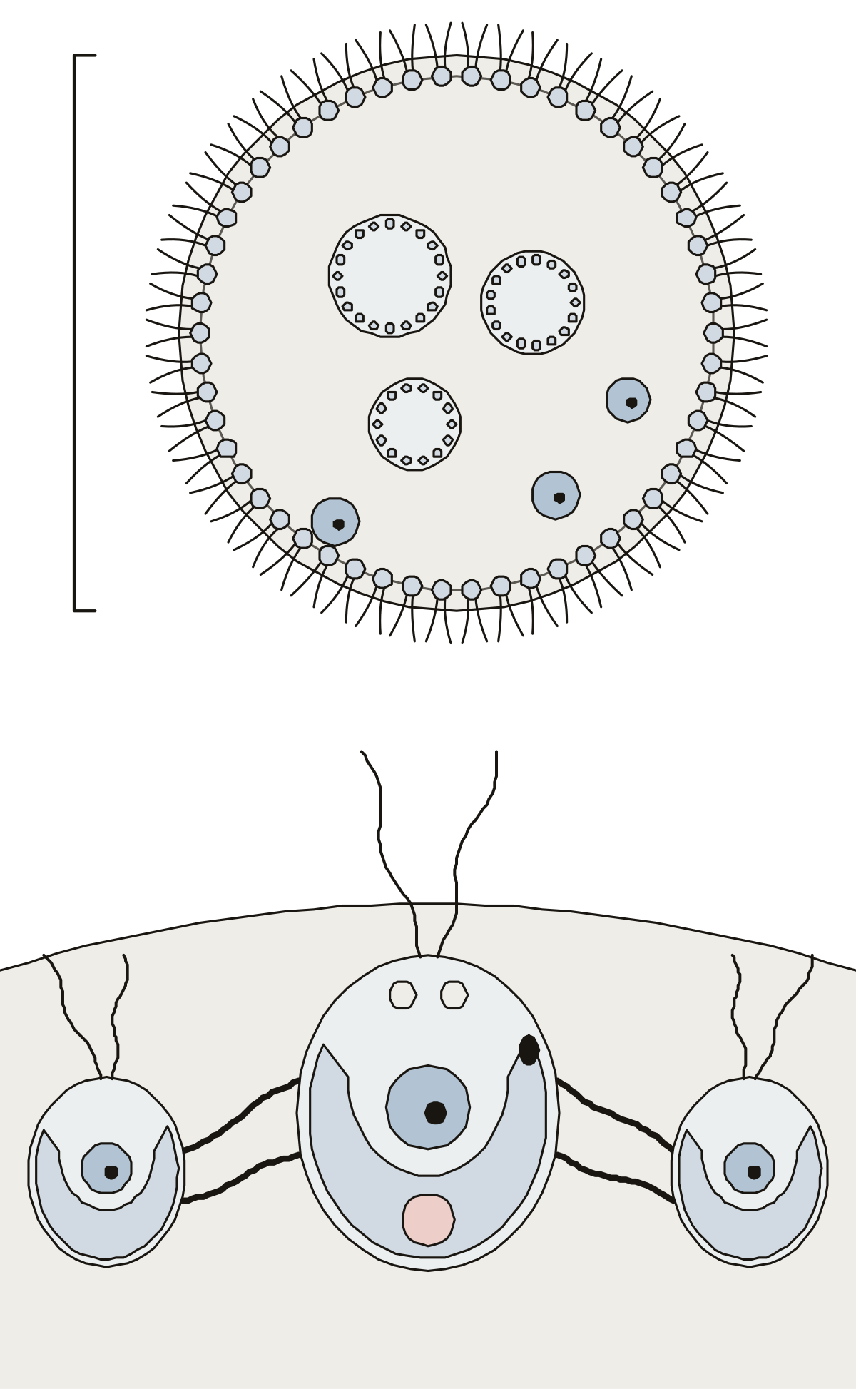 Volvox Diagram LabeledA Volvox colony in two views: somatic cells, gonidia, daughter colonies and gelatinous matrix, then one cell with its flagella, eyespot, chloroplast, nucleus and contractile vacuoles.
- Unlabeled Diagram of VolvoxThe Volvox drawing with nothing written on it, to print and label: the colony and three enlarged cells on one sheet, twelve parts and the answer key.
- Volvox Quiz DiagramA quiz from the first question: find the somatic cells, gonidia, daughter colonies and matrix on the colony, then seven parts of one cell.
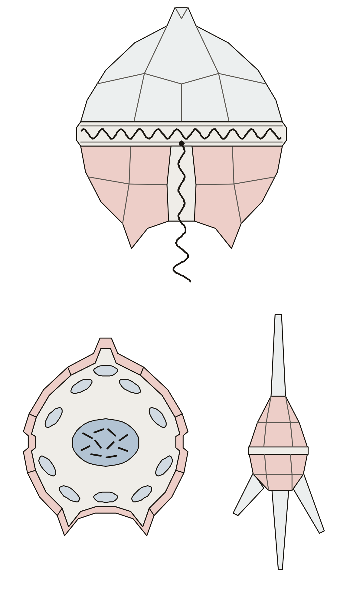 Dinoflagellate Diagram LabeledA dinoflagellate in two views: epitheca, hypotheca, cingulum, sulcus and the two flagella from outside, then the theca, nucleus and chloroplasts inside, and the horns of a horned cell.
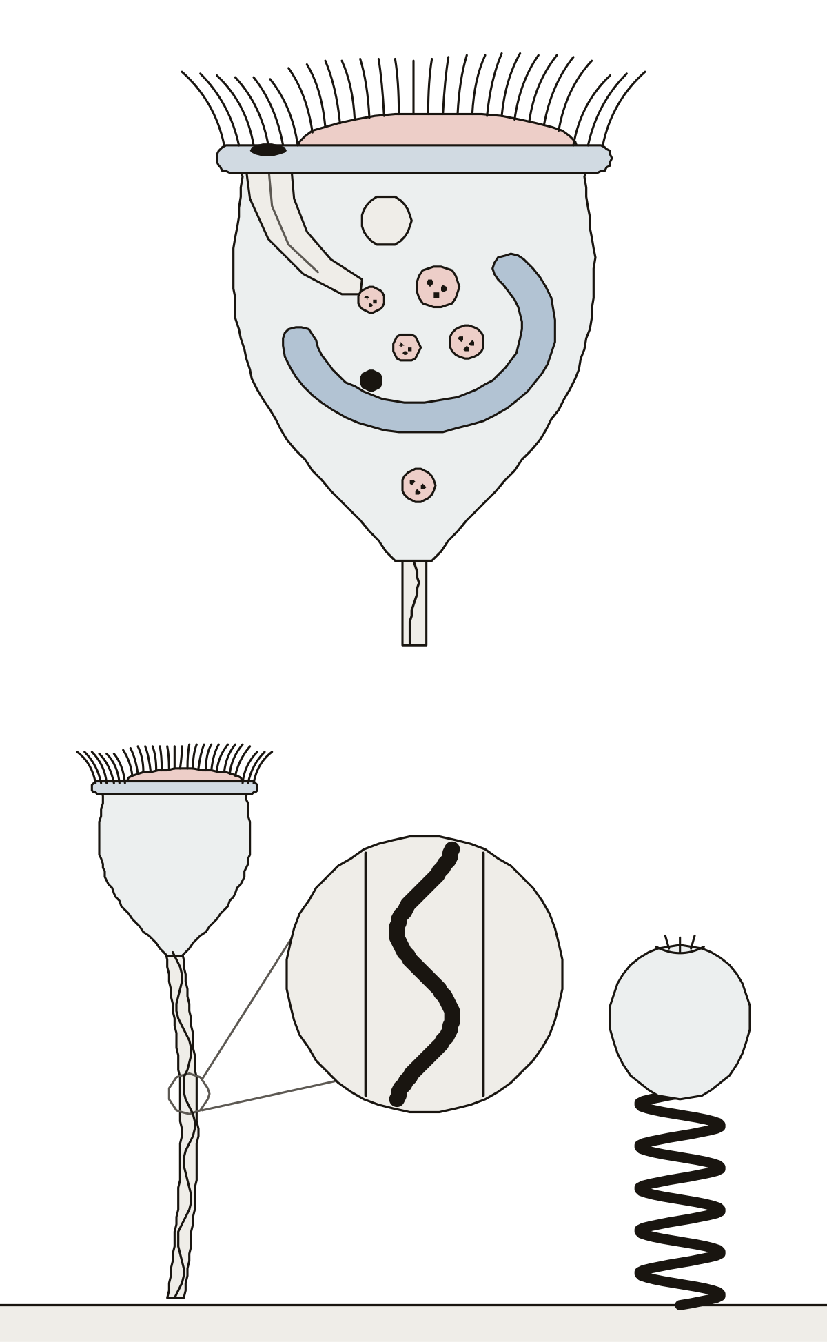 Vorticella LabeledA Vorticella in two views: the bell with its cilia, peristome, gullet, food vacuoles, two nuclei and contractile vacuole, then the stalk, its spasmoneme and the stalk coiled.
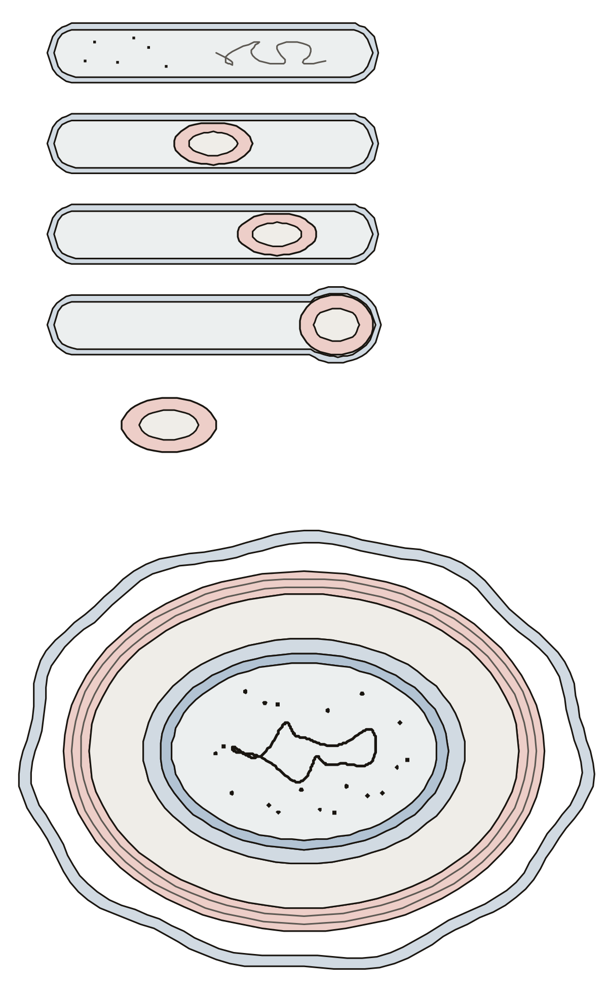 Endospore DiagramAn endospore in two views: the vegetative cell with central, subterminal and terminal endospores and a free spore, then the exosporium, spore coat, cortex, germ cell wall, inner membrane and core.
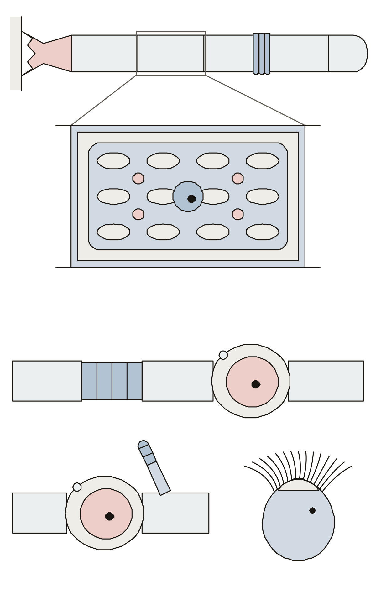 Oedogonium LabeledOedogonium in two views: the filament with its holdfast, caps, net-like chloroplast, pyrenoids and nucleus, then the oogonium, egg, antheridia, dwarf male and zoospore.
More about these tools
In Name it a ring lights up and you choose or type the name. Study turns every label on, with a line on what each part does. Missed parts come back at the end of the round, and every drawing prints as a blank worksheet or as a labeled answer key.
Start with a whole cell: plant, animal or bacteria. Then move in closer. The nucleus, mitochondrion, chloroplast, Golgi apparatus and cell membrane each have a larger drawing that shows the inner parts that are too small to tap on a whole cell. The drawings were made for this site, and names and facts were checked against open college textbooks on the date each page shows.
Keep exploring
- Animal body-part diagrams (94)
- Blank US maps (61)
- Blank maps (159)
- Bone labeling quizzes (49)
- Capitals quizzes (11)
- Car and engine part diagrams (54)
- Clothing, shoes and jewelry (45)
- Computer, phone and camera diagrams (43)
- Anatomy concept maps (1)
- Country map quizzes (28)
- Animal anatomy diagrams (100)
- DNA, cell division and cell processes (53)
- Science Quizzes
- All quizzes