Eye and Nervous System Diagrams: One Drawing per Page to Label
One drawing per page, a numbered ring on each part, to label on screen or to print blank.
74 pages
Nerve cells and nerves 9
A motor neuron from dendrites to axon terminals, the three shapes a neuron can take, and a nerve cut across to show the layers that bundle its axons; then the chemical synapse and the neuromuscular junction, where one cell signals the next, and the action potential as a graph.
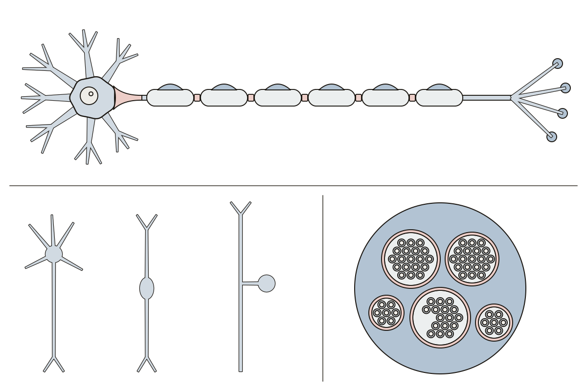 Labeled Neuron DiagramA motor neuron from dendrites to axon terminals, the three neuron shapes and a nerve cut across: 16 numbered parts to label or print blank.
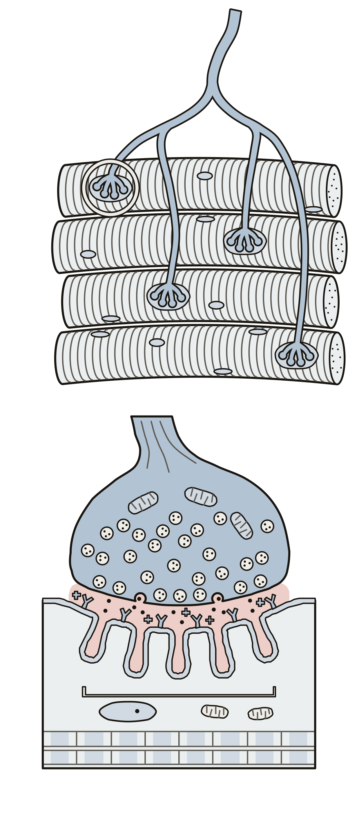 Neuromuscular Junction Labeled DiagramWhere a motor neuron meets a muscle fibre, placed and then enlarged: 12 parts to name. Motor neuron axon · Acetylcholine · Neuromuscular junction
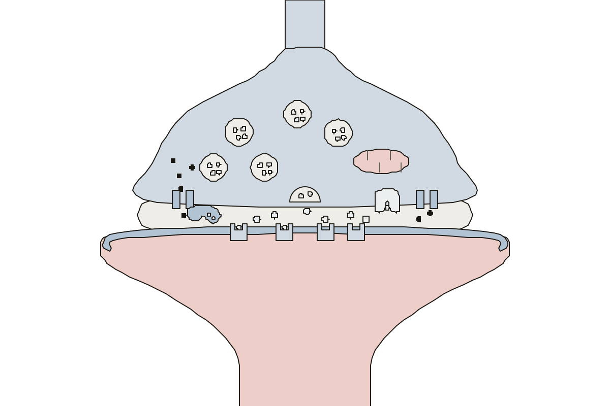 Chemical Synapse Diagram LabeledOne synapse from the sending bulb to the receiving cell: 12 parts to name. Neurotransmitter · Mitochondrion · Synaptic cleft
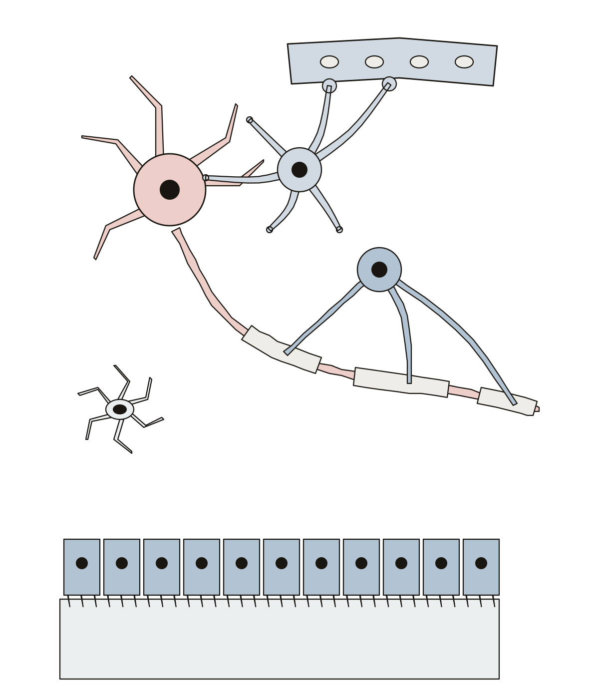 Glial Cells DiagramGlial cells of the CNS: 8 numbered parts, from the astrocyte, oligodendrocyte and myelin sheath to the microglia, ependymal cells, capillary and ventricle. Microglia · Neuron · Astrocyte
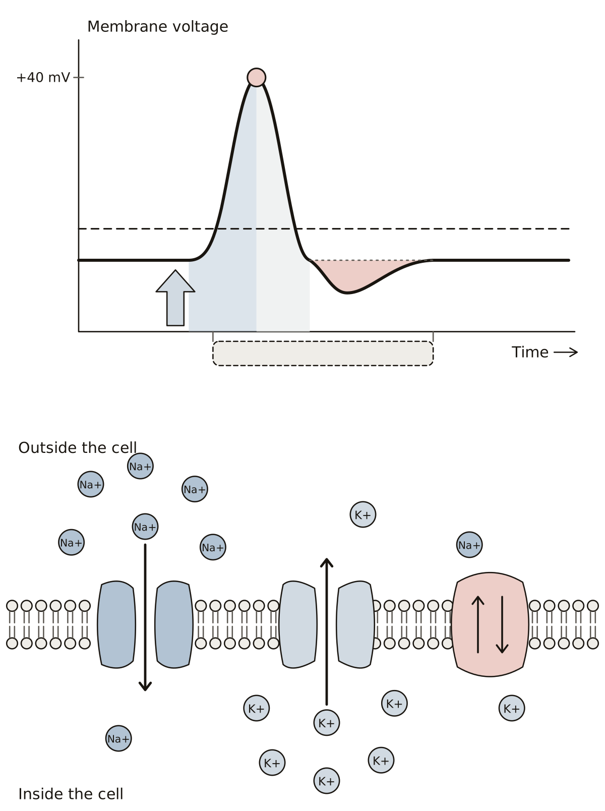 Action Potential DiagramResting potential, stimulus, threshold, depolarization, peak, repolarization, undershoot and refractory period on a graph; then the sodium channel, the potassium channel and the pump in the membrane.
- Action Potential Diagram UnlabeledThe action potential drawing with nothing written on it, to print and fill in: eight lines on the graph, five on the membrane, a word bank and an answer key.
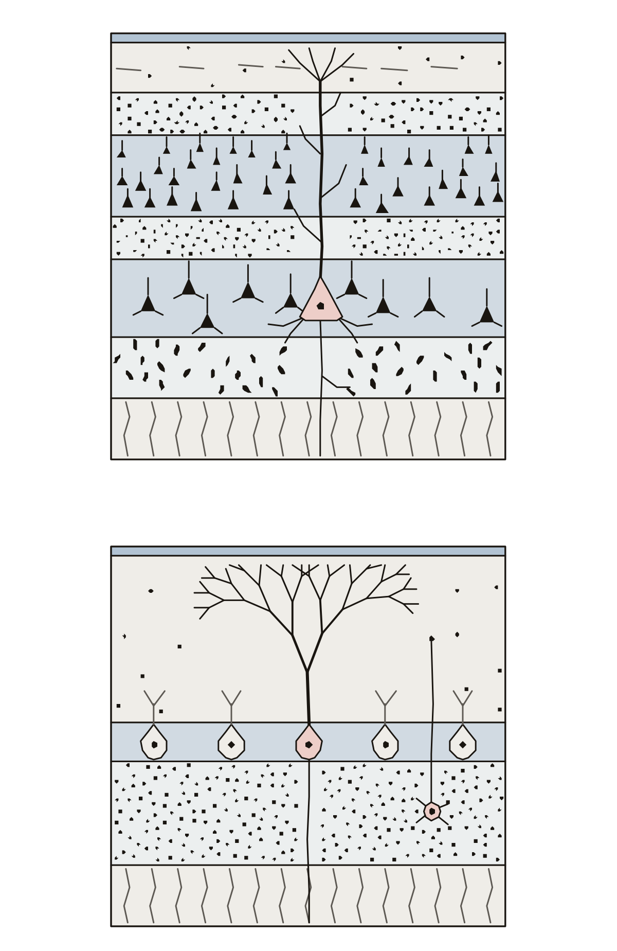 Brain Histology Labeled: Layers of the CortexBrain tissue as on a slide: the six layers of the cerebral cortex with one pyramidal cell and its apical dendrite, and the three layers of the cerebellar cortex with a Purkinje cell, its dendritic tree and a granule cell.
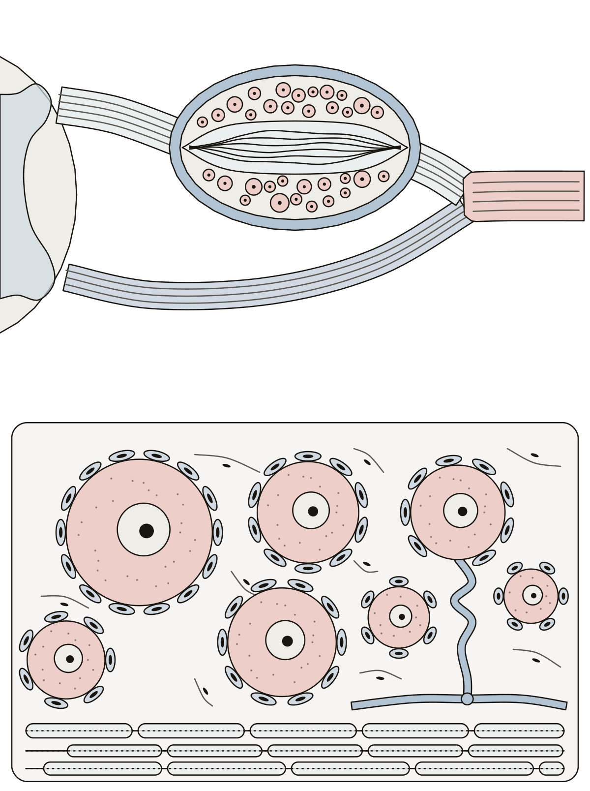 Dorsal Root Ganglion Histology LabeledDorsal root, ganglion, capsule, fiber bundles, ventral root and spinal nerve; then the cell body, nucleus, nucleolus, satellite cells, T-shaped process and myelinated fibers.
- Label the Dorsal Root Ganglion HistologyDorsal root ganglion histology as a worksheet: six numbered parts on the ganglion and its roots, seven on the cells enlarged, a line for each name, and a key.
Sense organs 17
The eye from the cornea to the optic nerve, with its retina cells in a close-up, and the eyelids and tear system in front of it; the retina layer by layer, and one rod beside one cone; the ear; the outer ear, the middle ear, the inner ear, the cochlea and a taste bud, each drawn large.
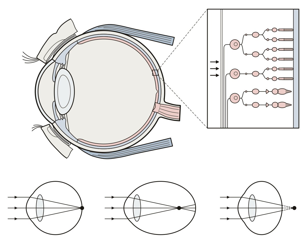 Label the EyeCornea to optic nerve on a side view of the eye, with rods and cones up close and a nearsighted eye beside a normal one. Label it on screen or print it blank.
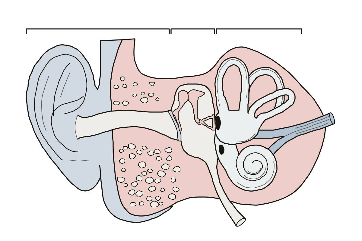 Label the EarOne section through the ear, outside to inside: 17 parts to name, labeled or blank. Oval window · Round window · Temporal bone
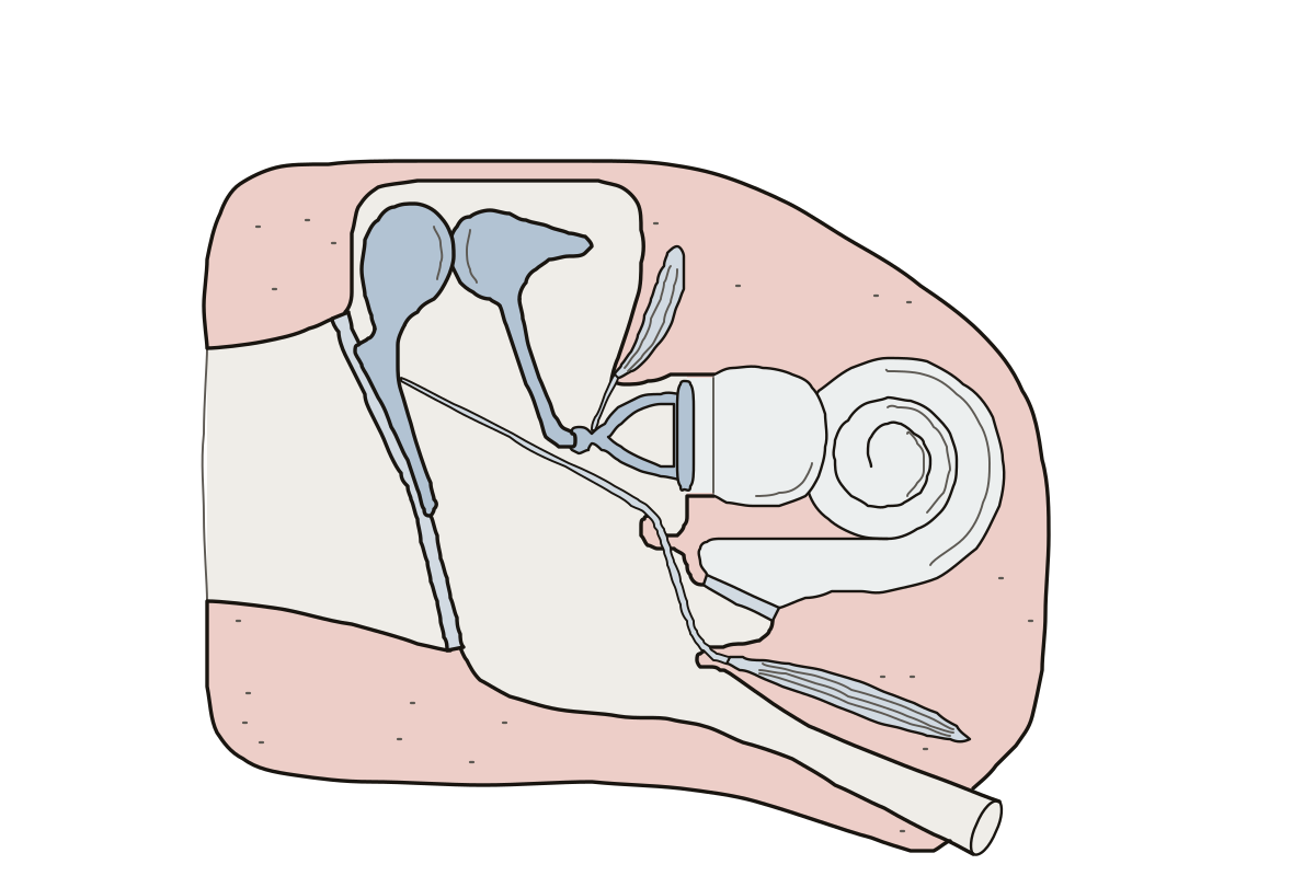 Middle Ear DiagramThe middle ear drawn large, bone by bone: 13 parts to name. Crura of the stapes · Footplate of the stapes · Stapedius
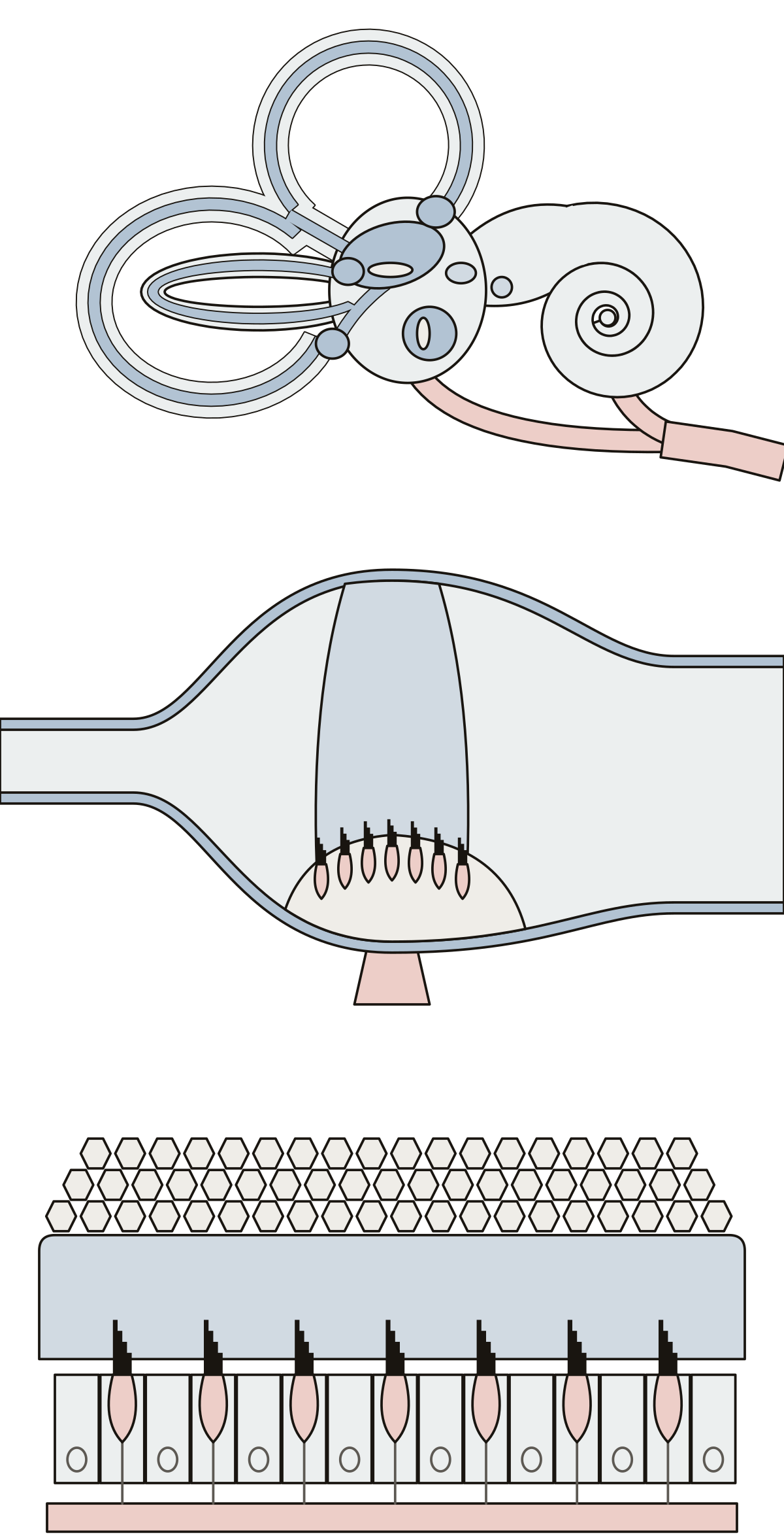 Inner Ear DiagramSemicircular canals and ducts, ampulla, cupula, utricle, saccule, macula, otolithic membrane, otoliths, hair cells, windows and nerves.
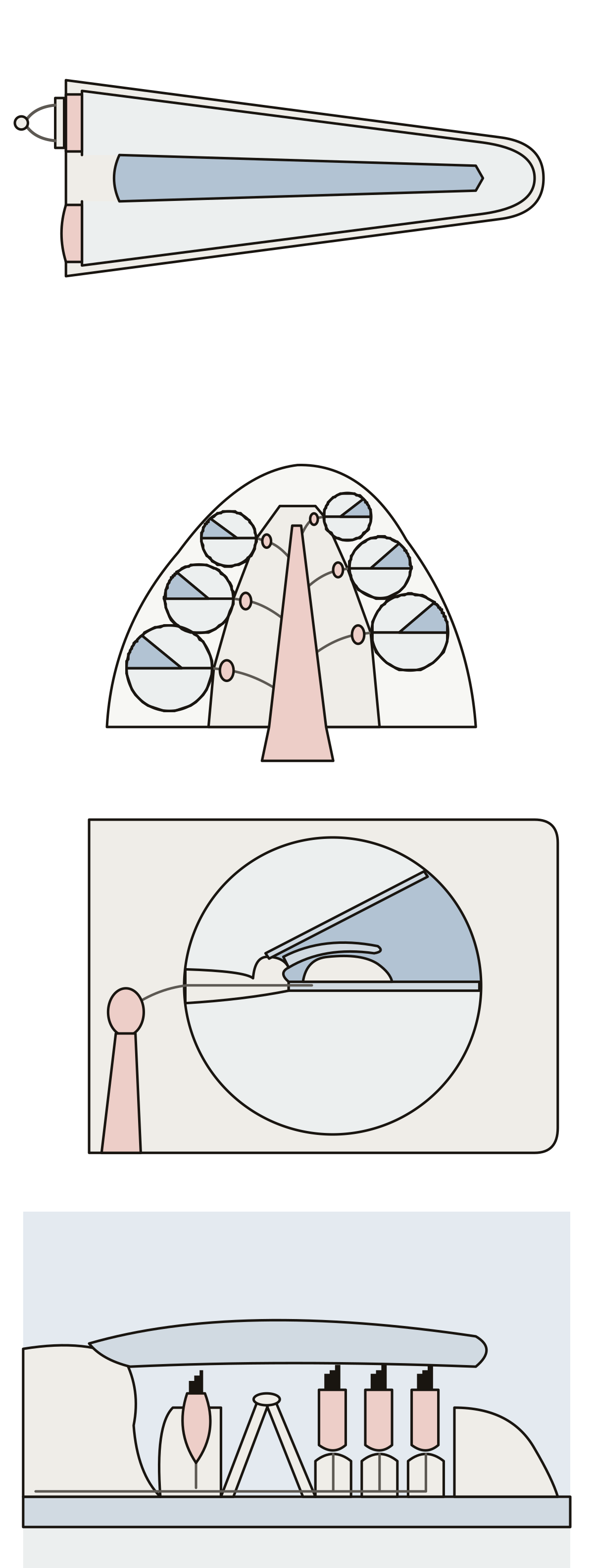 Cochlea DiagramScala vestibuli, cochlear duct, scala tympani, helicotrema, modiolus, spiral ganglion, basilar and tectorial membranes, inner and outer hair cells.
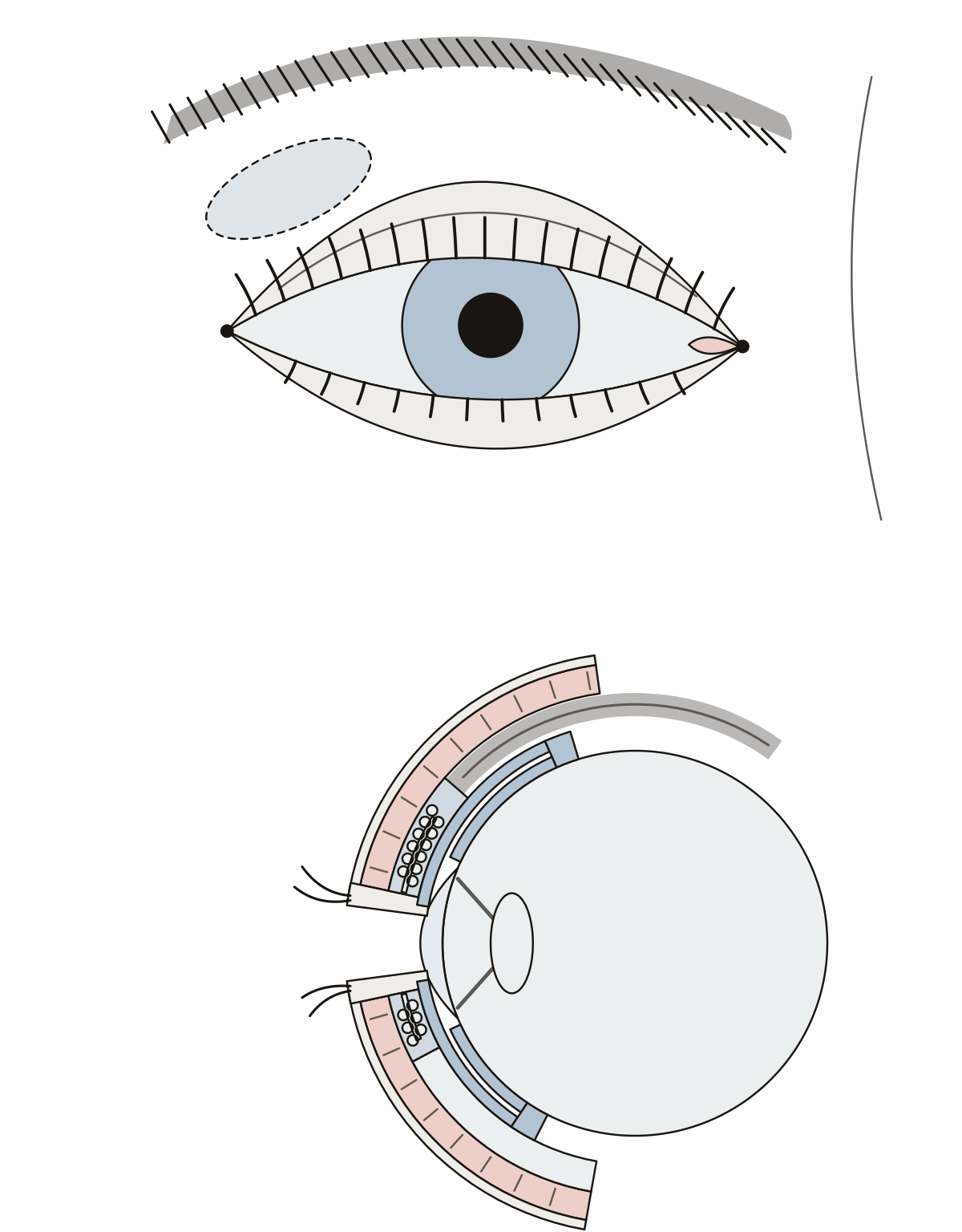 Accessory Structures of the Eye DiagramEyebrow, eyelids, eyelashes, canthi, lacrimal gland, orbicularis oculi, tarsal plate and glands, conjunctiva, fornix, levator palpebrae superioris.
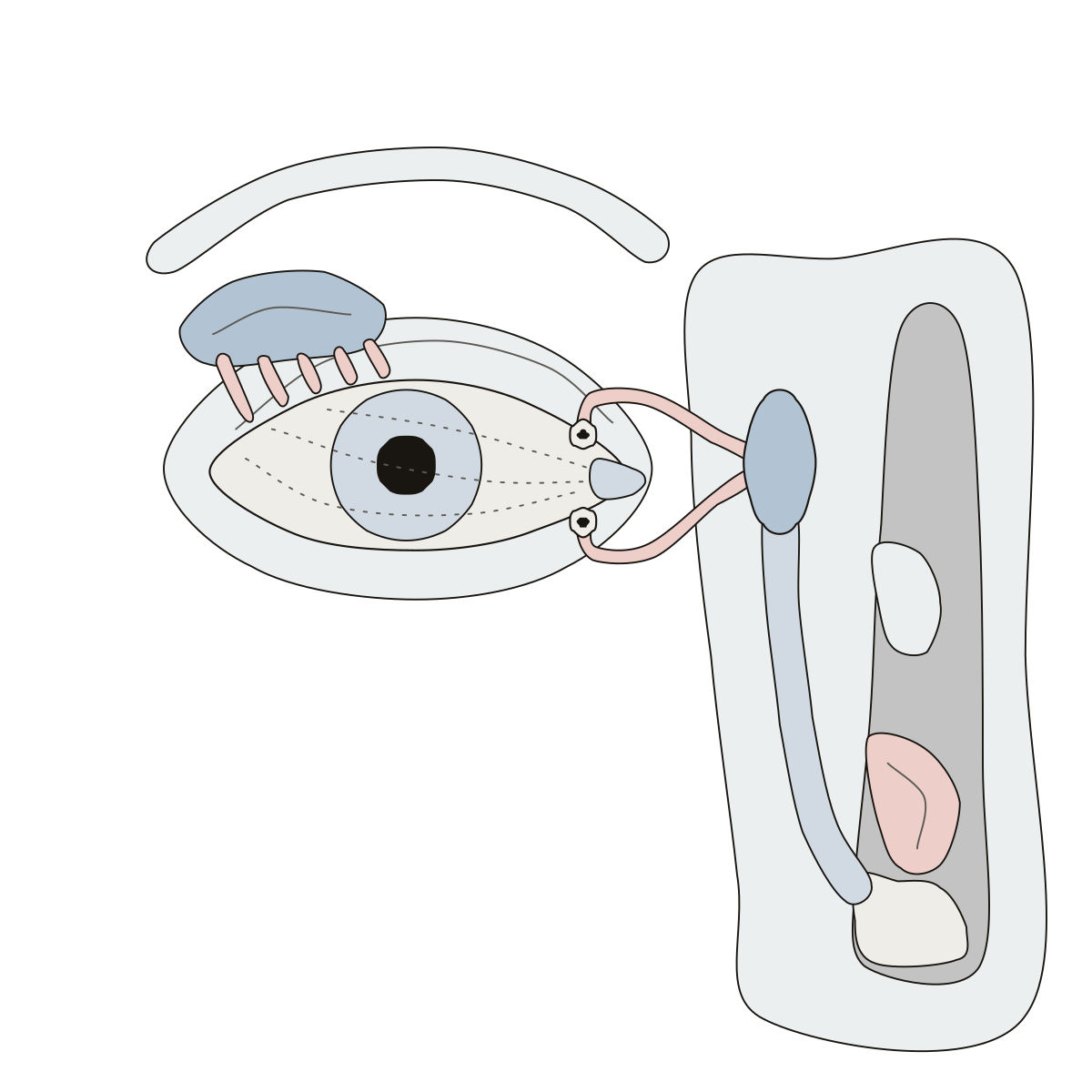 Lacrimal Apparatus DiagramWhere tears are made and where they go: lacrimal gland, puncta, canaliculi, lacrimal sac and nasolacrimal duct, numbered in the order a tear meets them.
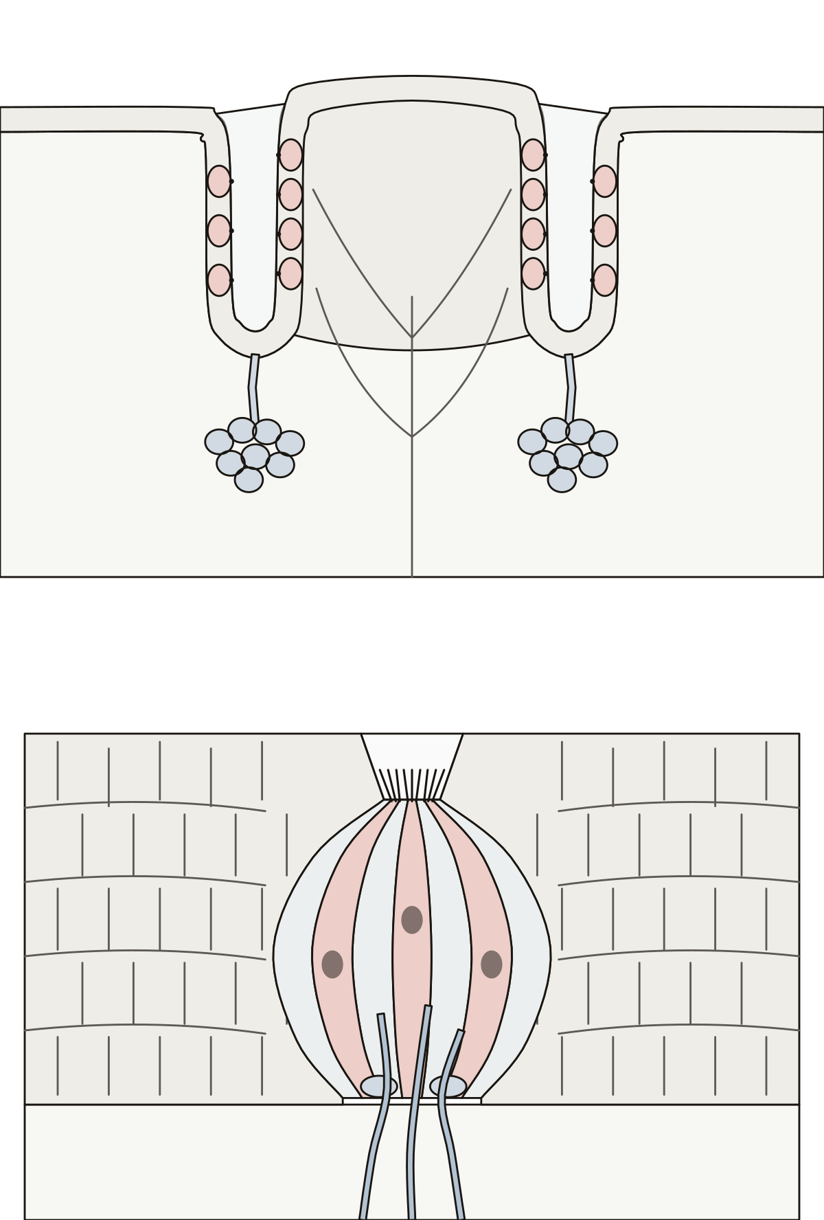 Taste Bud DiagramOne taste bud enlarged, with its taste pore, gustatory hairs, gustatory cells, supporting cells and nerve fibers, and a vallate papilla cut through to show the buds in its walls.
- Taste Buds Diagram UnlabeledThe taste bud drawing with nothing written on it, to print and fill in: a vallate papilla cut open, one bud enlarged, a word bank for each and an answer key.
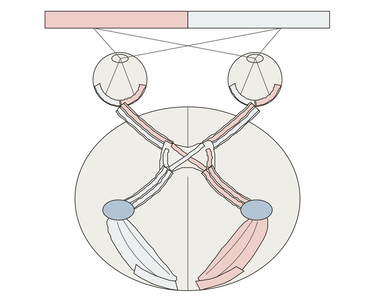 Visual Pathway DiagramThe two half fields, the nasal and temporal halves of the retina, optic nerve, optic chiasm, optic tract, lateral geniculate nucleus, optic radiation and primary visual cortex.
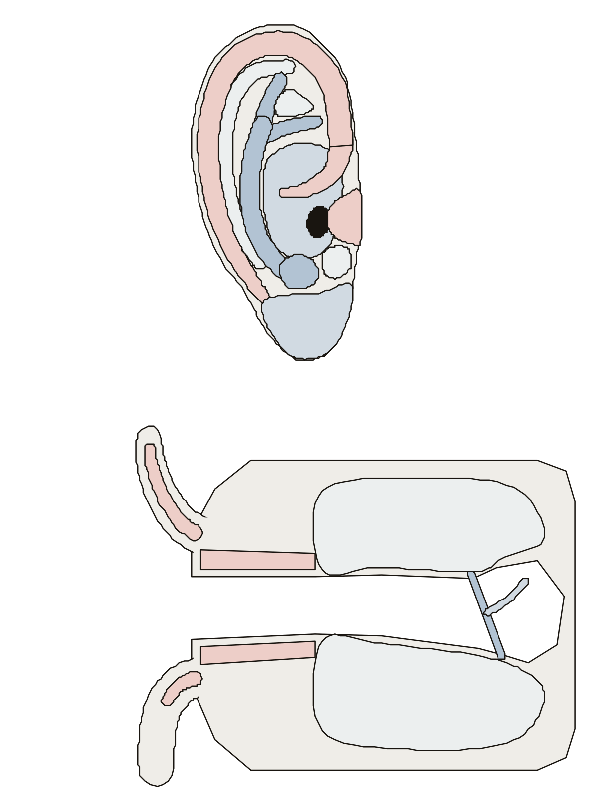 Outer Ear DiagramHelix, antihelix and its crura, triangular fossa, scapha, concha, tragus, antitragus and lobule on a right ear; then the auricular cartilage, the two parts of the ear canal and the eardrum in a cut.
- Outer Ear Anatomy QuizA quiz from the first screen on the outer ear drawing: find twelve folds and hollows on the auricle, then the cartilage, the two parts of the canal and the eardrum in a cut.
- Unlabeled Diagram of Outer EarThe outer ear drawing with nothing written on it, to print and fill in: twelve lines round the auricle, four on the ear canal cut open, a word bank and an answer key.
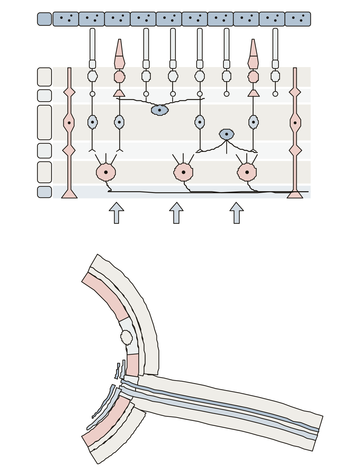 Retina DiagramRods, cones, bipolar, horizontal, amacrine, Müller and ganglion cells in their layers, light coming from below; then the macula, fovea, optic disc and the central retinal artery and vein.
- Retina Diagram UnlabeledBoth retina drawings without their names, for printing: fifteen lines on the layers and cells, six where the nerve leaves the eye, with the word bank and the key.
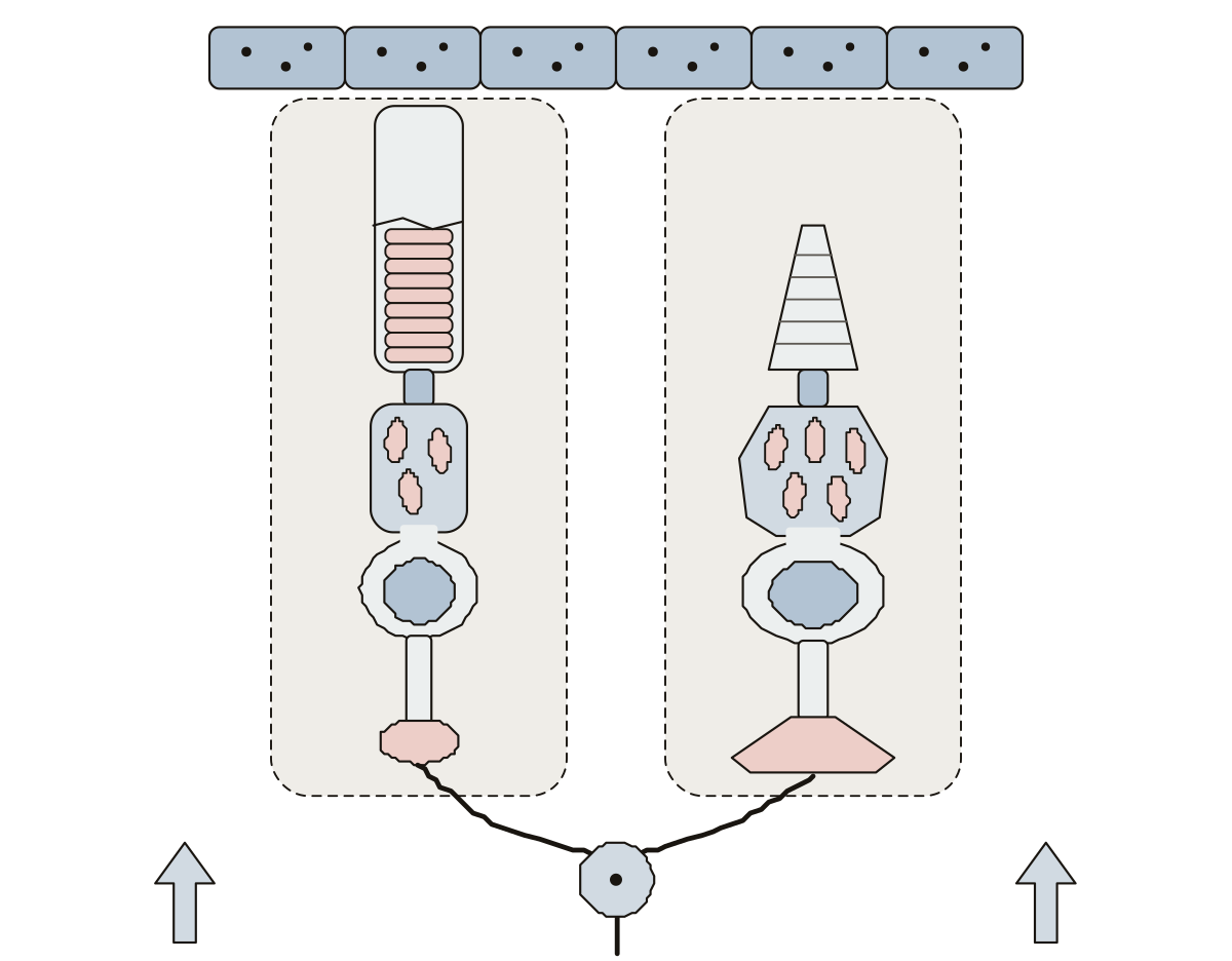 Rod and Cone Cells DiagramA rod and a cone standing side by side: outer segment, membrane discs, connecting cilium, inner segment, mitochondria, nucleus, axon and synaptic terminal, with the pigment epithelium above and a bipolar cell below.
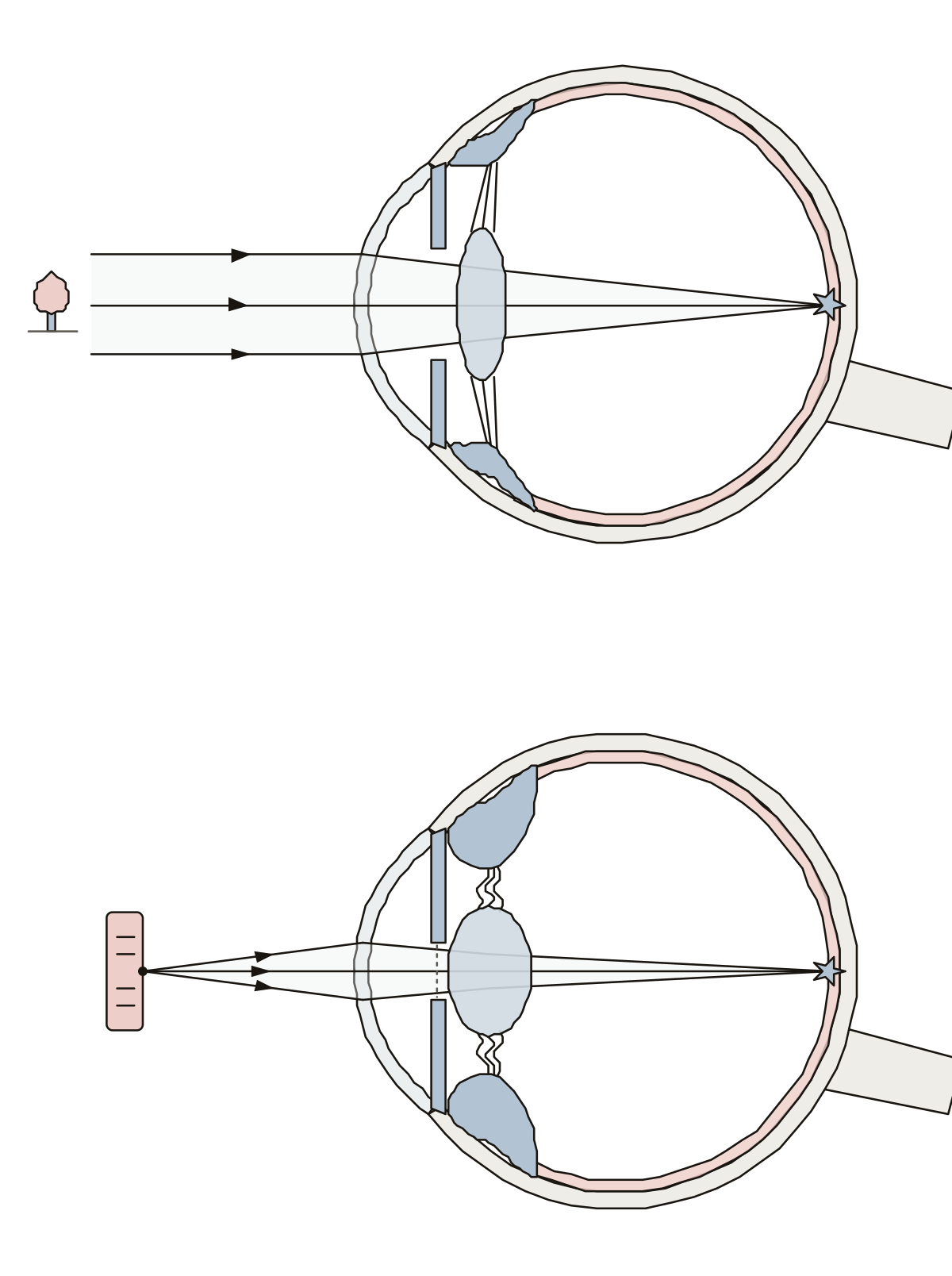 Accommodation of the Eye DiagramRelaxed and contracted ciliary muscle, taut and slack suspensory ligaments, flattened and rounded lens, light from a far and a near object, the narrowed pupil and the focus on the retina.
Brain maps and cranial nerves 31
The whole brain from the side and cut down the middle, lobes to brainstem; the cerebellum cut down the middle and from above; the cortex's functional areas, from the motor strip to Broca's and Wernicke's areas; the somatosensory cortex as a strip map, toes to pharynx, with the motor map beside it; the twelve cranial nerves as numerals that draw a face; and the four ventricles as a see-through cast. Then come the limbic system, the basal ganglia, a horizontal section, and the whole brain from the front, from behind and from above.
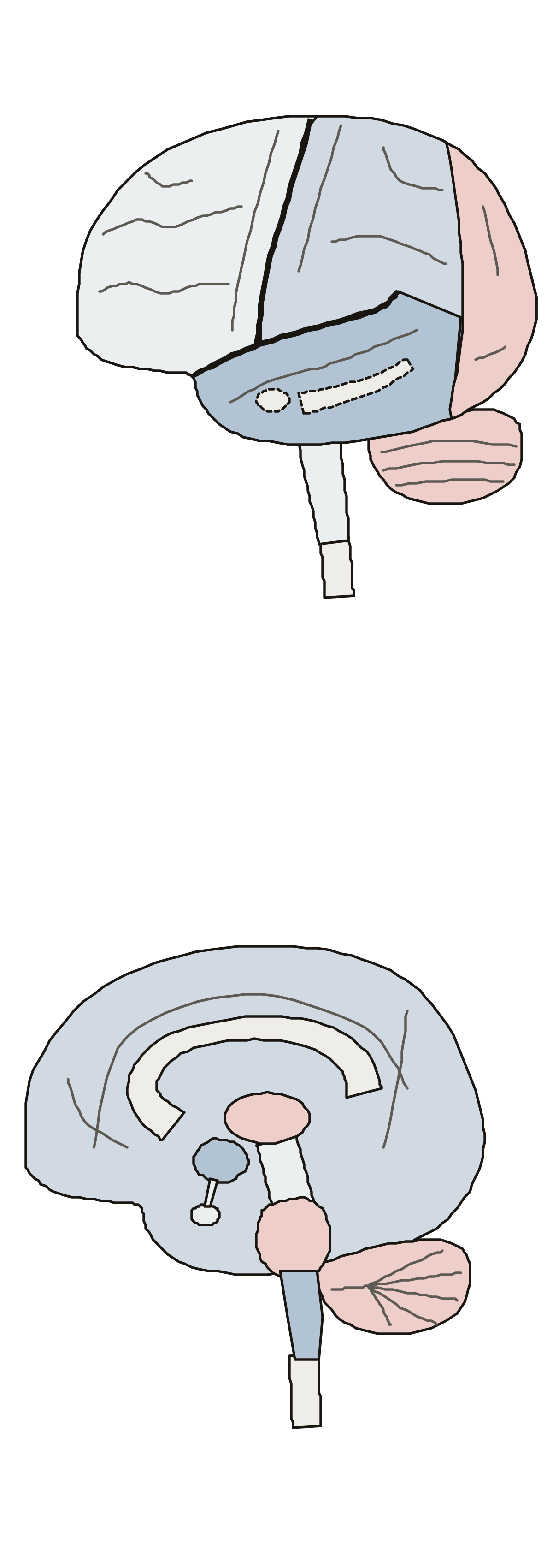 Brain Labeling QuizThe brain from the side and cut down the middle: 17 parts to name. Parietal lobe · Pituitary gland · Thalamus
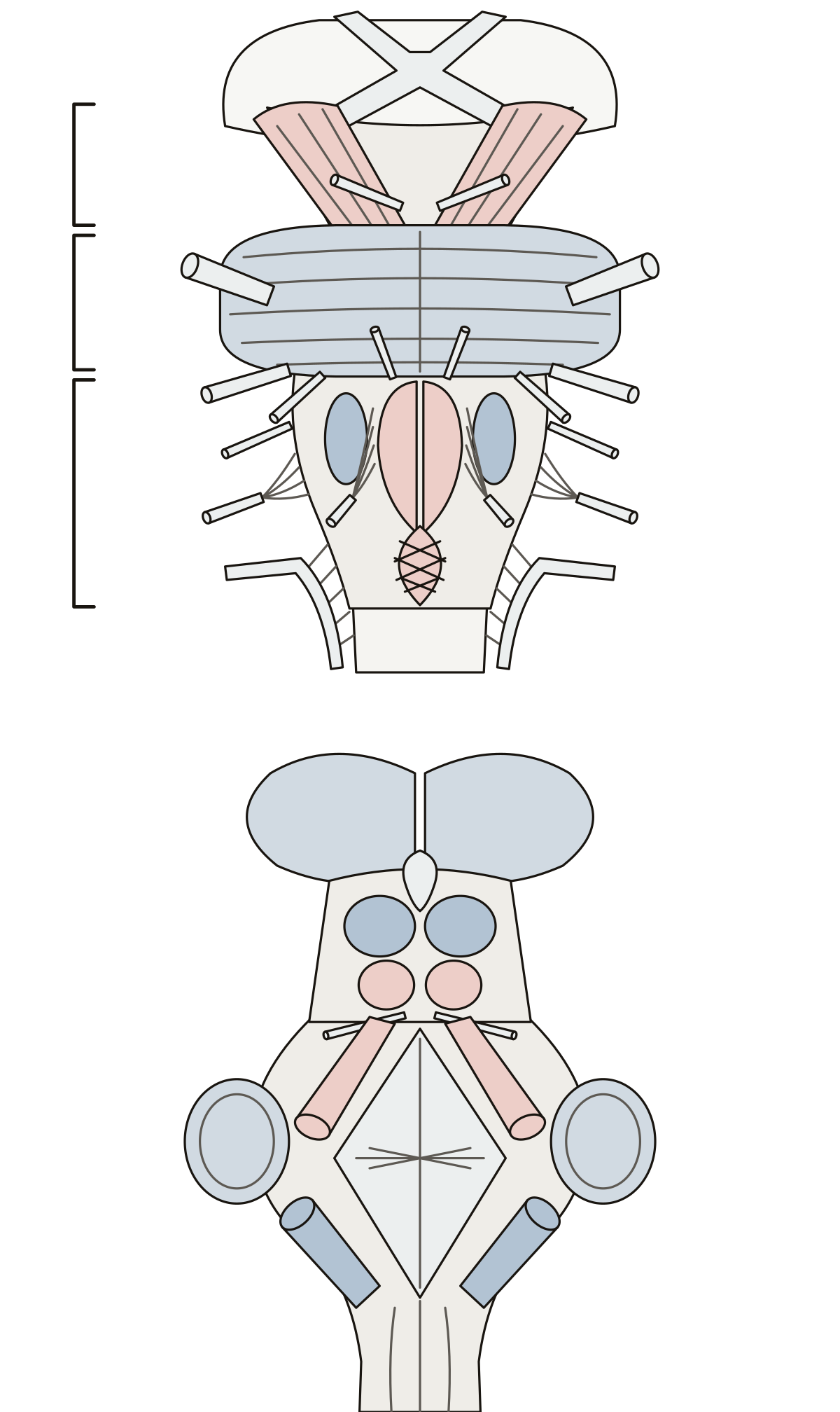 Brain Stem Model Labeled, Front and BackMidbrain, pons and medulla, the pyramids and olive, the colliculi, the three cerebellar peduncles and the roots of cranial nerves III to XII.
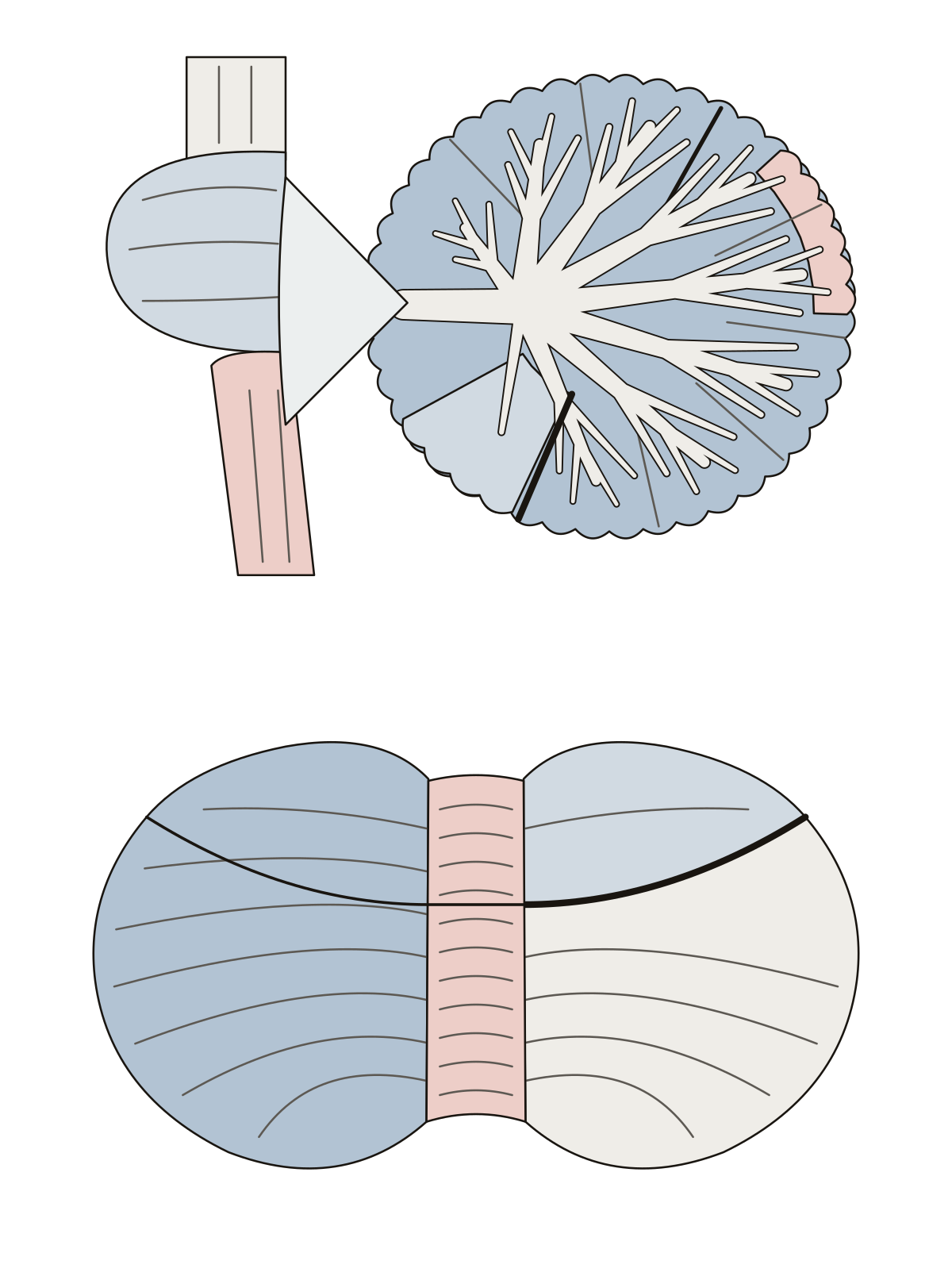 Cerebellum DiagramArbor vitae, folia, cerebellar cortex and the flocculonodular lobe in a cut down the middle; vermis, hemisphere, anterior and posterior lobes and the primary fissure from above.
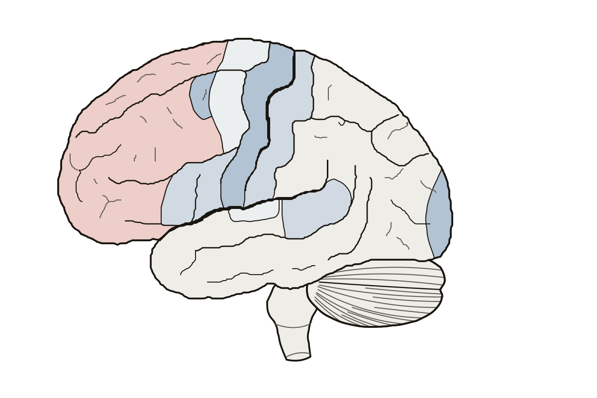 Functional Areas of the Cerebral CortexThe left hemisphere, area by area: 11 regions and grooves to name. Frontal eye field · Primary visual cortex · Primary somatosensory cortex
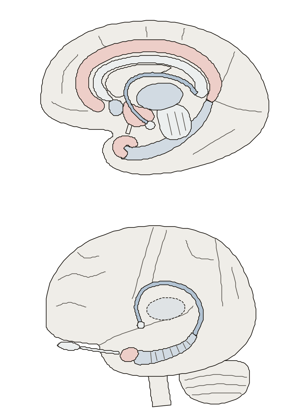 Limbic System DiagramCingulate gyrus, fornix, mammillary body, septal area, parahippocampal gyrus and uncus on the inner face of the brain; hippocampus, amygdala and olfactory bulb seen through its side.
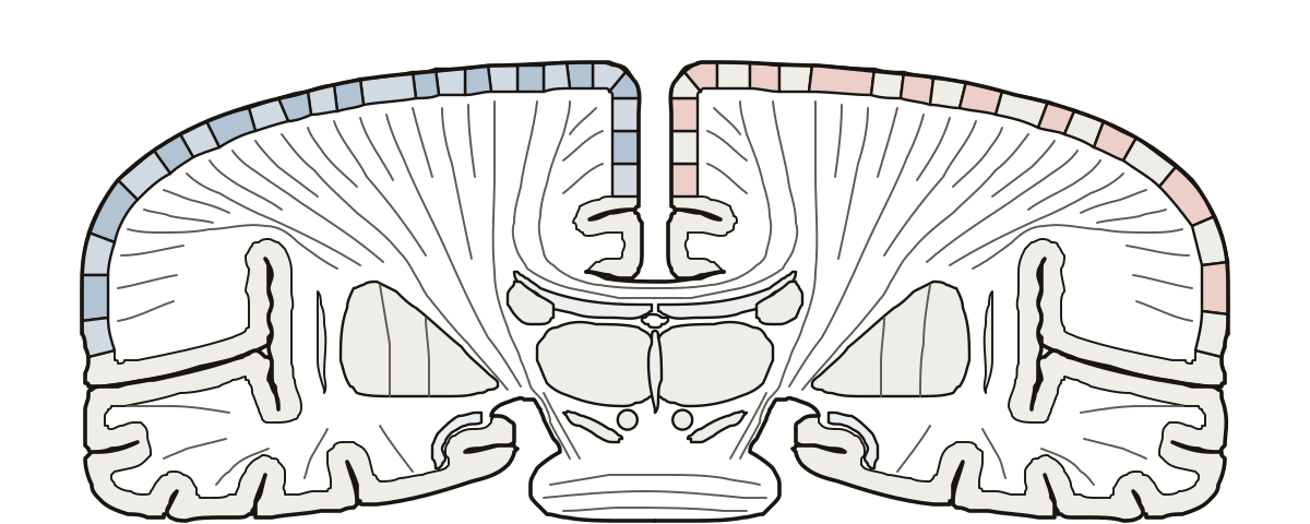 Somatosensory Cortex MapThe sensory homunculus as a strip map: 25 body parts in order along the postcentral gyrus, toes to pharynx, with the motor map of the precentral gyrus as a second view. Index finger · Nose · Hip
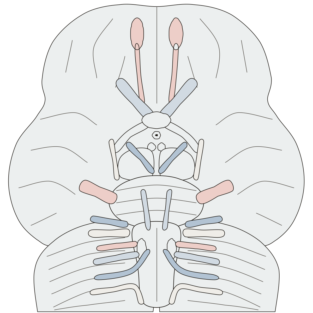 Cranial Nerves Labeling QuizA 12 cranial nerves quiz on the underside of the brain: tap each nerve where it leaves, from the olfactory bulb to the hypoglossal nerve on the medulla.
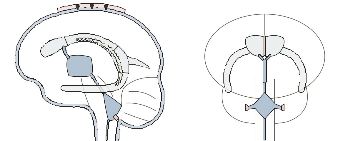 Brain Ventricle Model LabeledThe four ventricles of the brain as a see-through cast, from the side and from the front, with rings 1 to 9 in the order the cerebrospinal fluid flows.
- Brain Ventricles Diagram UnlabeledThe brain ventricles drawing with nothing written on it, to print and fill in: a side view and a front view, a word bank for each and an answer key.
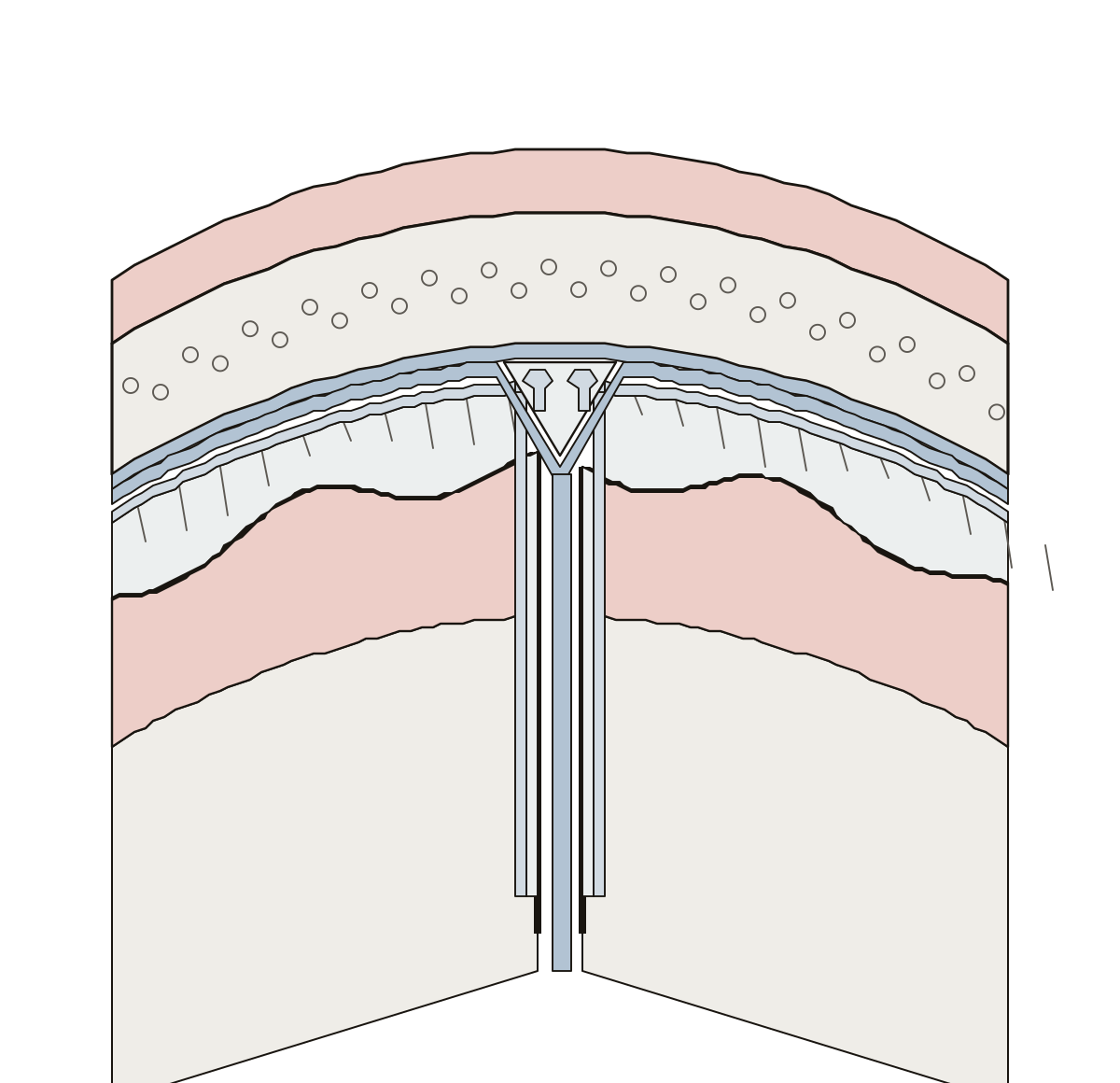 Layers of the MeningesThe meninges in a slice: skull, dura (endosteal and meningeal layers), superior sagittal sinus, falx cerebri, arachnoid, subarachnoid space, pia mater, cortex. Falx cerebri · Sagittal sinus · Cortex
- Unlabeled Meninges DiagramThe meninges drawing without its names, for printing: nine lines between the bone and the brain, with the word bank and the key. Skull · Falx cerebri · Pia mater
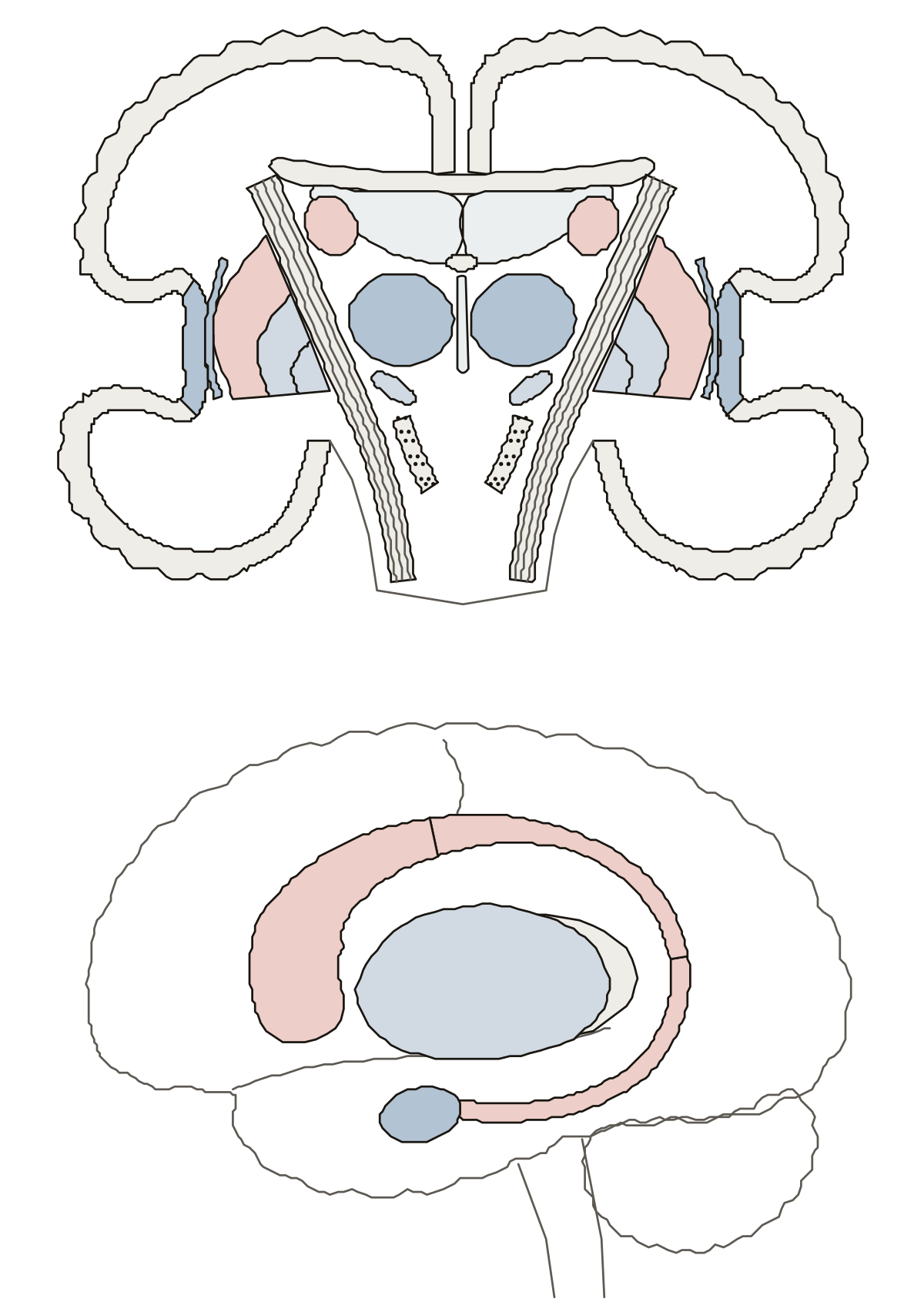 Basal Ganglia Anatomy QuizCaudate nucleus, putamen, both parts of the globus pallidus, internal capsule, subthalamic nucleus and substantia nigra in a cut from ear to ear; head, body and tail of the caudate nucleus from the side.
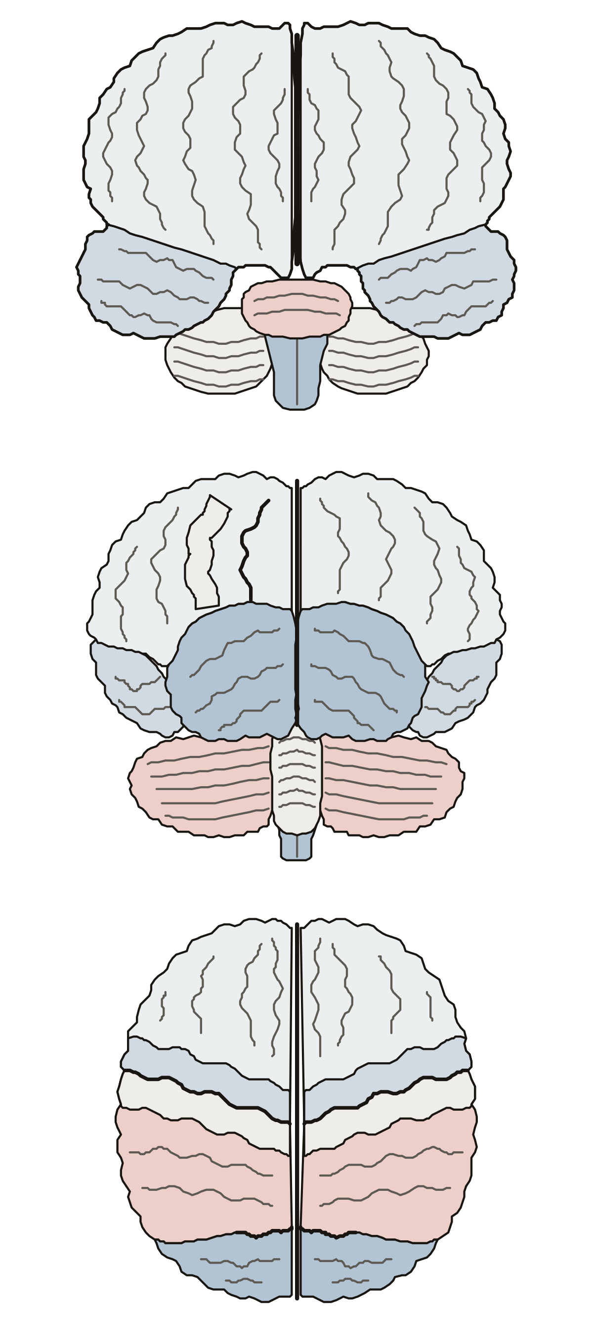 Anterior Brain Labeled, With Back and Top ViewsLeft and right hemispheres, longitudinal fissure, frontal and temporal lobes, pons and medulla from the front; parietal and occipital lobes, cerebellum and vermis from behind; central sulcus and the gyri beside it from above.
- Label the Hemispheres of the BrainThe brain from the front and from the back as a worksheet: both hemispheres, the fissure between them, the four lobes and the cerebellum, numbered, with a key.
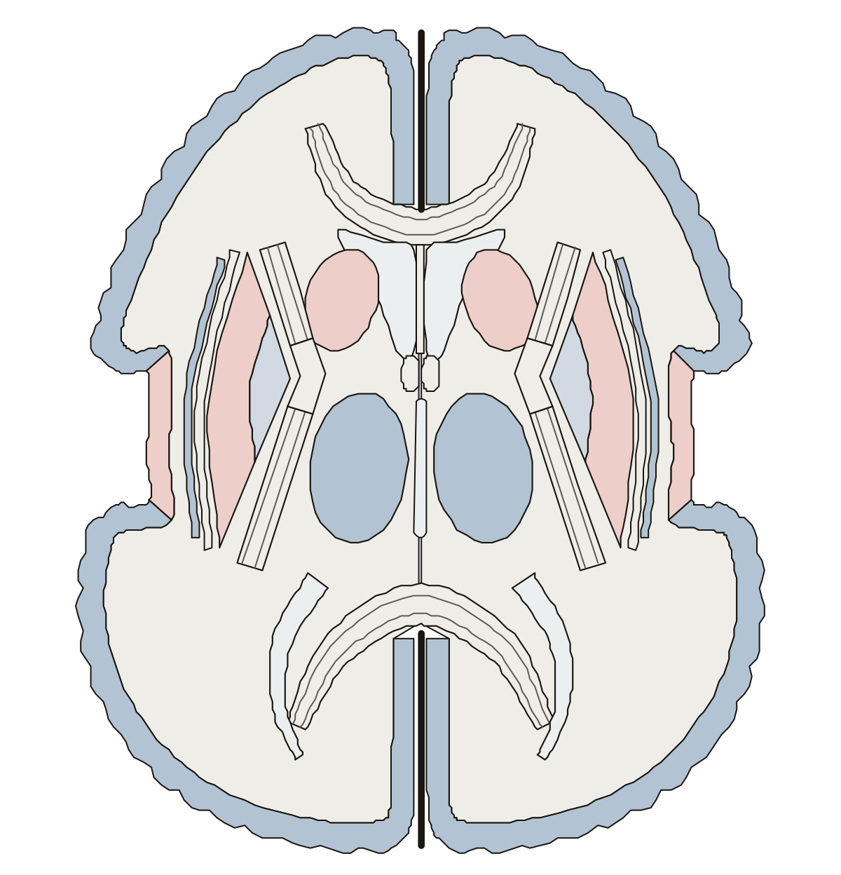 Axial Brain Labeled: a Horizontal SectionA horizontal section through the thalamus: the three parts of the internal capsule, caudate head, putamen, globus pallidus, claustrum, both ends of the corpus callosum and the horns of the lateral ventricle.
- Brain Axial Section Diagram UnlabeledThe horizontal section of the brain without its names, for printing: fifteen lines, five names that go on a shape, with the word bank and the key.
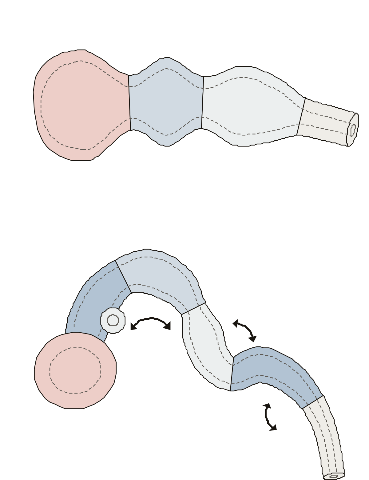 Label the Portions of the Prenatal BrainProsencephalon, mesencephalon and rhombencephalon in a row; then telencephalon, diencephalon, metencephalon and myelencephalon on the bent tube, with the optic vesicle and the three flexures.
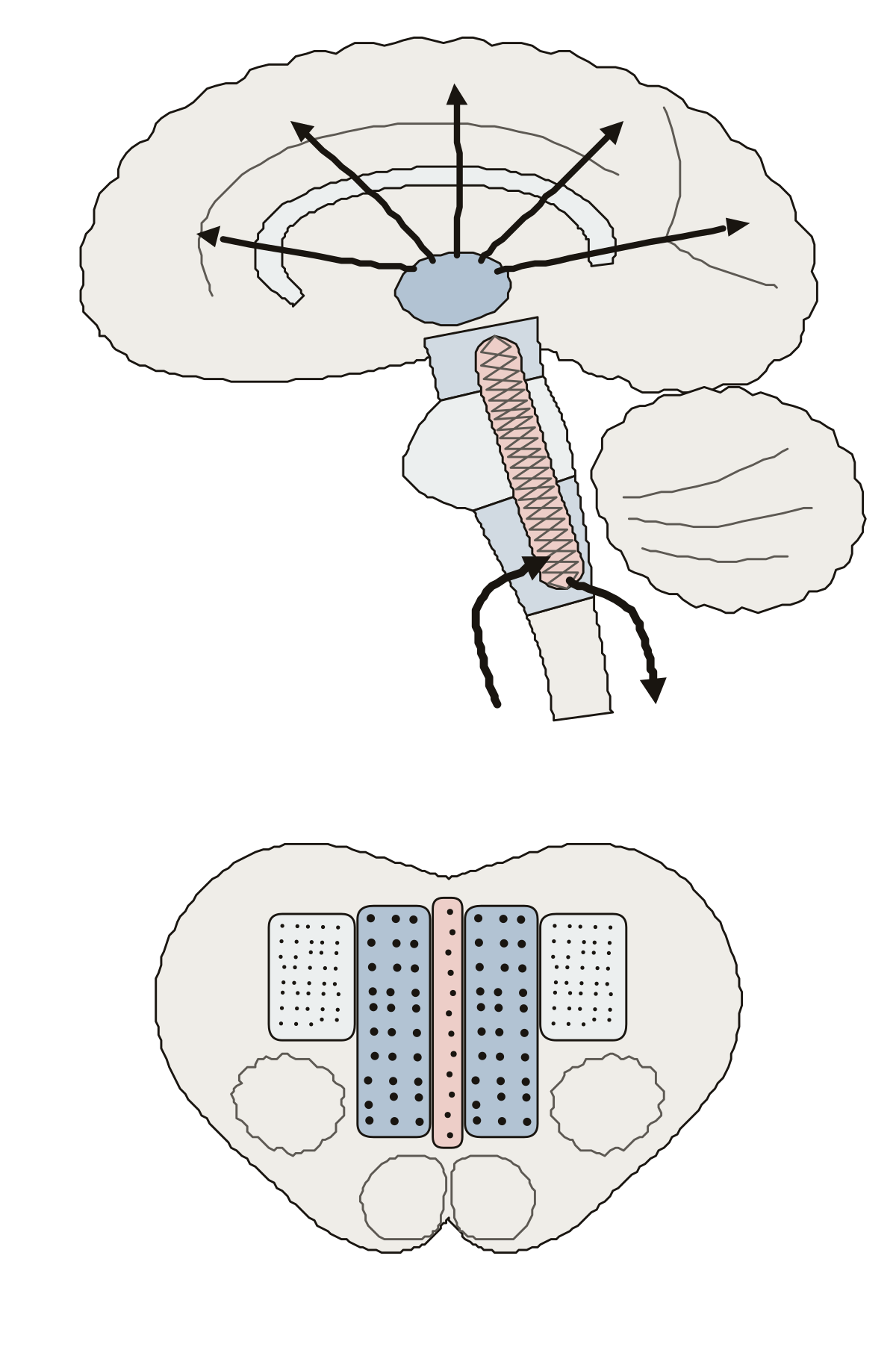 Brain Diagram Labeled Reticular FormationThe reticular formation as the core of the midbrain, pons and medulla oblongata, the thalamus, the paths up to the cortex and down the spinal cord, and the median, medial and lateral columns.
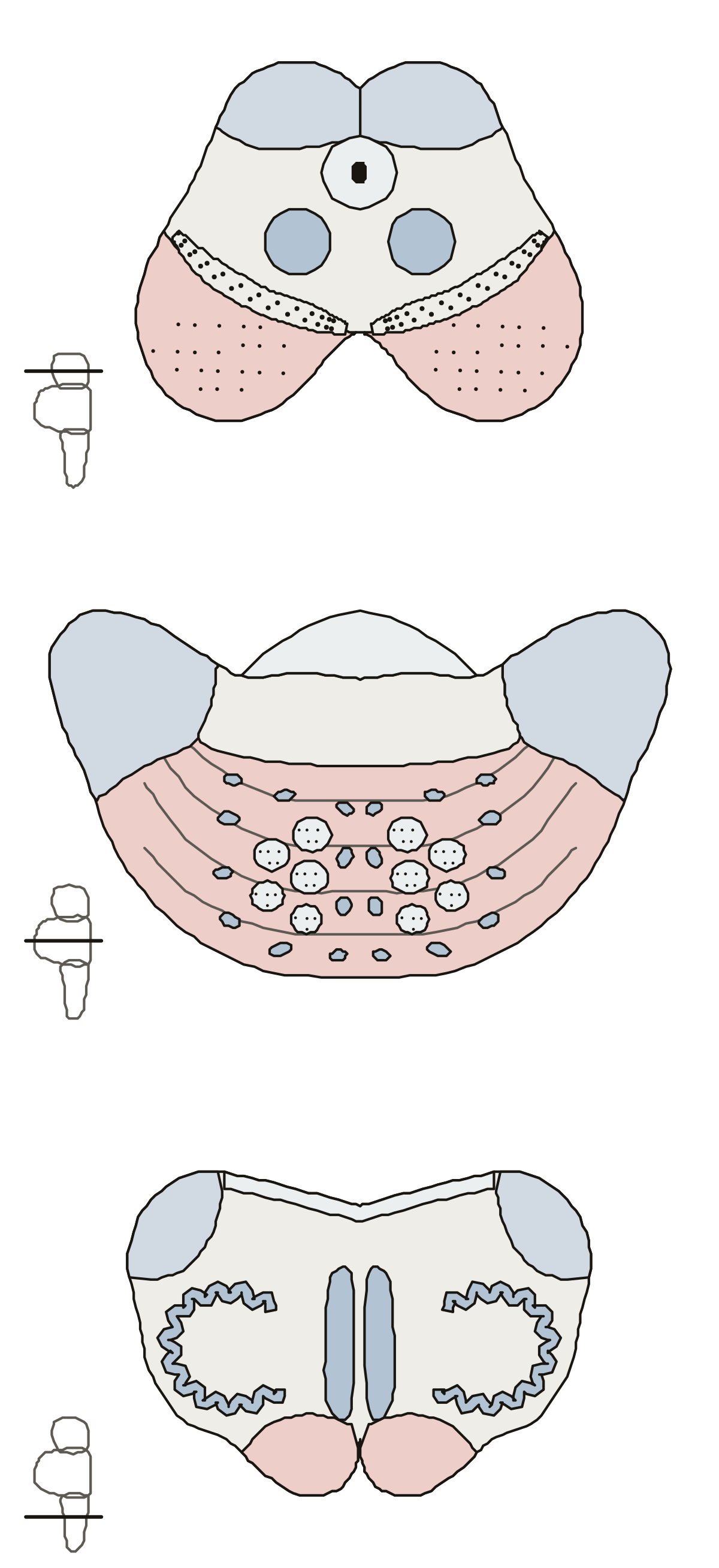 Brain Stem Cross Section LabeledThe midbrain at the superior colliculi, the pons and the medulla at the olive: aqueduct, red nucleus, substantia nigra, crus cerebri, pontine nuclei, peduncles, olivary nucleus, medial lemniscus and pyramids.
- Brain Stem Cross Section Diagram UnlabeledThe three brain stem cross sections with nothing written on them, to print and fill in: midbrain, pons and medulla, a word bank for each slice and an answer key.
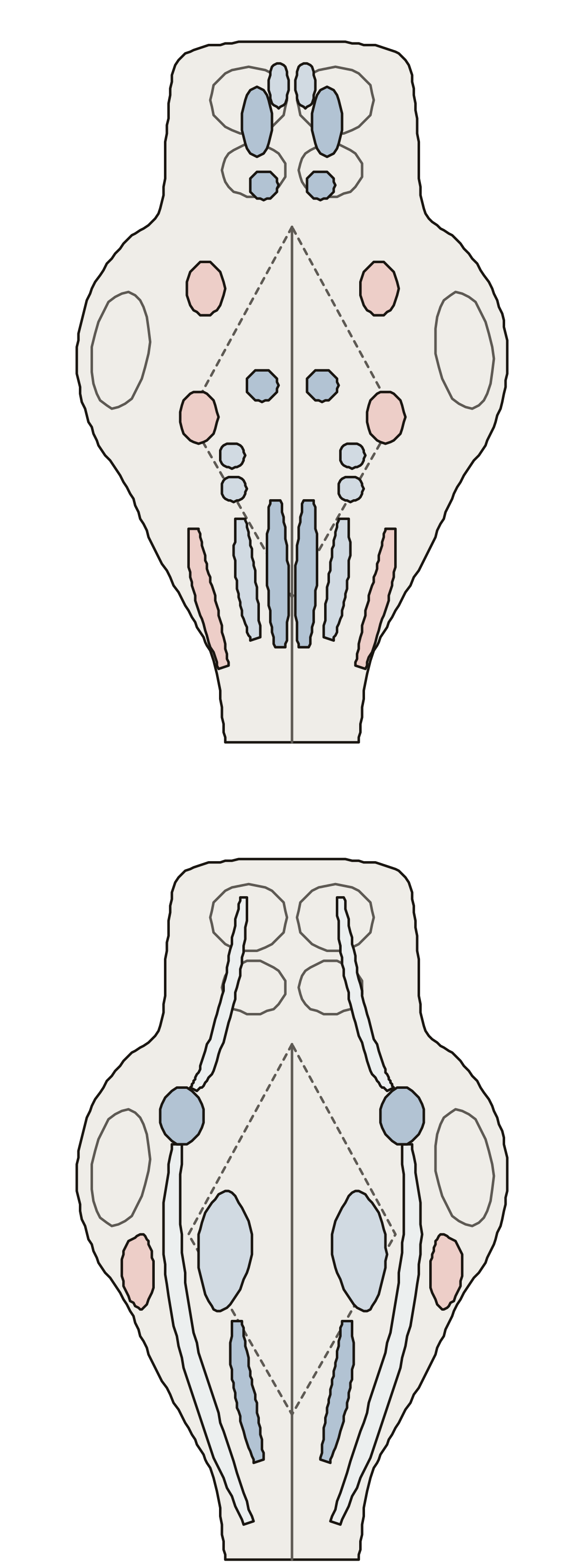 Cranial Nerve Nuclei QuizEleven motor nuclei and six sensory nuclei of the cranial nerves on the brainstem seen from behind, each with the nerve it serves.
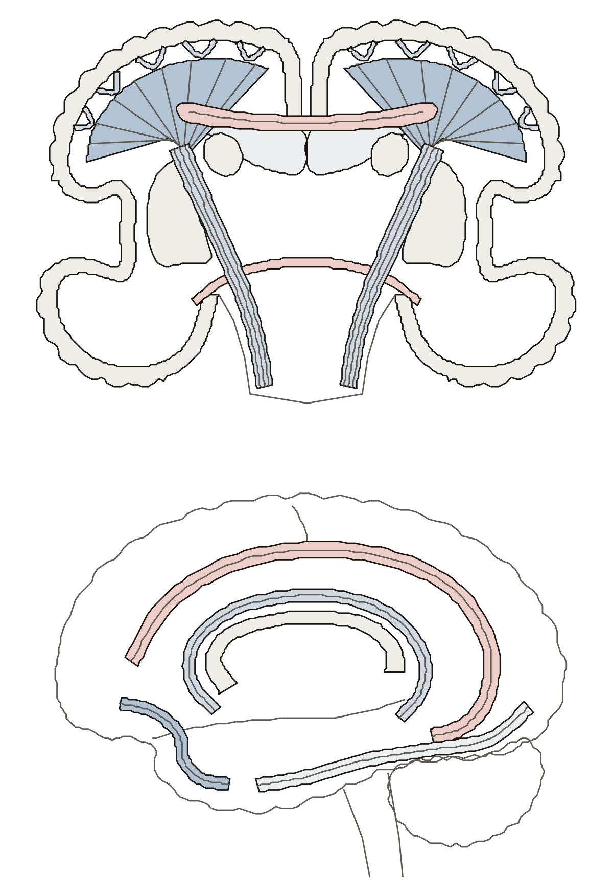 Brain Tracts Labeled: The White Matter of the BrainCorpus callosum and anterior commissure, corona radiata and internal capsule, short association fibers, and the superior longitudinal, uncinate and inferior longitudinal fasciculi with the cingulum.
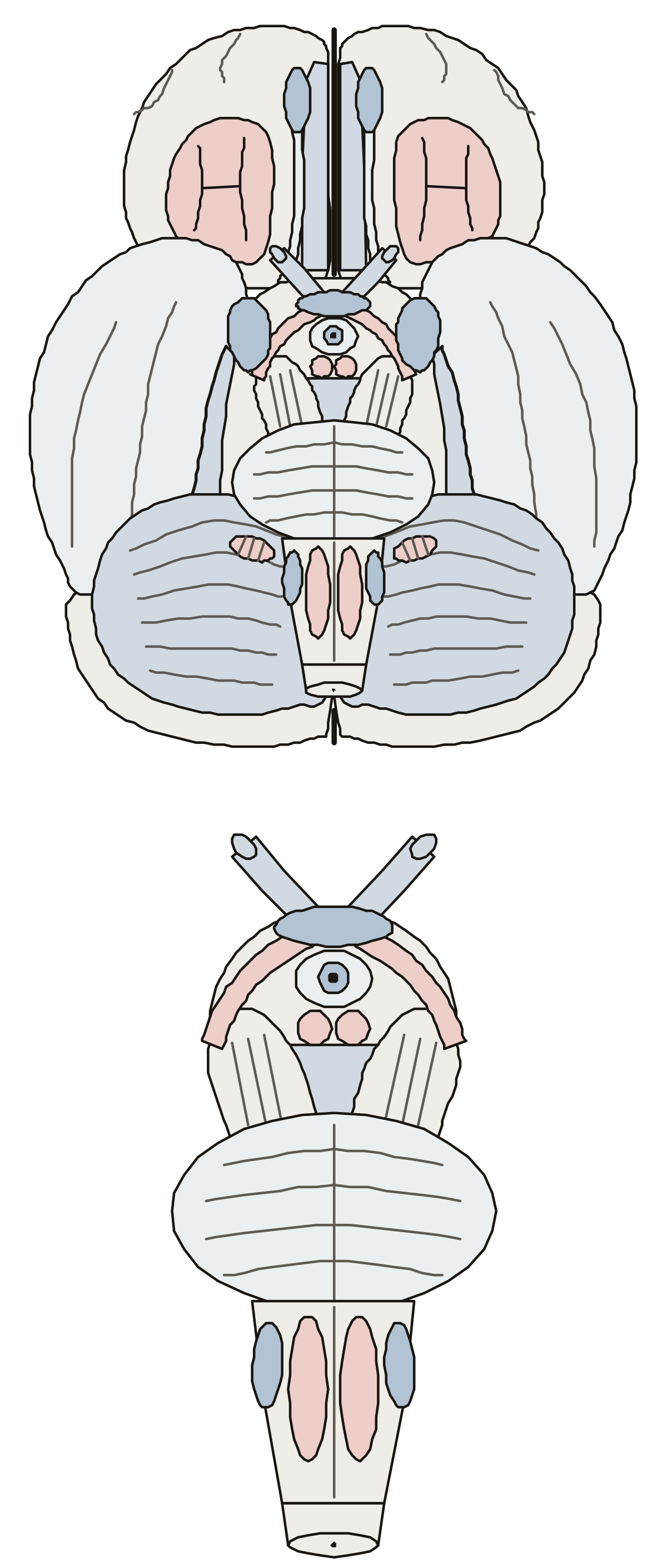 Human Brain Inferior View Labeled: the UndersideThe whole brain from below, then its midline close up: olfactory bulb and tract, straight gyrus, orbital gyri, uncus, optic nerve, chiasm and tract, pituitary stalk, mammillary bodies, cerebral peduncles, pons, pyramids, olives and cerebellum.
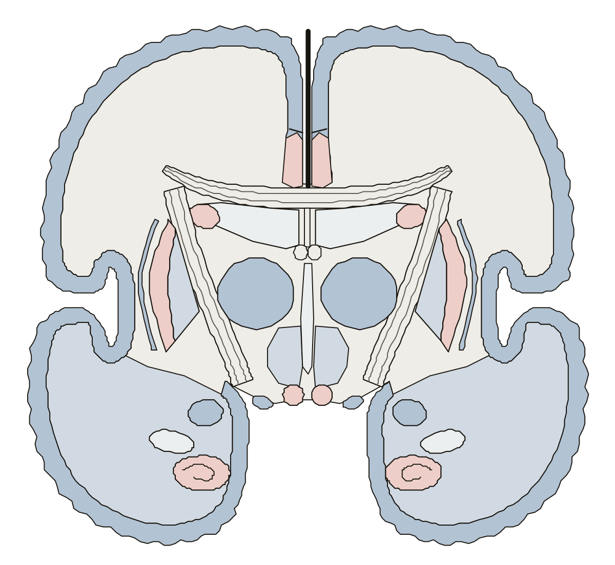 Coronal Section of Brain With Labels: a Frontal SliceA coronal slice through the thalamus: cingulate gyrus, body of the corpus callosum, lateral and third ventricles, thalamus, caudate, internal capsule, putamen, globus pallidus, hypothalamus, mammillary bodies, hippocampus and amygdala.
- Coronal Section of Brain Label QuizA quiz from the first screen on the coronal section drawing: seven parts down the midline, five deep masses and bands, then seven at the edge, in the temporal lobe and on the base.
- Label the Brain Coronal ViewThe brain in coronal view as a worksheet: nineteen numbered structures, from the longitudinal fissure to the optic tract, a line for each name, and a key.
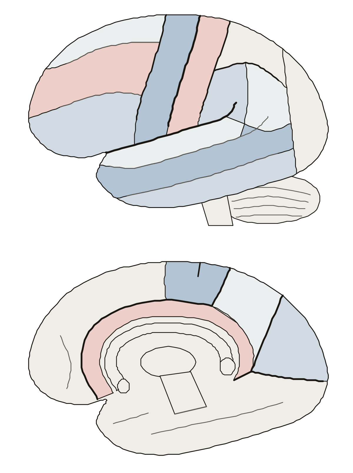 Brain Gyrus and Sulcus Labeled, Side and Inner FaceOne hemisphere from the side and on its inner face: central, lateral, precentral, postcentral and intraparietal sulci, the frontal and temporal gyri, supramarginal and angular gyri, cingulate gyrus, paracentral lobule, precuneus, cuneus and calcarine sulcus.
- Label Gyri and Sulci of the Brain QuizA quiz from the first screen on the gyri and sulci drawing: find the central and lateral sulci, the frontal and temporal gyri, and the cingulate gyrus, precuneus and cuneus on the inner face.
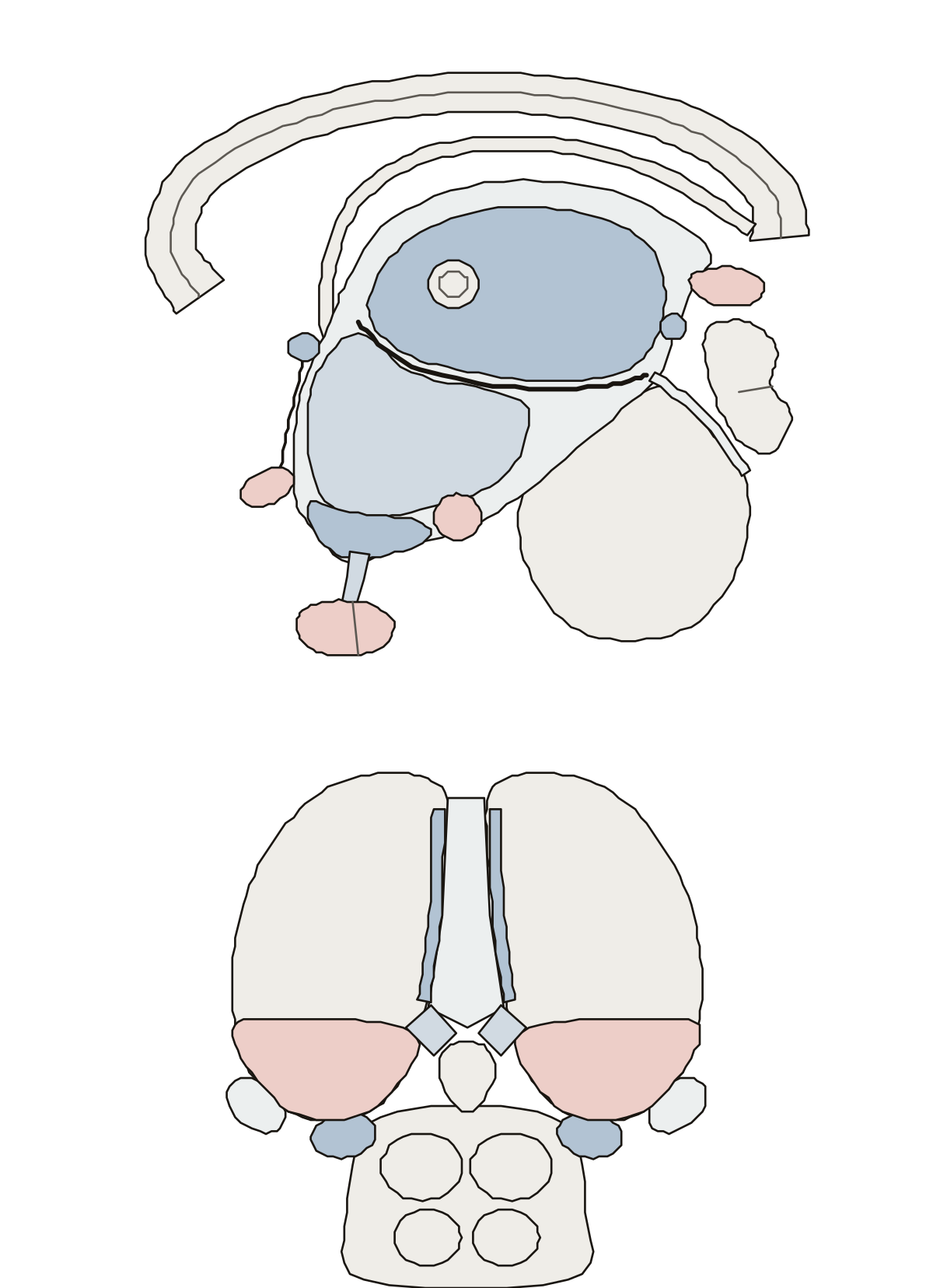 Diencephalon Labeling QuizThe diencephalon in two views: thalamus, hypothalamus, third ventricle, pituitary and pineal glands, optic chiasm, mammillary body, tuber cinereum, the two commissures, pulvinar and the geniculate bodies.
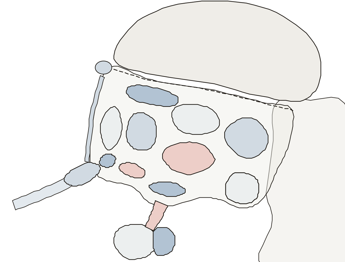 Hypothalamus DiagramParaventricular, supraoptic, suprachiasmatic, anterior, dorsomedial, ventromedial, arcuate and posterior nuclei, the preoptic area and mammillary body, with the optic chiasm, infundibulum, pituitary gland and thalamus.
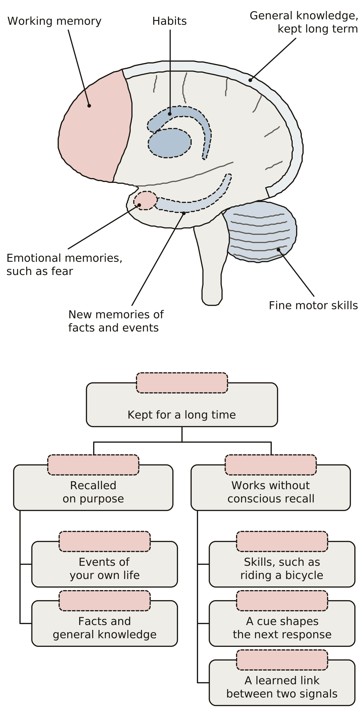 Types of Memory DiagramHippocampus, amygdala, cerebellum, prefrontal cortex, basal ganglia and cerebral cortex, each beside the kind of memory it serves; then long-term memory split into explicit and implicit, down to episodic, semantic, procedural, priming and conditioning.
The spinal cord, its roots and the autonomic nerves 17
The spinal cord cut across, horns, columns, roots and meninges, beside the whole cord from its enlargements to the cauda equina; the reflex arc, and a muscle spindle with its stretch reflex; the brachial and sacral plexuses from roots to nerves; the dermatome map: 28 rings from C2 to S5, each on the key point of one spinal nerve root; and the parasympathetic division as a chart, from the brainstem and sacral cord to the organs. Three more pages draw the tracts of the cord, the branches of one spinal nerve and the autonomic plexuses.
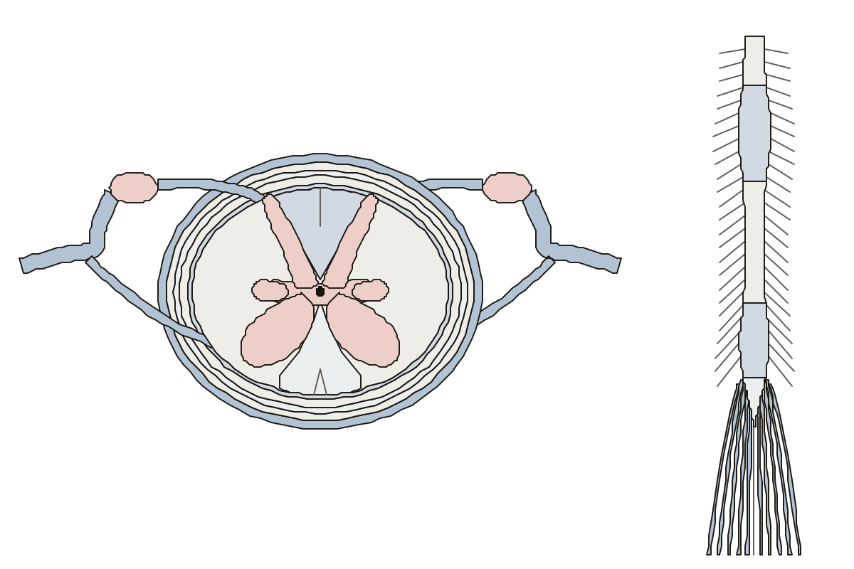 Label the Spinal CordThe cord cut across and seen whole: 18 parts to name, labeled or blank. Central canal · Ventral root · Lateral column
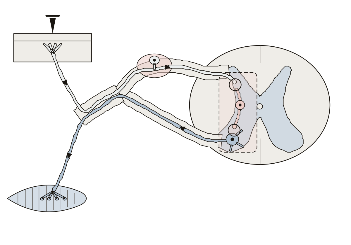 Reflex Arc Diagram LabeledReceptor, sensory neuron, integration center, motor neuron and effector, with the dorsal root ganglion, the two roots, the interneuron and the gray and white matter of the cord.
- Unlabeled Reflex Arc DiagramThe reflex arc drawing with nothing written on it, to print and fill in: thirteen names from receptor to effector, a word bank and an answer key.
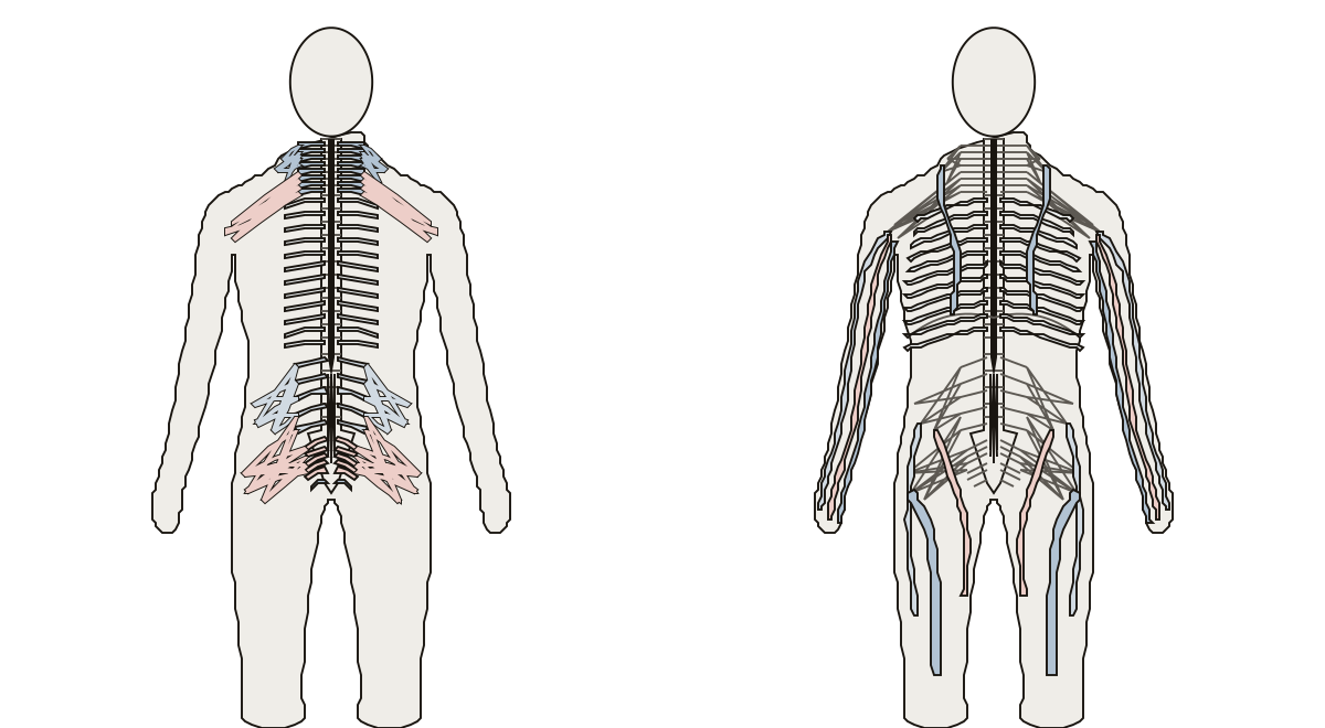 Spinal Nerves and Plexuses DiagramCervical, thoracic, lumbar, sacral and coccygeal nerves, the cervical, brachial, lumbar and sacral plexuses, and the phrenic, intercostal, radial, median, ulnar, femoral, obturator and sciatic nerves.
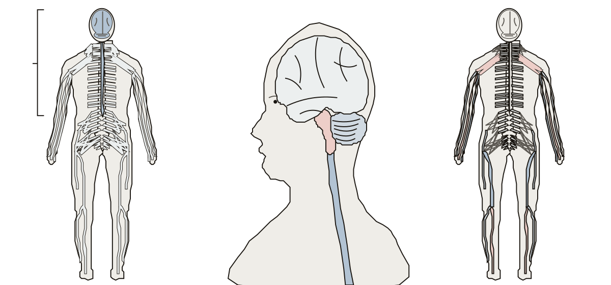 Nervous System Anatomy QuizThe central and peripheral nervous systems, the brain, spinal cord, cerebrum, cerebellum and brainstem, the spinal nerves, the brachial plexus and seven long nerves of the limbs.
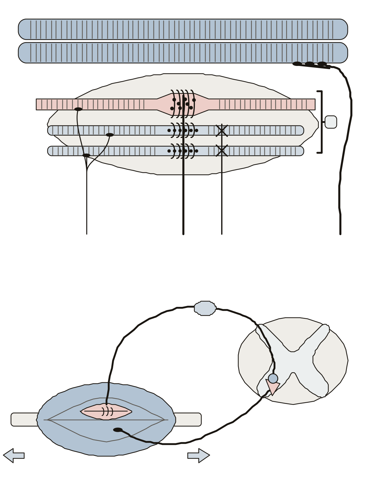 Muscle Spindle DiagramNuclear bag and nuclear chain fibers, the primary (Ia) and secondary (II) sensory endings, the gamma and alpha motor neurons, and the stretch reflex from the spindle to the spinal cord and back.
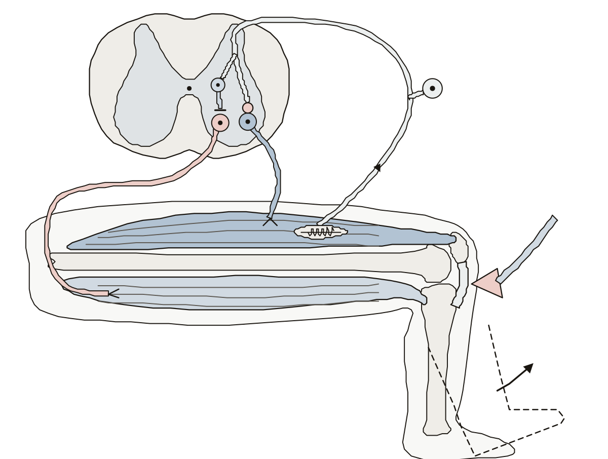 Patellar Reflex Arc Diagram LabeledReflex hammer, patellar ligament, quadriceps, muscle spindle, sensory neuron, synapse, motor neuron and the kick; then the inhibitory interneuron, the motor neuron to the hamstrings and the hamstring muscles.
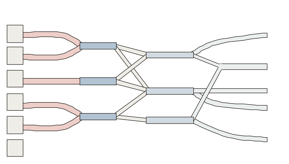 Brachial Plexus Diagram LabeledRoots, trunks, divisions, cords, branches: 13 parts in the usual schematic. Ulnar nerve · Axillary nerve · Roots
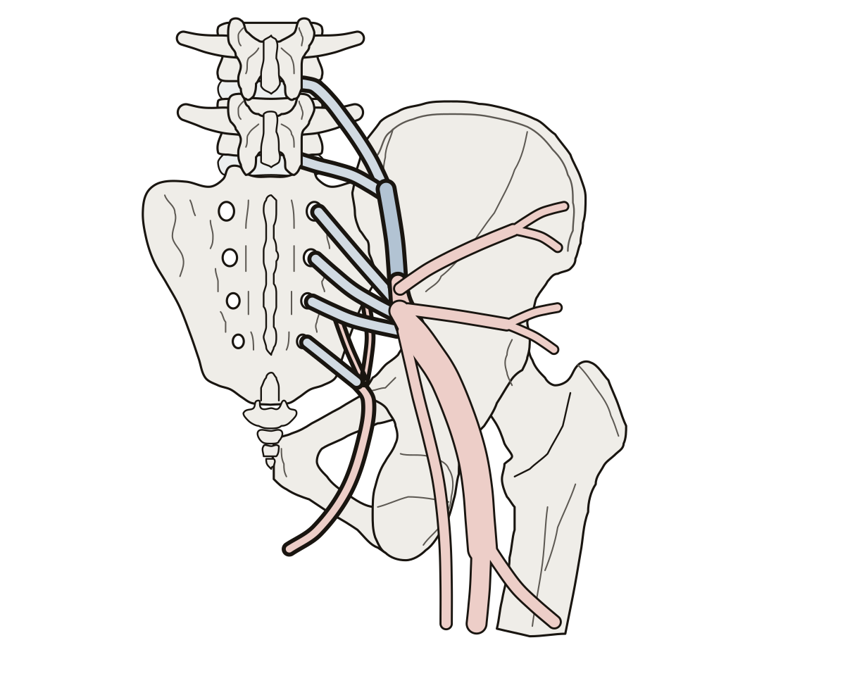 Sacral Plexus DiagramRoots L4, L5 and S1 to S4, lumbosacral trunk, superior and inferior gluteal nerves, sciatic, tibial, common fibular, posterior femoral cutaneous and pudendal nerves.
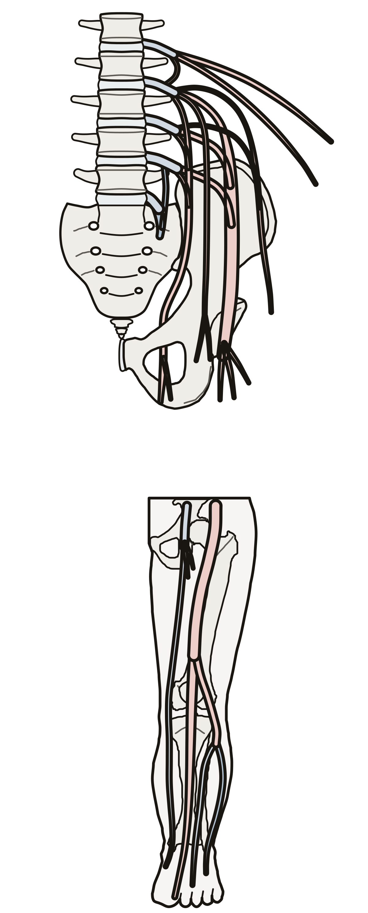 Lumbosacral Plexus DiagramRoots L1 to L4, iliohypogastric, ilioinguinal, genitofemoral, lateral femoral cutaneous, femoral and obturator nerves, the lumbosacral trunk, then the sciatic, tibial, fibular and saphenous nerves.
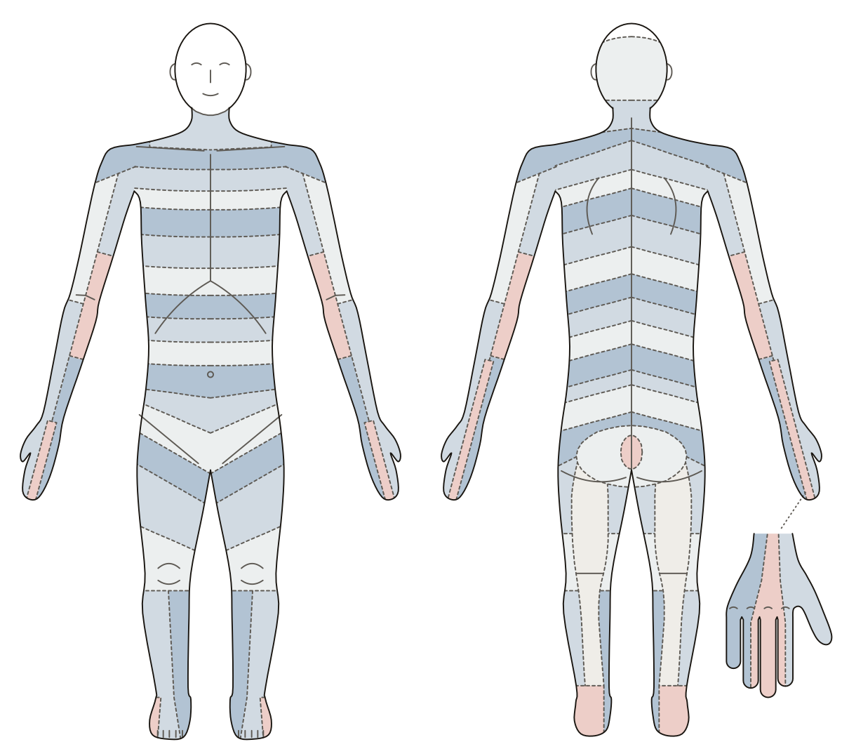 Nerve Root MapA dermatome map of the whole body, front and back: 28 rings from C2 to S5, each on the key point of its nerve root, with seven landmarks as a short set.
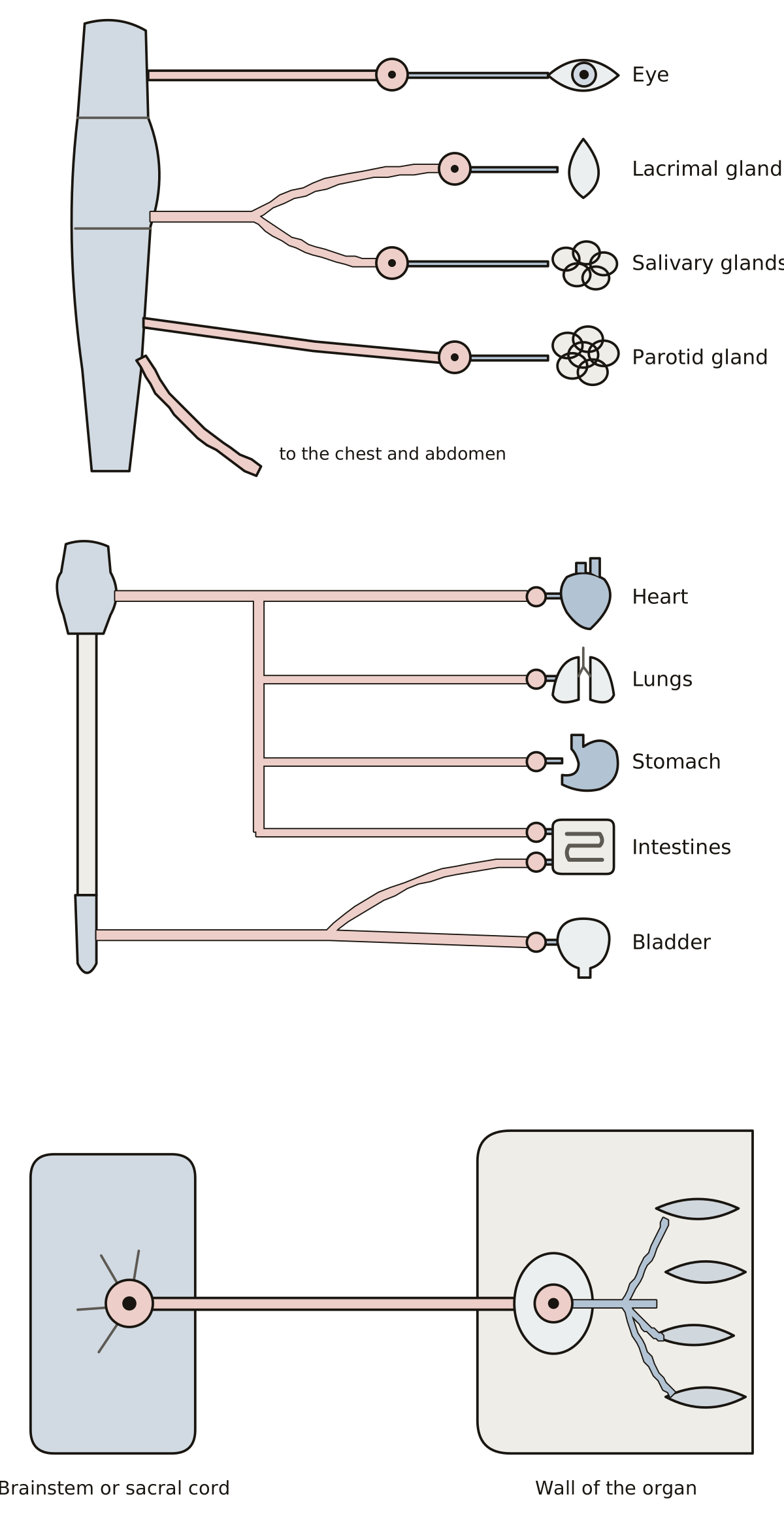 Parasympathetic Nervous System DiagramOculomotor, facial, glossopharyngeal and vagus nerves, ciliary, pterygopalatine, submandibular and otic ganglia, pelvic splanchnic nerves, terminal ganglia.
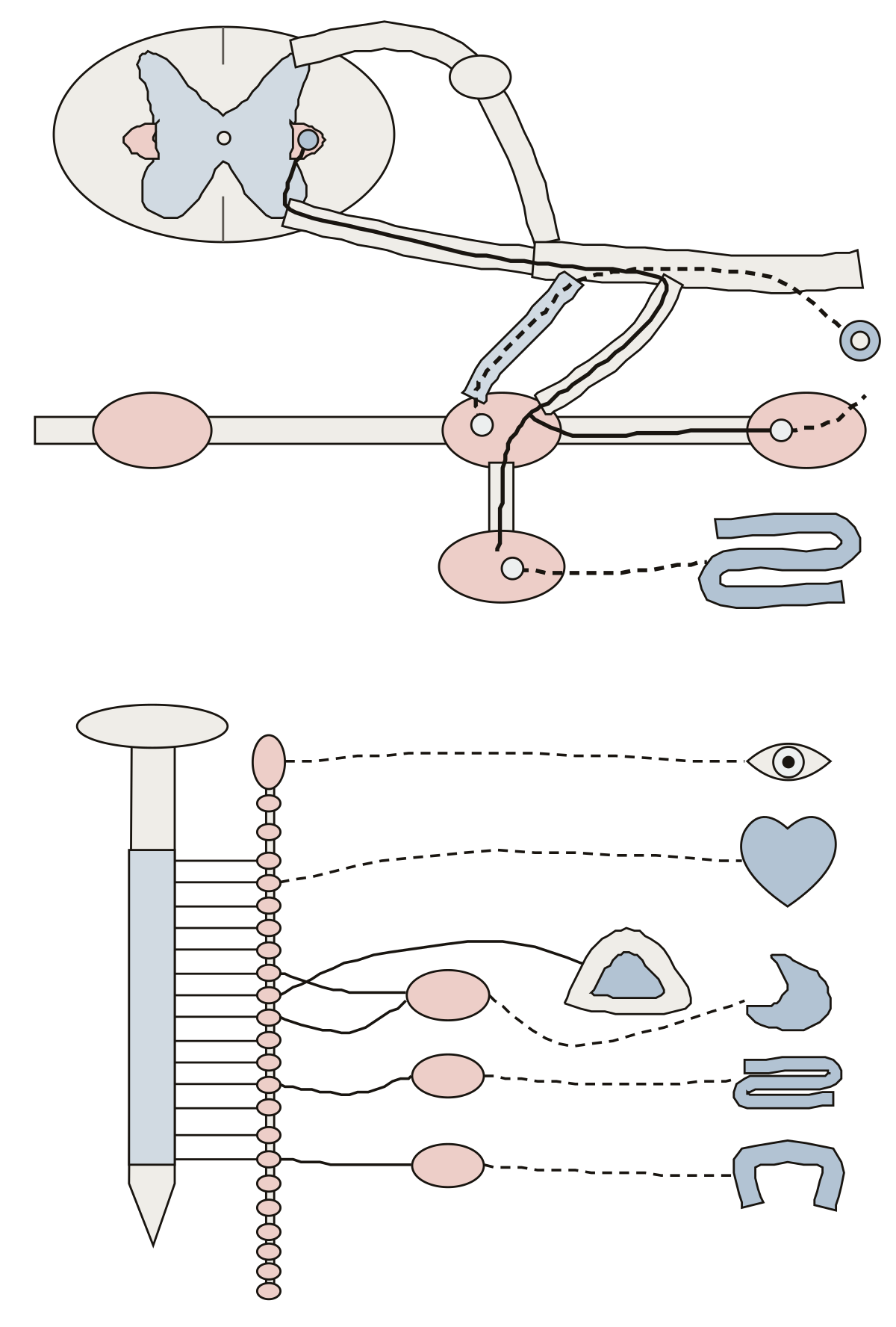 Sympathetic Nervous System DiagramLateral horn, preganglionic and postganglionic neurons, white and gray rami, the sympathetic trunk and its ganglia, a splanchnic nerve, the collateral ganglia and the adrenal medulla.
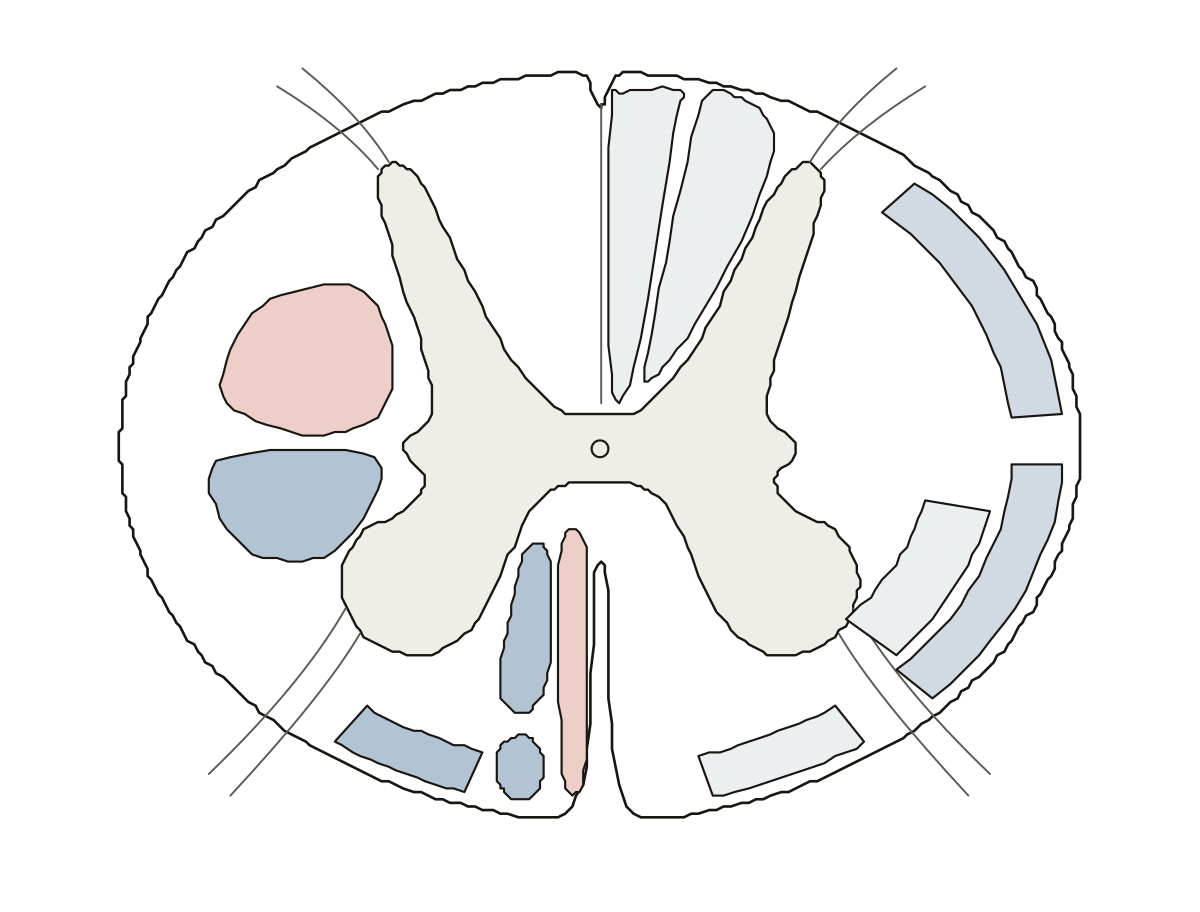 Spinal Cord Tracts DiagramThe corticospinal, rubrospinal, reticulospinal, vestibulospinal and tectospinal tracts on one half of a cross-section; the fasciculus gracilis and cuneatus, the spinothalamic and the spinocerebellar tracts on the other.
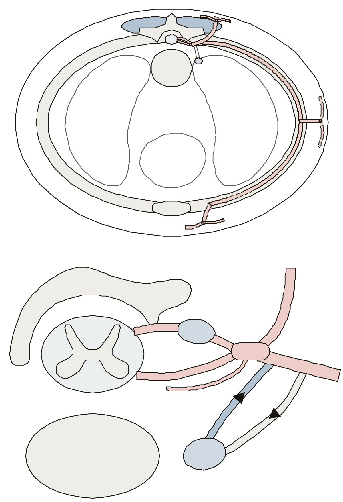 Spinal Nerve Branches DiagramDorsal and ventral rami and the lateral and anterior cutaneous branches on a chest cut across; the roots, the dorsal root ganglion, the meningeal branch and the white and gray rami communicantes close up.
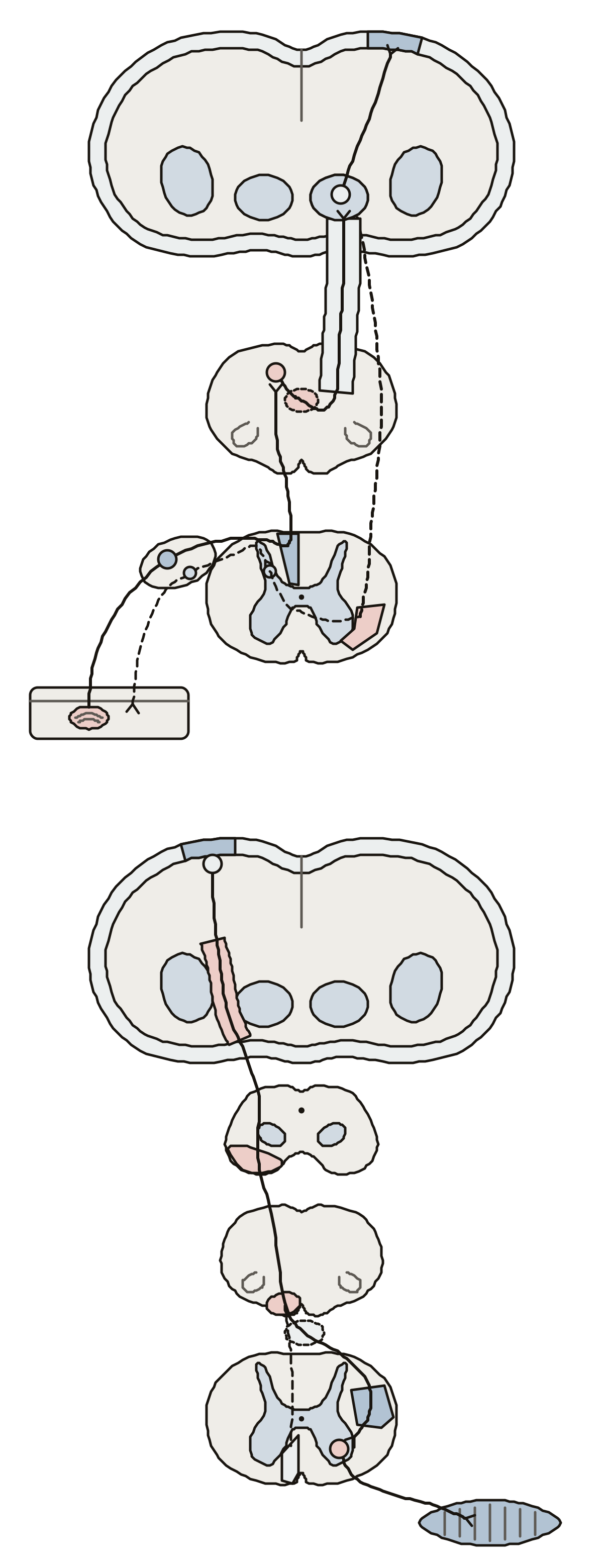 Ascending and Descending Pathways DiagramReceptor, first-, second- and third-order neuron, dorsal root ganglion, fasciculus gracilis, medial lemniscus, thalamus and somatosensory cortex; then the upper motor neuron through the internal capsule, cerebral peduncle and pyramid to the lower motor neuron.
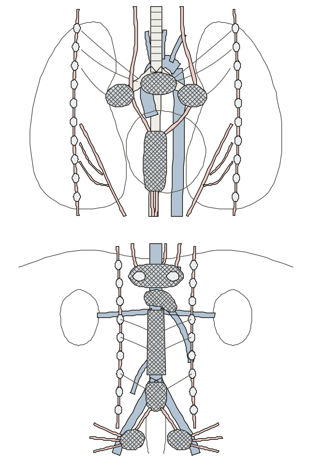 Autonomic Plexuses DiagramCardiac, pulmonary and esophageal plexuses with the vagus nerve and sympathetic trunk in the chest; celiac, superior mesenteric, aortic and both hypogastric plexuses with the splanchnic nerves below.
This set holds the site’s drawings of the eye and the nervous system. The eye comes first: a side view with 25 numbered rings, a close-up of the rods and cones in the retina, and three small eyes that compare normal, near and far sight.
More about these tools
The ear follows in one section from the auricle to the cochlea, with 17 numbered rings. Maps of the brain and the nerves join the list one at a time, each when its drawing has been checked against its part list.
Every page is one drawing. Find it names a part for you to tap, Name it lights a ring for you to name, and Study shows every label with a line about the part. After a round, one press replays the parts you missed, and each drawing prints blank or labeled. These pages teach names and places and give no medical advice.
Keep exploring
- Animal body-part diagrams (94)
- Blank US maps (61)
- Blank maps (159)
- Bone labeling quizzes (49)
- Capitals quizzes (11)
- Car and engine part diagrams (54)
- Cell and organelle diagrams (63)
- Clothing, shoes and jewelry (45)
- Computer, phone and camera diagrams (43)
- Anatomy concept maps (1)
- Country map quizzes (28)
- Animal anatomy diagrams (100)
- Human Body Quizzes
- All quizzes