Human Body System Diagrams: Label Them on Screen or on Paper
Each page is one drawing with a numbered ring on every part, to label on screen or on paper.
137 pages
Whole systems 36
One organ system on a body outline. The digestive system has a simple set of 10 main organs for younger classes and a full set of 25. Two pages stay inside the abdomen: the peritoneum cut front to back, and the veins of the hepatic portal system. Two quizzes open the chest and the abdomen, a cross section cuts the abdomen level, and one page draws the heart of an embryo as a tube.
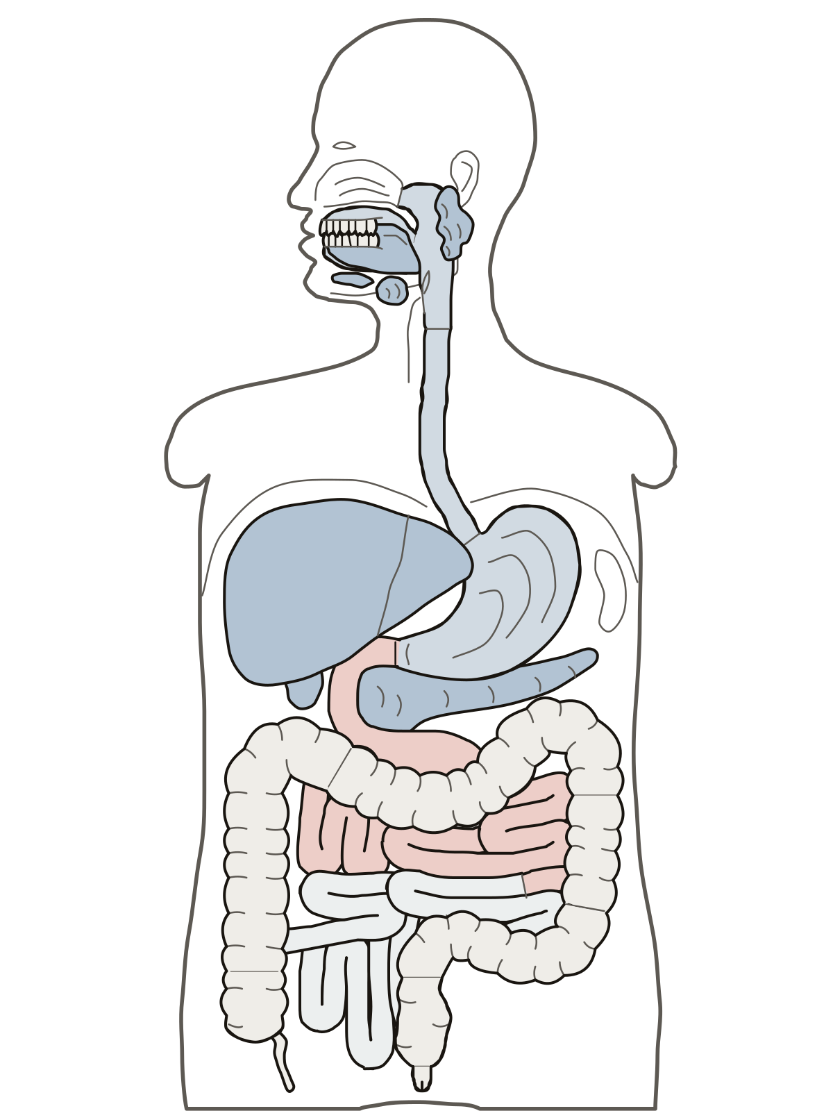 Digestive System Diagram LabeledA labeled diagram of digestive system organs from mouth to anus: 25 parts to find or name, a simple set of 10, and a blank sheet to print.
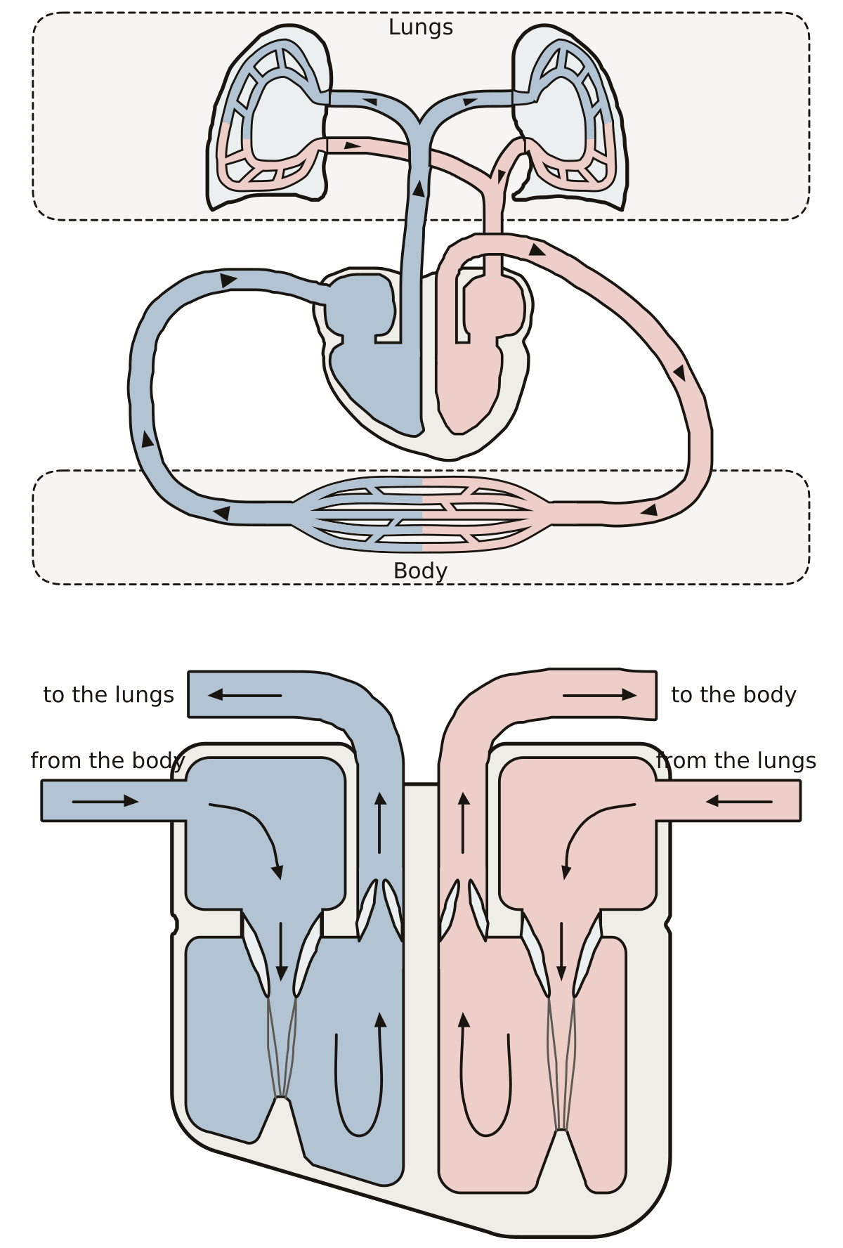 Cardiovascular System Diagram Labeled: the Path of BloodThe path of blood as a loop of pipes through the heart, lungs and body, and the heart as a box diagram with its four chambers and four valves.
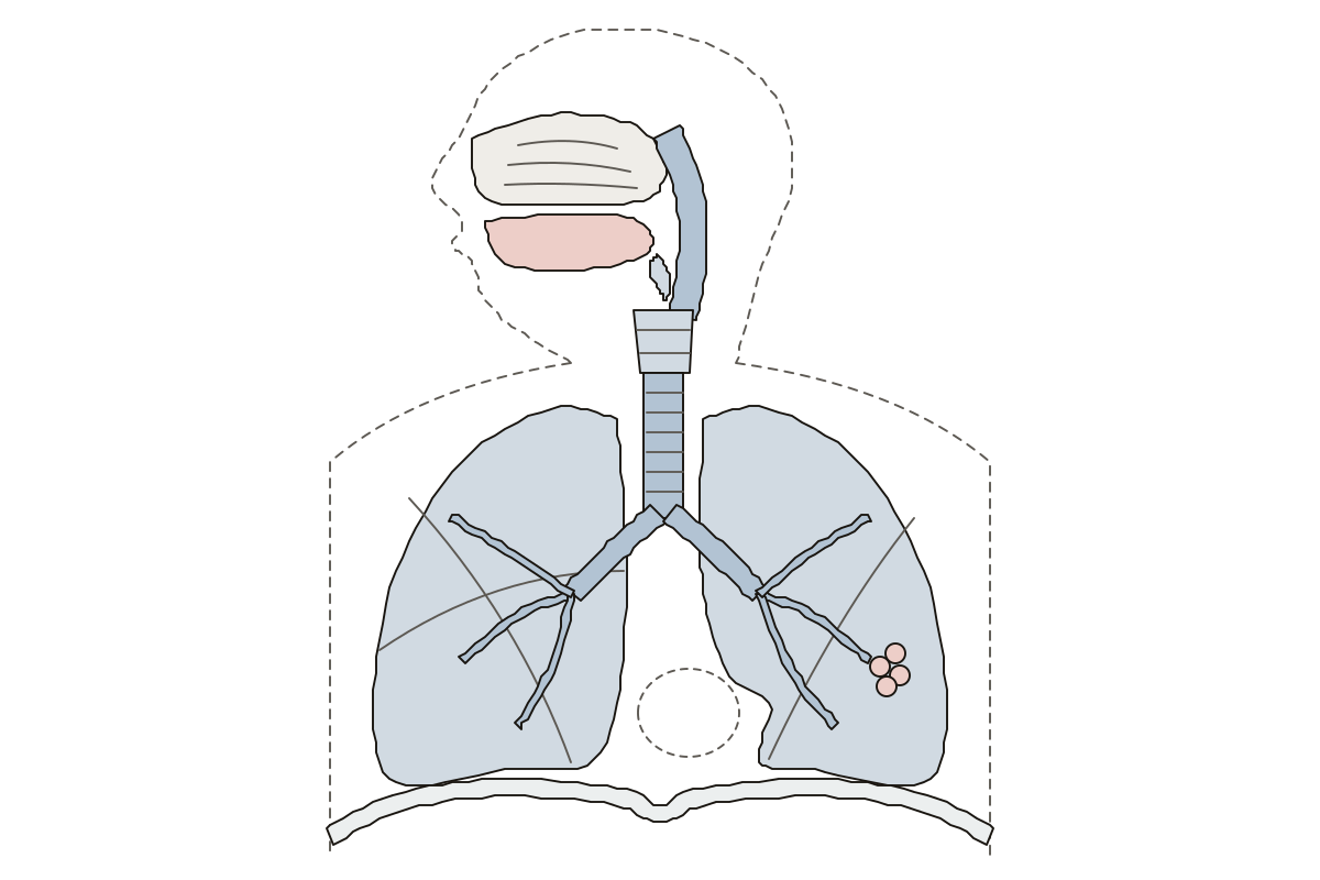 Human Respiratory System DiagramThe airway from the nose to the lungs: 12 parts to name, labeled or blank. Trachea · Epiglottis · Left lung
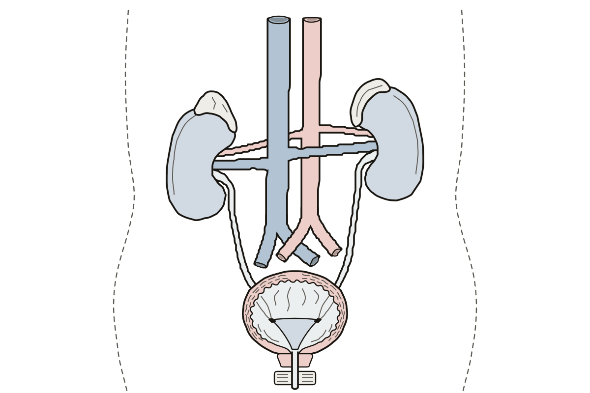 Urinary System Diagram LabeledThe urinary system from the front, bladder cut open: 14 parts to name. Kidneys · Trigone · Ureters
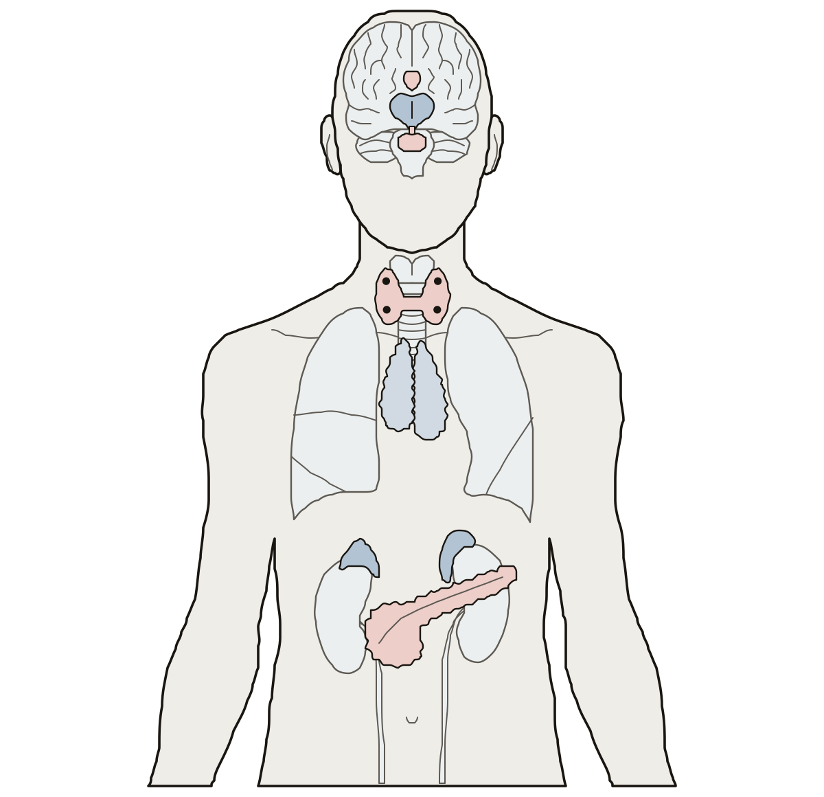 Endocrine System DiagramThe endocrine system on a body outline: pituitary, thyroid, adrenal glands and pancreas, then hypothalamus, pineal, parathyroids and thymus. Adrenal glands · Parathyroid glands · Hypothalamus
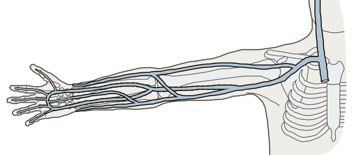 Label the Veins of the Upper LimbDigital veins and palmar arches, radial and ulnar, cephalic, basilic, median cubital, brachial, axillary, subclavian and jugular.
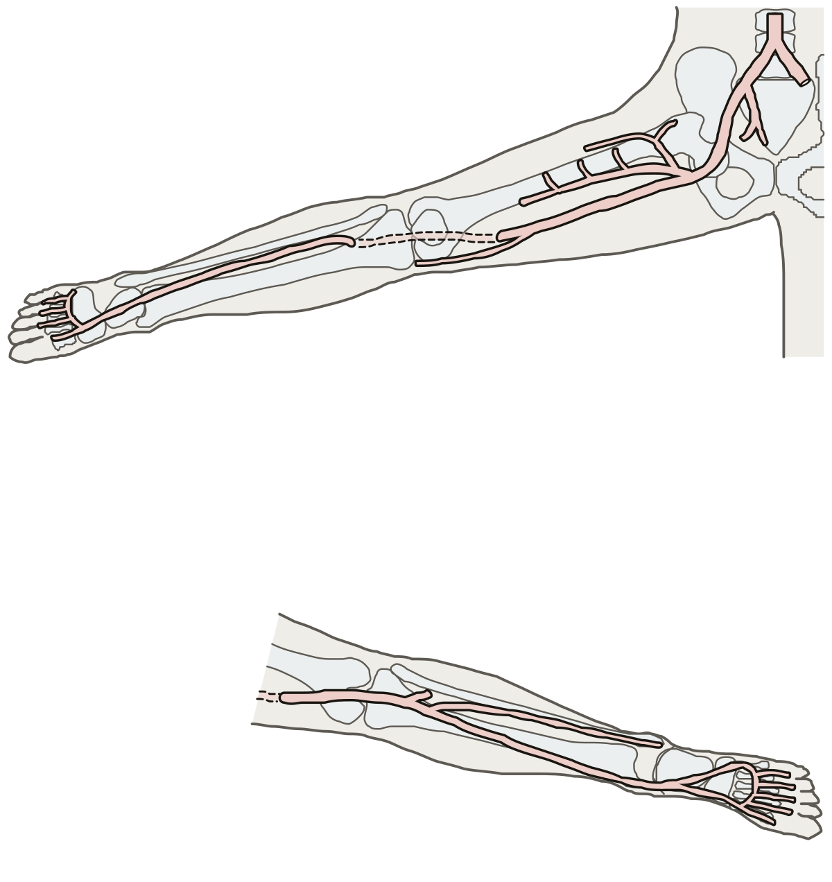 Lower Limb Arteries DiagramIliac, femoral, deep femoral, circumflex, genicular, popliteal, tibial, fibular, dorsalis pedis, plantar arteries and arches.
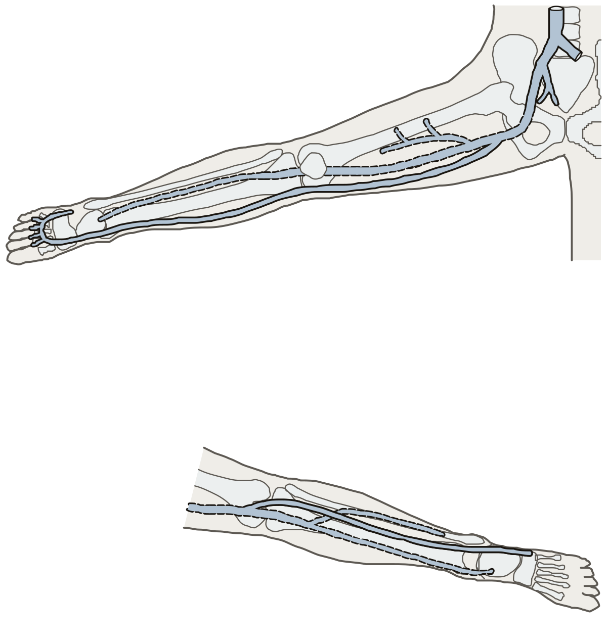 Lower Limb Veins DiagramDorsal venous arch, great and small saphenous, tibial, fibular, popliteal, femoral, deep femoral, iliac veins and vena cava.
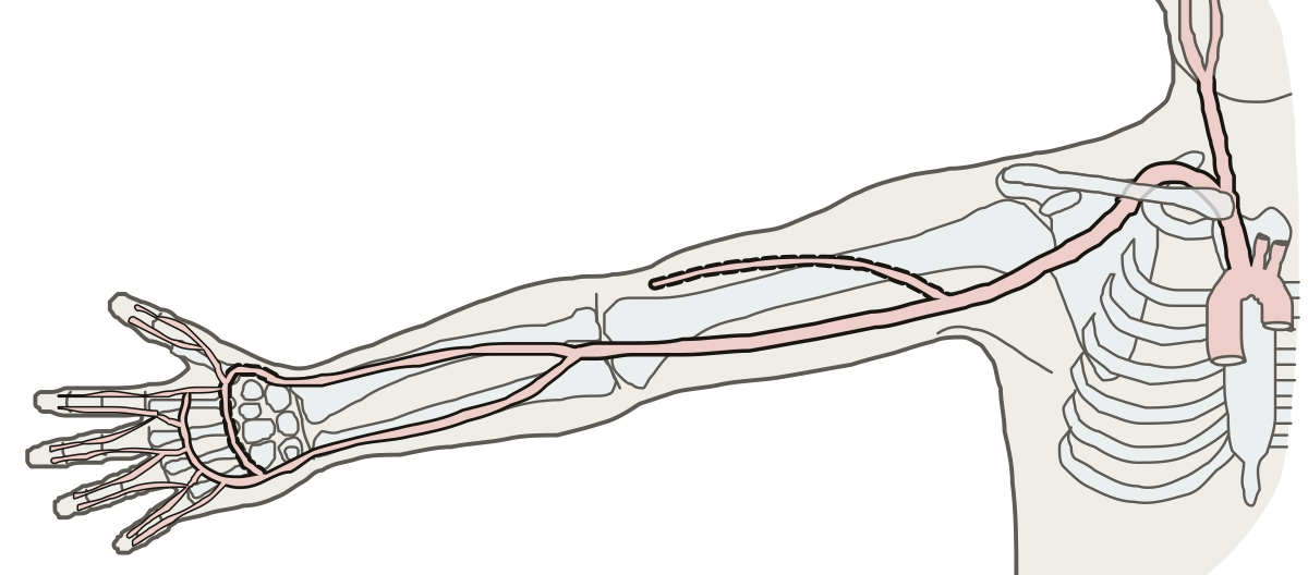 Label the Arteries of the Upper LimbBrachiocephalic trunk, common carotid, subclavian, axillary, brachial, deep brachial, radial, ulnar, palmar arches and digital arteries.
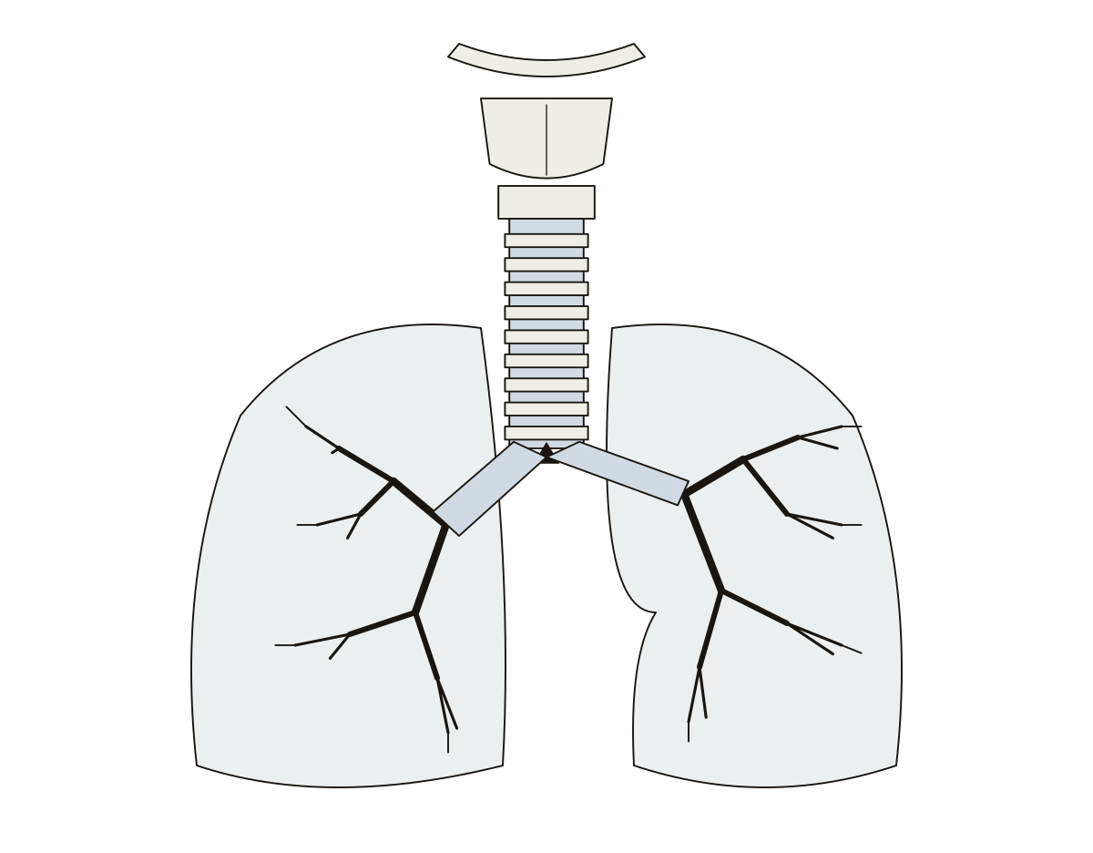 Lower Respiratory Tract DiagramHyoid, thyroid and cricoid cartilages, trachea and its C-rings, carina, primary and tertiary bronchi, bronchioles, terminal bronchioles. Tracheal cartilages (C-rings) · Trachea (windpipe)
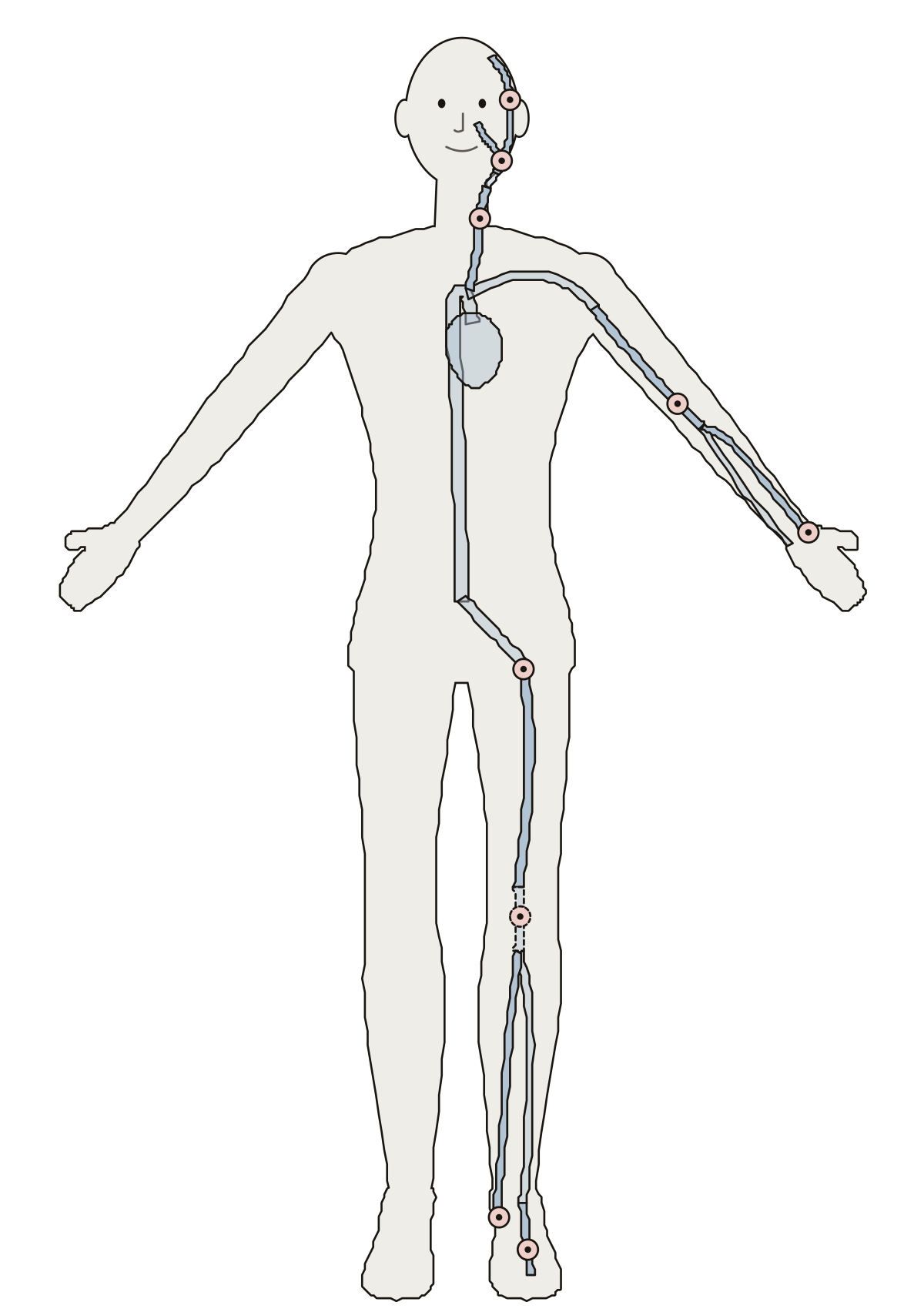 Pulse Points DiagramPulse sites on a standing figure: temporal, facial, carotid, brachial, radial, femoral, popliteal, posterior tibial and dorsalis pedis arteries.
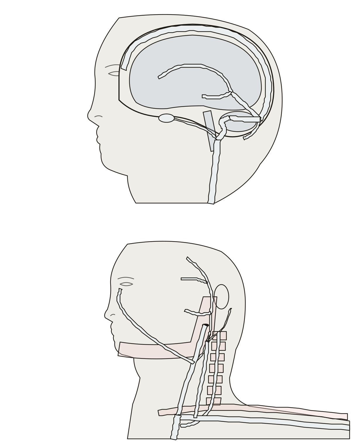 Label the Veins of the Head and NeckSagittal, straight, transverse, sigmoid and cavernous sinuses; facial, temporal and maxillary veins; jugular, vertebral, subclavian and brachiocephalic veins.
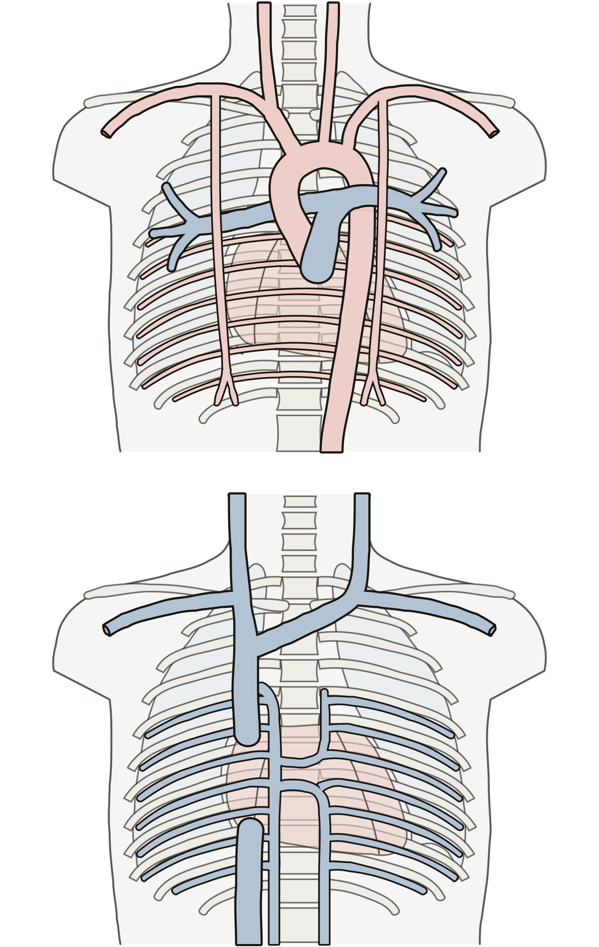 Thoracic Blood Vessels DiagramAorta, aortic arch and its branches, intercostal and internal thoracic arteries, pulmonary trunk; venae cavae, brachiocephalic, azygos and hemiazygos veins.
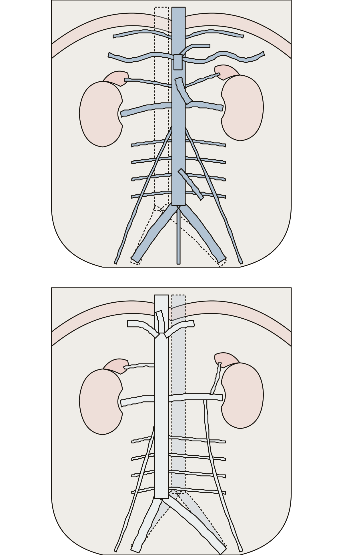 Abdominal Blood Vessels DiagramAbdominal aorta, celiac trunk and its three branches, mesenteric, renal, gonadal and lumbar arteries; inferior vena cava, hepatic, renal and iliac veins.
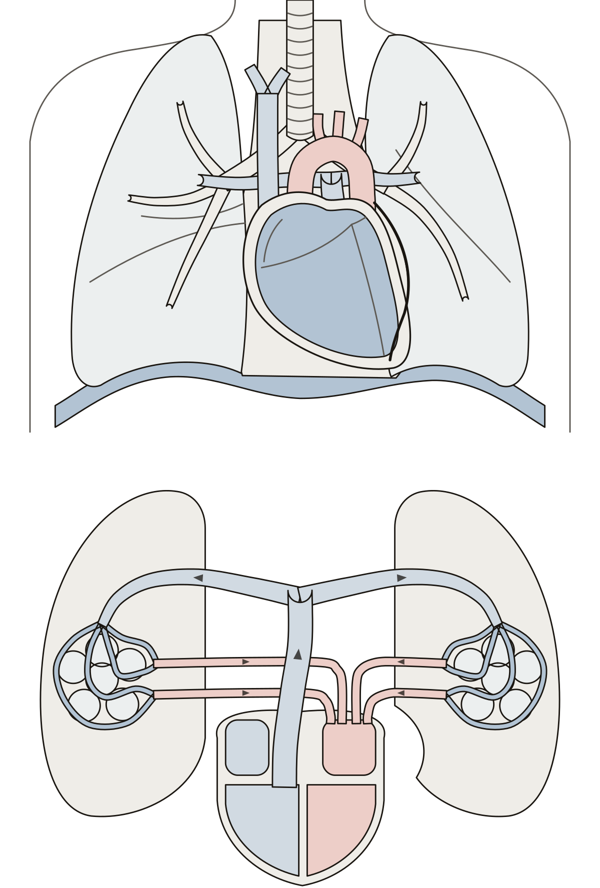 Heart and Lungs DiagramTrachea, bronchi, both lungs, the heart in its pericardium, the aorta and the superior vena cava, then the pulmonary trunk, arteries, capillaries and veins.
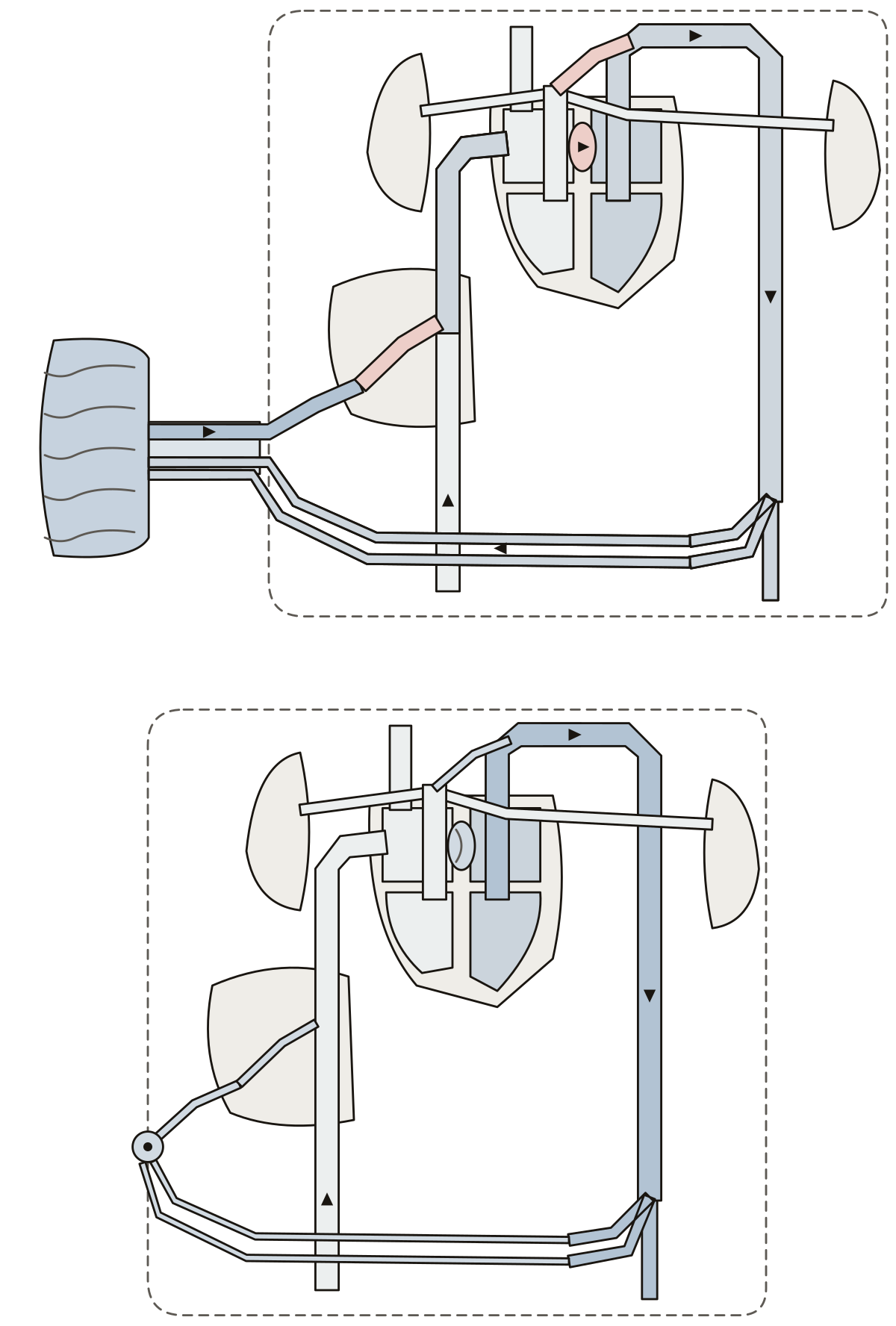 Fetal Heart DiagramThe placenta, umbilical vein and arteries, ductus venosus, foramen ovale and ductus arteriosus before birth, and the fossa ovalis and ligaments they become.
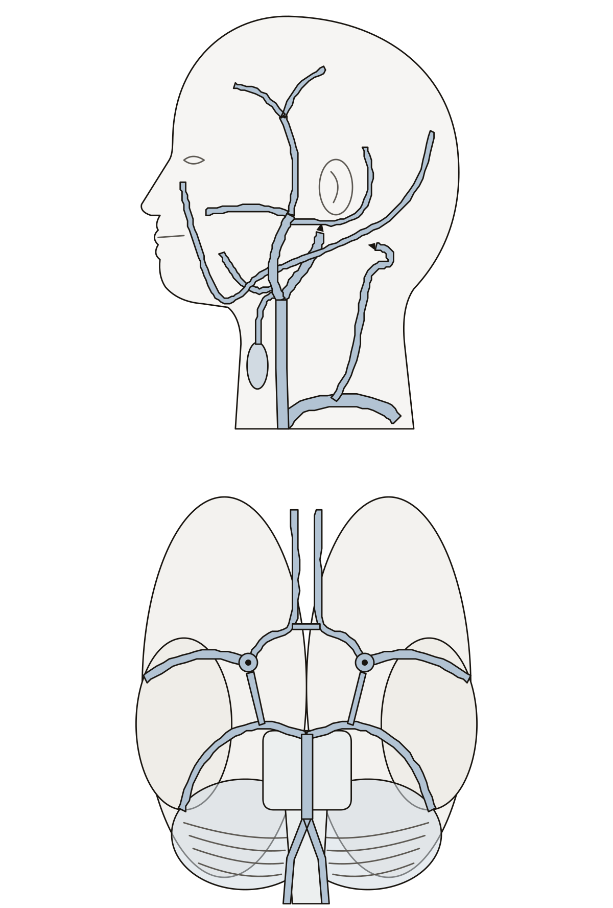 Label the Arteries of the Head and NeckThe common, internal and external carotid arteries, the vertebral artery and the branches to the face and scalp from the side, and the circle of Willis under the brain.
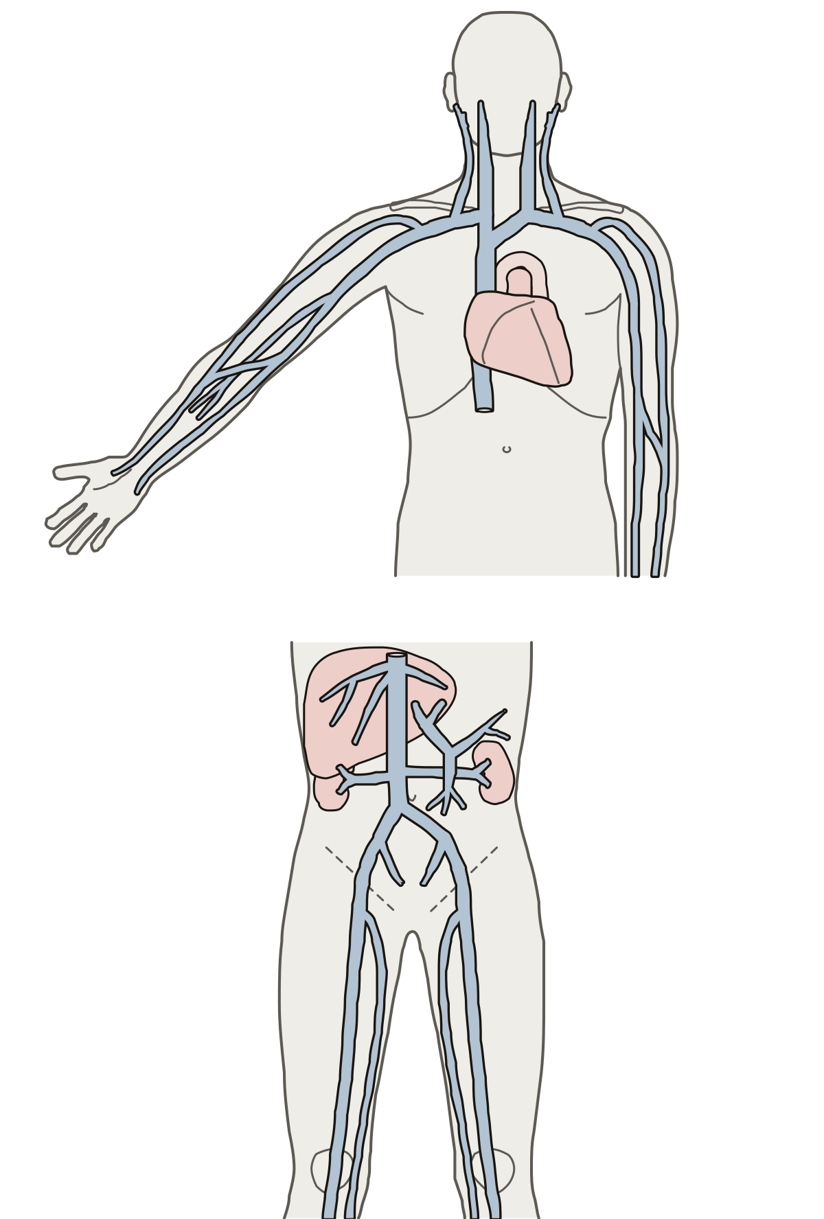 Major Veins of the Body LabeledThe jugular, subclavian, brachiocephalic, cephalic and basilic veins, the superior and inferior vena cava, and the renal, hepatic, hepatic portal, iliac, femoral and great saphenous veins.
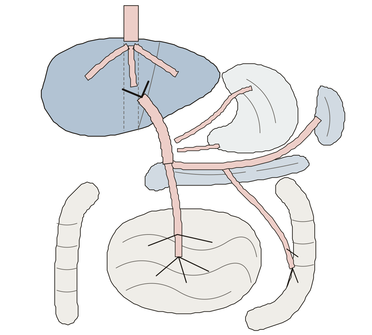 Hepatic Portal System DiagramHepatic portal, splenic, superior and inferior mesenteric and gastric veins, hepatic veins, inferior vena cava and five organs.
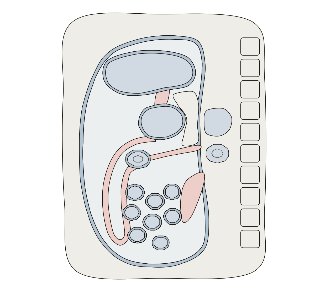 Peritoneum DiagramParietal and visceral peritoneum, the peritoneal cavity, greater and lesser omentum, mesentery, transverse mesocolon and the lesser sac.
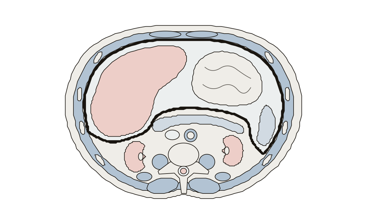 Abdomen Cross Section DiagramLiver, stomach, spleen, pancreas, kidneys, abdominal aorta, inferior vena cava, vertebra, spinal cord, rib, rectus abdominis, psoas major, erector spinae, parietal peritoneum and peritoneal cavity.
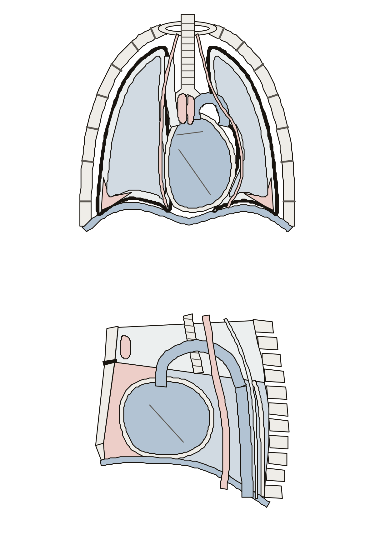 Thorax Anatomy QuizLungs, trachea, diaphragm, pericardium, aortic arch, superior vena cava, thymus, parietal pleura, pleural cavity, costodiaphragmatic recess, phrenic nerve, the four parts of the mediastinum, sternal angle, esophagus, descending aorta, thoracic duct and azygos vein.
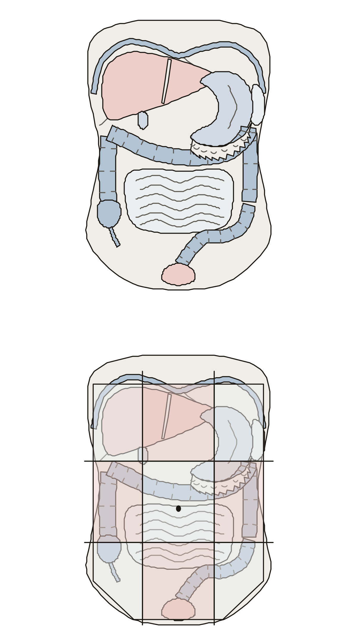 Abdominal Anatomy QuizDiaphragm, liver, falciform ligament, gallbladder, stomach, spleen, greater omentum, the four parts of the colon, cecum, appendix, small intestine, urinary bladder, and the nine regions of the abdomen.
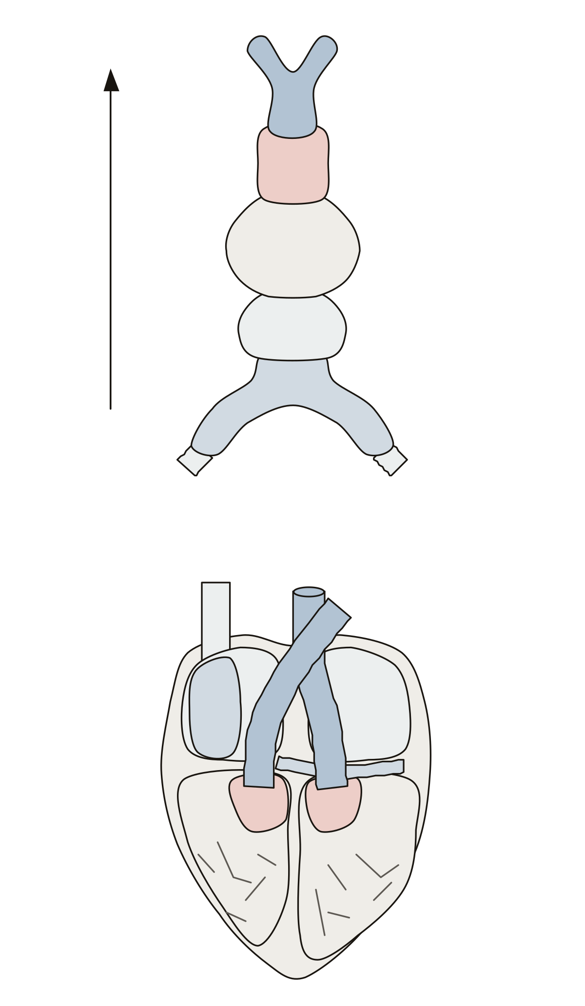 Primitive Heart Tube DiagramTruncus arteriosus, bulbus cordis, primitive ventricle, primitive atrium and sinus venosus, then six parts of the finished heart they form.
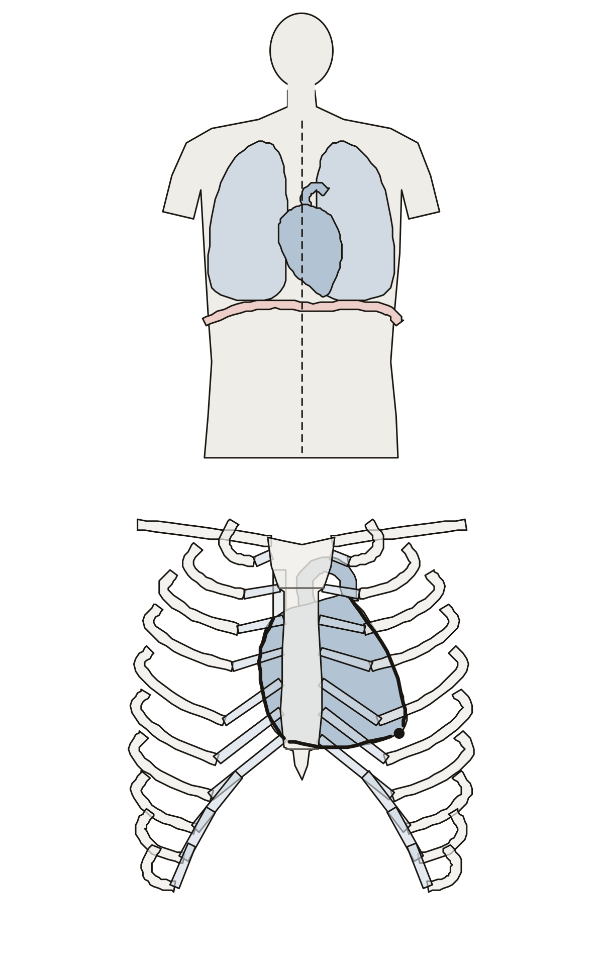 Heart Location in Body DiagramThe heart in the body between the lungs and on the diaphragm, then behind the sternum, ribs and cartilages with its apex and right, left and inferior borders.
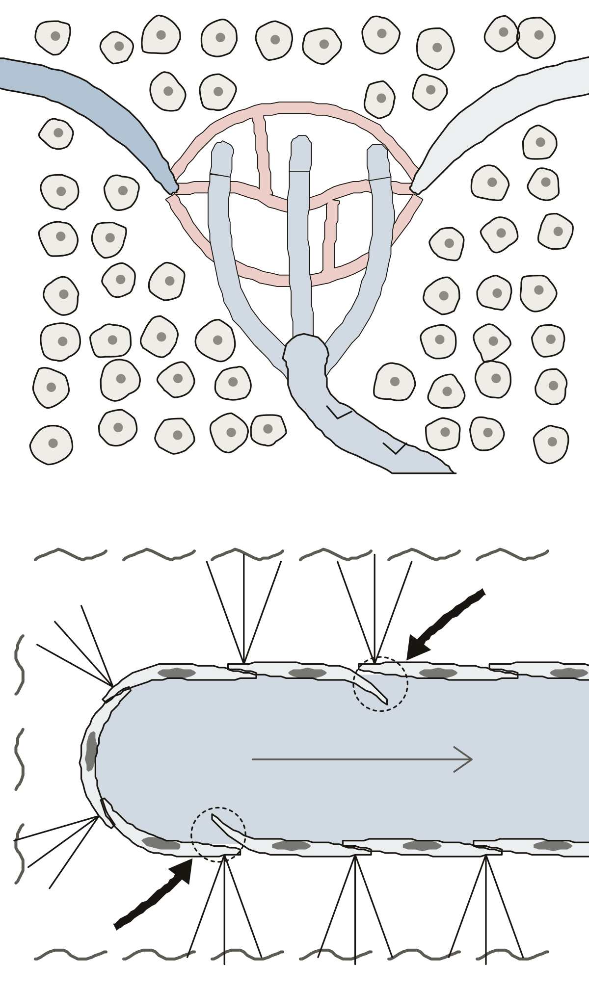 Lymphatic Capillary DiagramLymphatic capillaries starting as closed ends beside an arteriole, blood capillaries and a venule, then the wall enlarged: endothelial cells, their one-way valves, anchoring filaments, interstitial fluid and lymph.
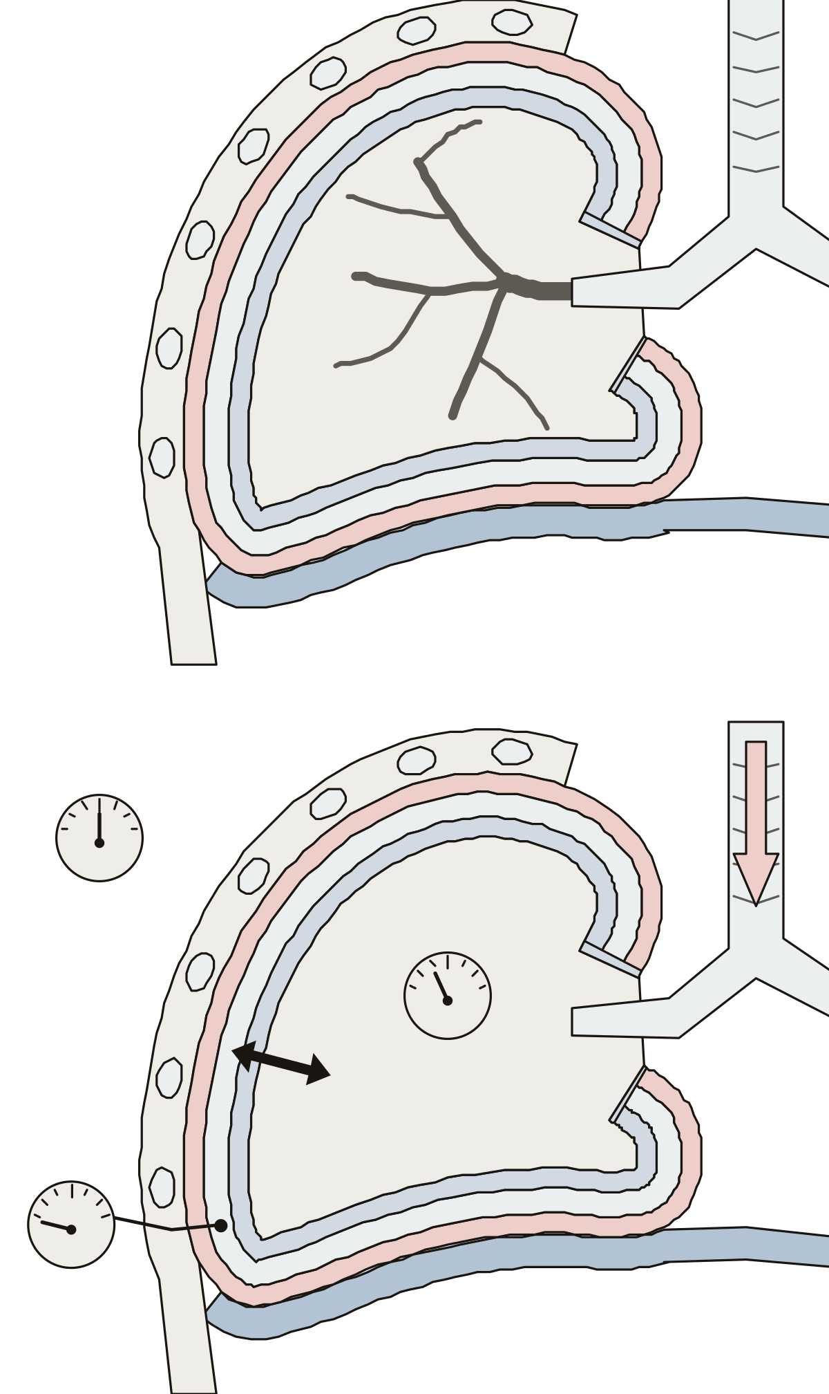 Pleural Membranes and Pressures DiagramThe parietal pleura, pleural cavity and visceral pleura around one lung, with the chest wall and diaphragm, then atmospheric, intrapulmonary, intrapleural and transpulmonary pressure and the air flowing in.
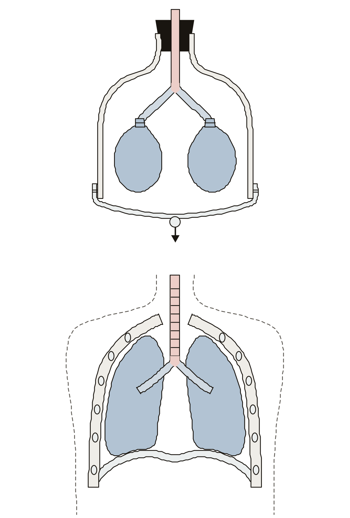 Bell Jar Model LabeledBell jar, stopper, glass tube, Y-tube branches, balloons and rubber sheet, then the chest wall, trachea, bronchi, lungs, diaphragm and thoracic cavity. Rubber stopper · Thoracic cavity · Thoracic wall
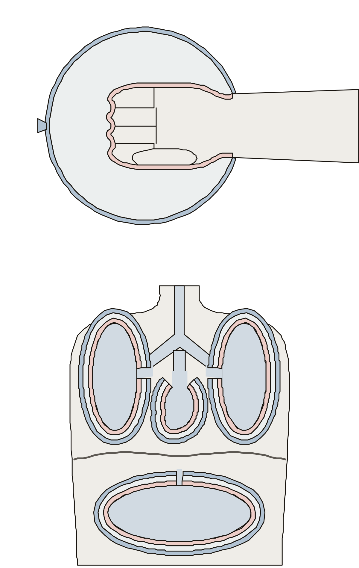 Serous Membranes LabeledVisceral layer, parietal layer and serous cavity on a fist in a balloon, then the parietal and visceral pleura, pericardium and peritoneum with the pleural, pericardial and peritoneal cavities.
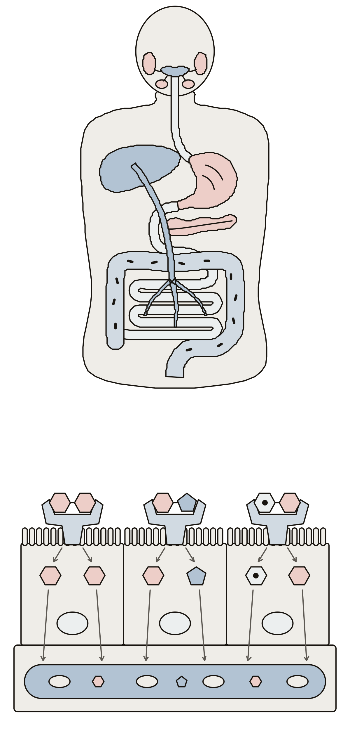 Carbohydrate Digestion DiagramSalivary amylase, stomach acid, pancreatic amylase, brush border enzymes, hepatic portal vein, liver and gut bacteria on the tract; maltase, sucrase, lactase and their sugars at the brush border.
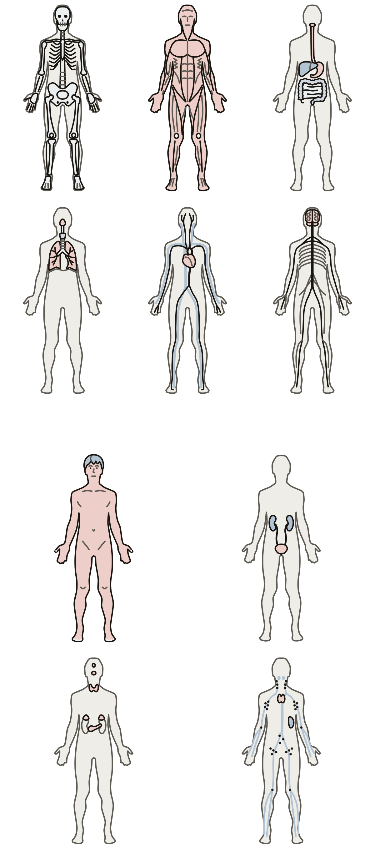 Organ System LabeledSkeletal, muscular, digestive, respiratory, cardiovascular, nervous, integumentary, urinary, endocrine and lymphatic systems, each drawn inside the same small body outline.
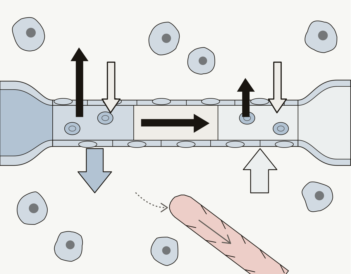 Capillary Exchange DiagramA capillary between an arteriole and a venule: arterial and venous ends, hydrostatic and colloid osmotic pressure, net filtration, net reabsorption, interstitial fluid and a lymphatic capillary.
- Serous Membrane Anatomy QuizA quiz from the first question: name the parietal layer, the visceral layer and the cavity of the pleura, the pericardium and the peritoneum.
- Veins in the Body Diagram Without LabelsThe major veins drawing with nothing written on it, to print and fill in: fifteen lines from the neck to the thigh, a word bank for each half and an answer key.
- Label the Serous Membranes of the BodyThe serous membranes as a worksheet: nine numbered layers and cavities around a lung, the heart and the abdominal organs, a line for each name, and a key.
- Label the Bell Jar ModelThe bell jar model of the lungs as a worksheet: six numbered parts, balloons to stopper, a line for each name, and a key. Rubber stopper · Bell jar · Rubber sheet
Organs, close up 49
One organ or gland per drawing, enlarged until its regions and layers are big enough to tap, from the heart and its valves to the kidney and the thyroid. The heart is also drawn from outside and the kidney with its blood vessels; the spleen and the teeth have pages, and one shows where the electrodes of a 12-lead ECG sit.
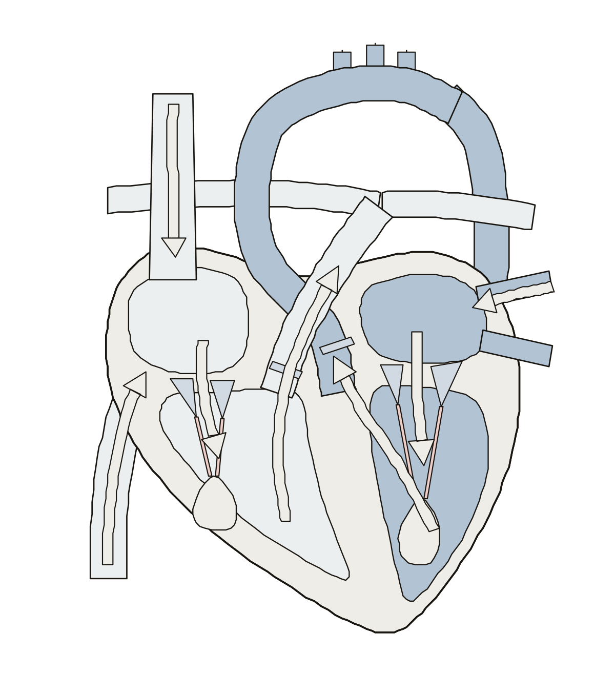 Heart DiagramThe heart cut open: 4 chambers, 4 valves, septum, aorta, venae cavae, pulmonary trunk, arteries and veins, plus blood flow. Tricuspid valve · Left ventricle · Pulmonary trunk
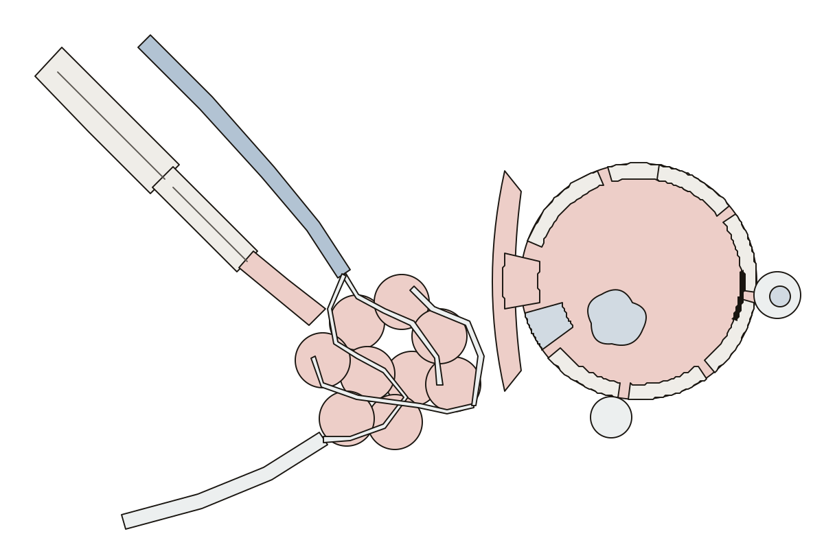 Alveoli Diagram LabeledTerminal and respiratory bronchioles, alveolar duct and sac, arteriole, venule, capillaries, type I and II cells, macrophage and the respiratory membrane. Alveolar pore · Respiratory membrane · Type II alveolar cell
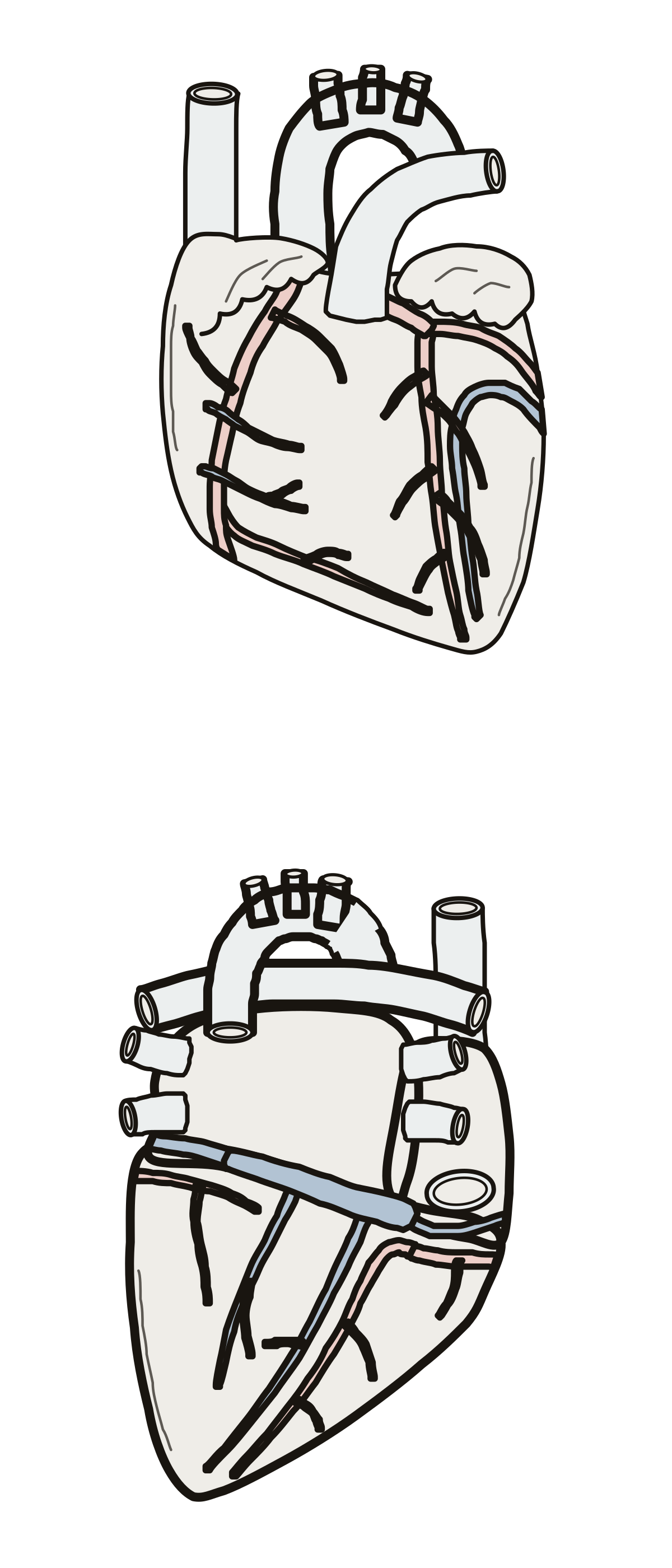 Label the Cardiac Veins and Coronary ArteriesThe heart's own blood supply, front and back: 12 vessels to name. Circumflex artery · Right coronary artery · Marginal artery
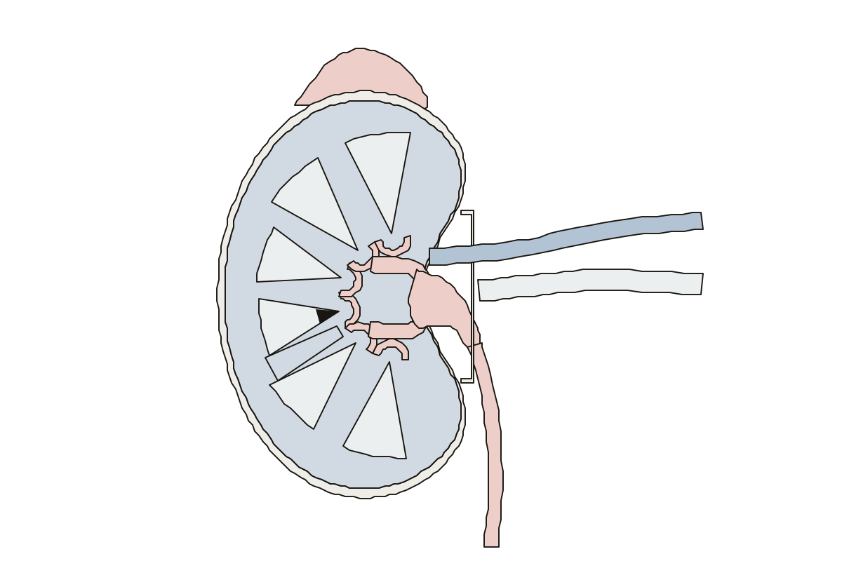 Label the KidneyOne kidney cut open from the front: 14 parts to name, labeled or blank. Renal artery · Renal hilum · Major calyx
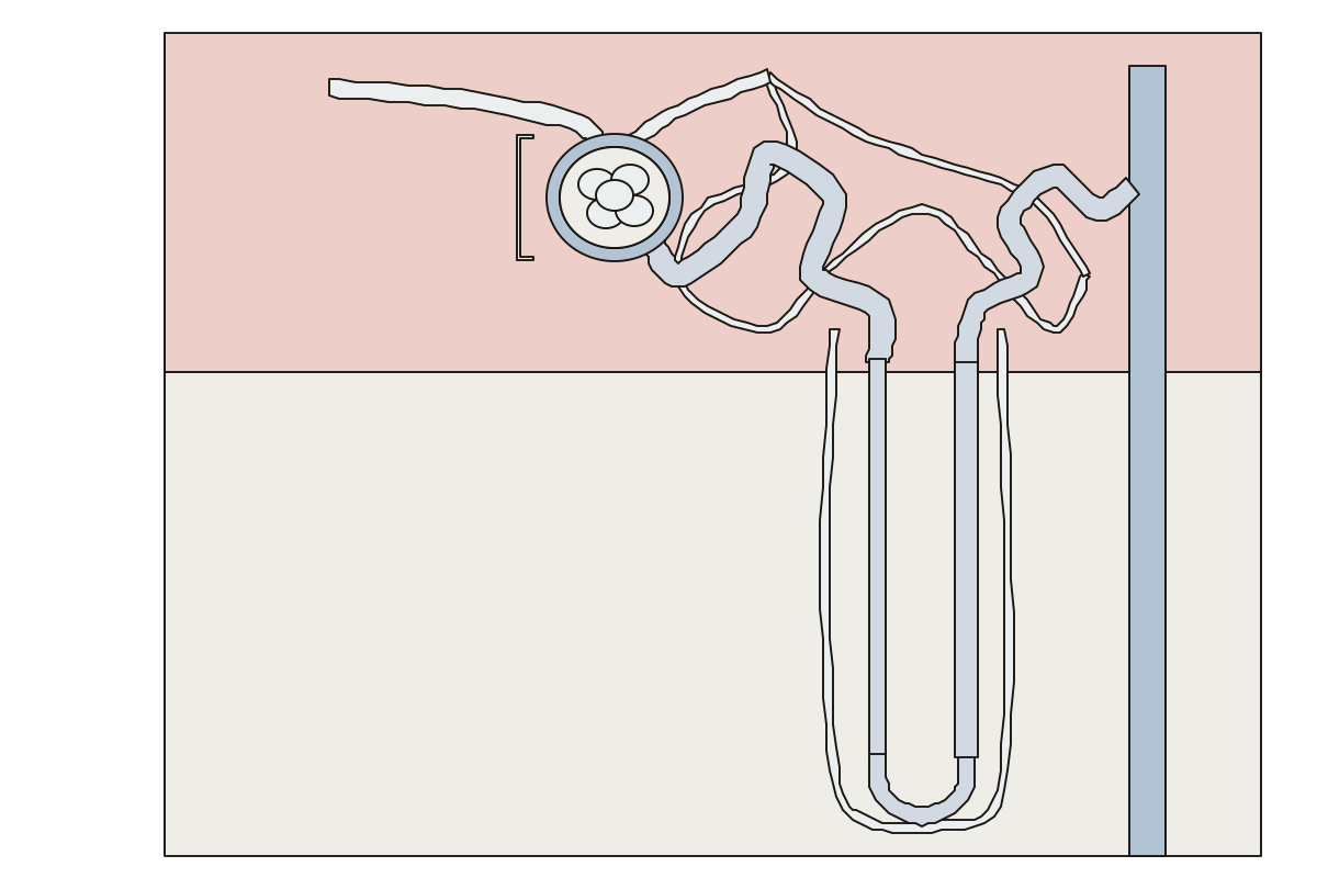 Nephron Labeled DiagramOne nephron across the cortex and medulla: 15 parts to name, labeled or blank. Loop of Henle · Descending limb · Bowman's capsule
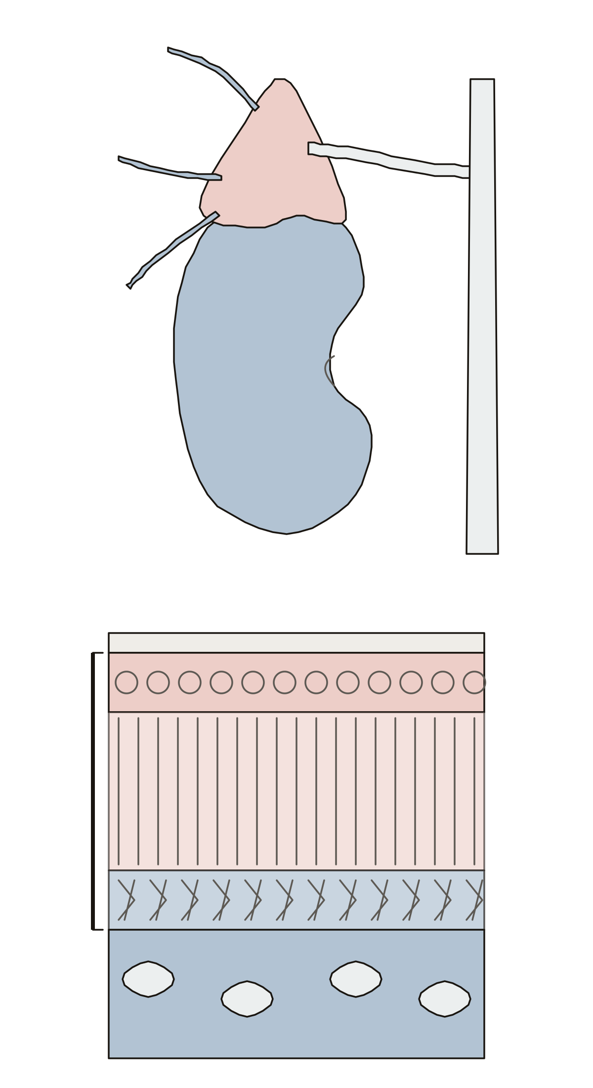 Parts of the Adrenal GlandThe adrenal gland: on the kidney with its vein and arteries, then capsule, cortex, zona glomerulosa, fasciculata and reticularis, and medulla. Glomerulosa · Reticularis · Medulla
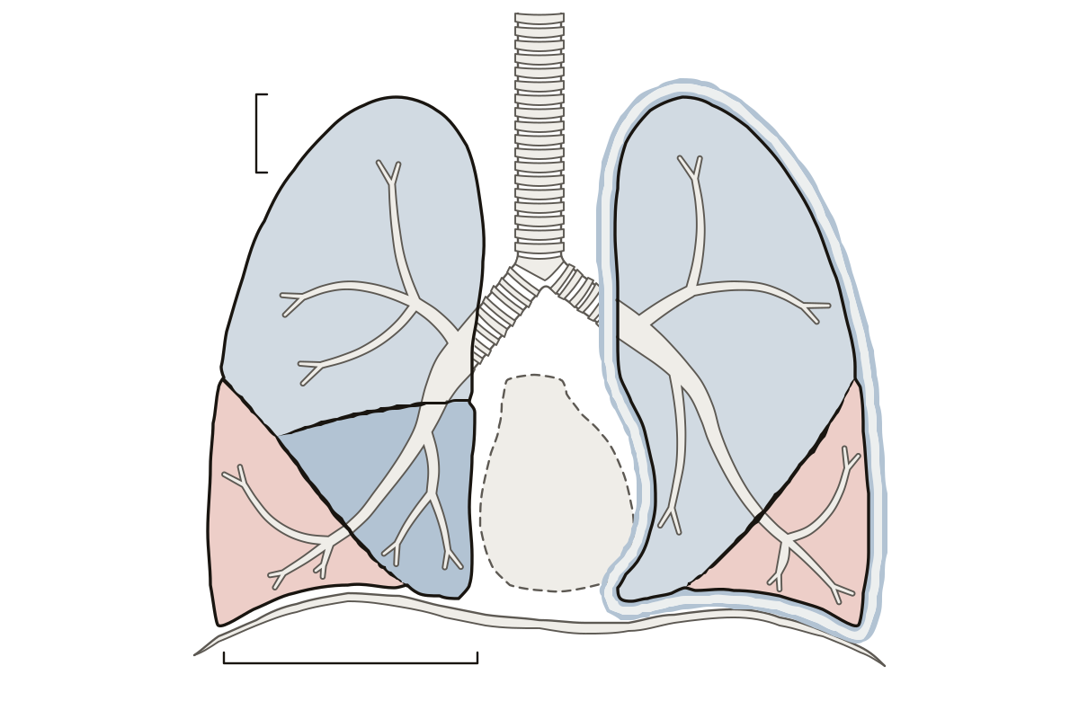 Label the LungsBoth lungs from the front: lobes, fissures and pleura, 13 parts to name. Cardiac notch · Horizontal fissure · Left superior lobe
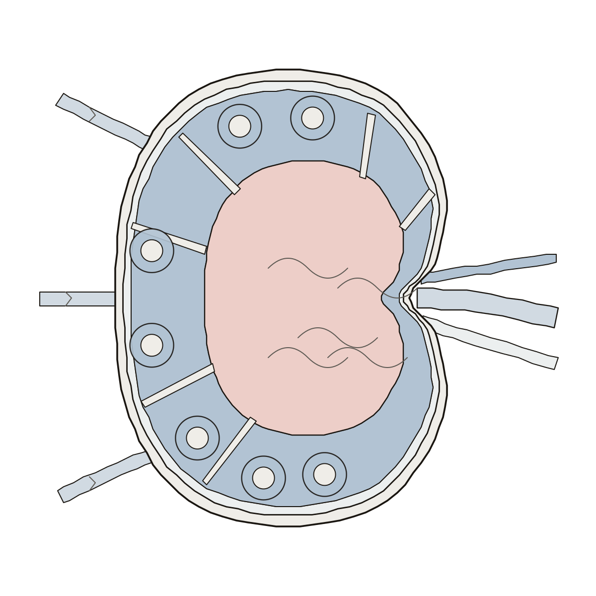 Lymph Node DiagramA lymph node cut in half: capsule, trabeculae, lymph sinus, cortex, follicles, germinal centres, medulla, afferent and efferent vessels, artery and vein. Capsule · Medulla · Blood vessels
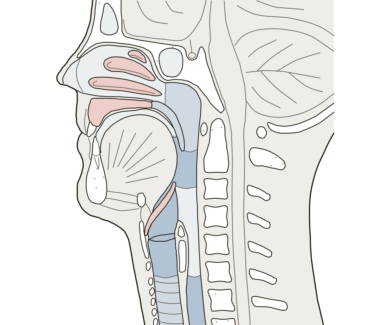 Upper Respiratory Tract DiagramThe head and neck cut down the middle: 12 parts of the upper airway. Nasal conchae · Esophagus · Hard palate
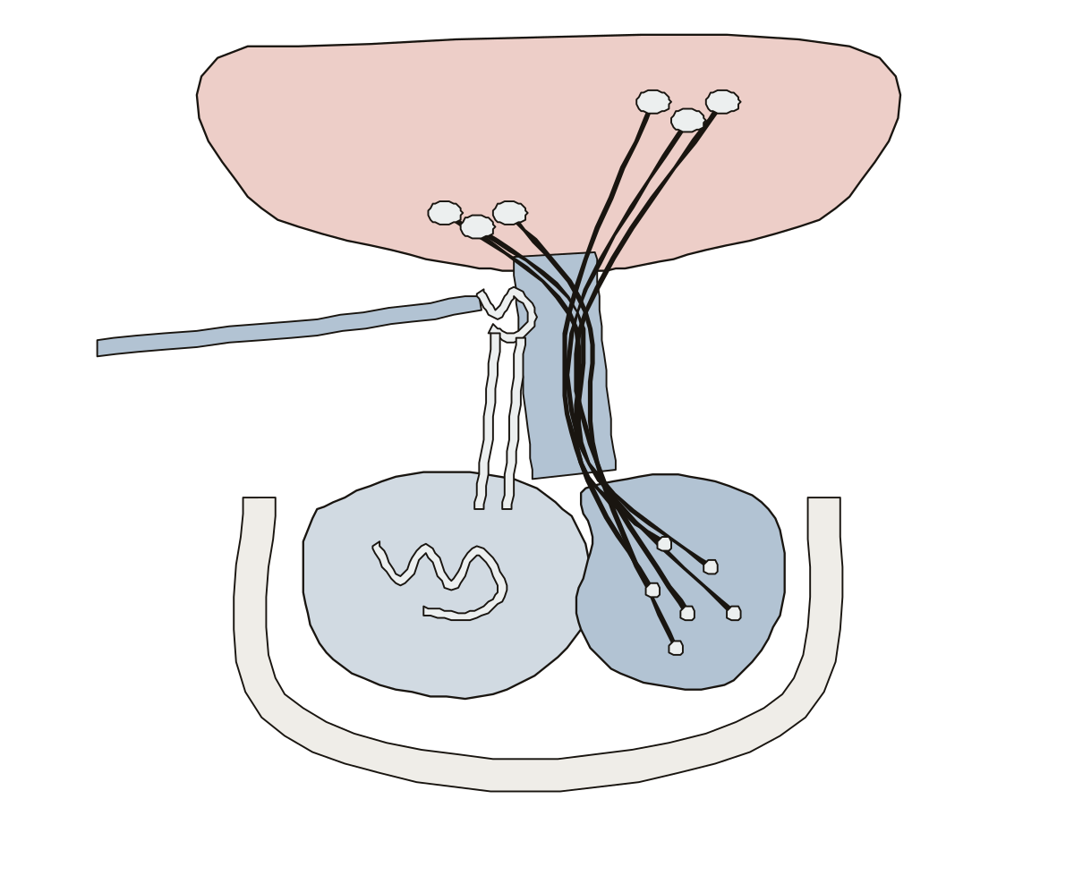 Pituitary Gland Diagram LabeledThe pituitary and hypothalamus, nerve route and blood route: 11 parts. Primary capillary plexus · Secondary capillary plexus · Neurosecretory cells
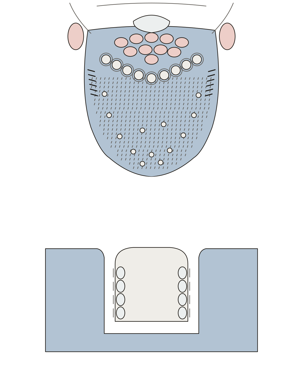 Tongue DiagramTip, filiform, fungiform, vallate and foliate papillae, lingual and palatine tonsils, epiglottis and taste buds.
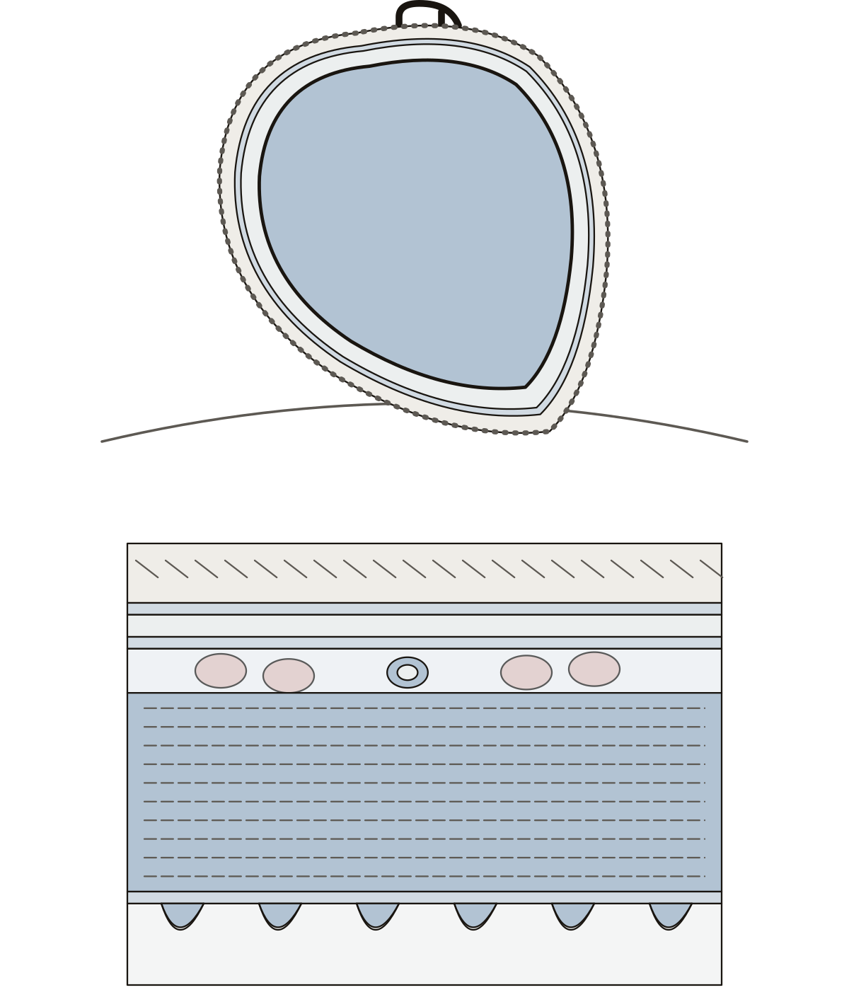 Pericardium and Heart Wall DiagramFibrous pericardium, parietal pericardium, pericardial cavity, epicardium, coronary vessel, myocardium, endocardium, trabeculae carneae. Visceral pericardium (epicardium)
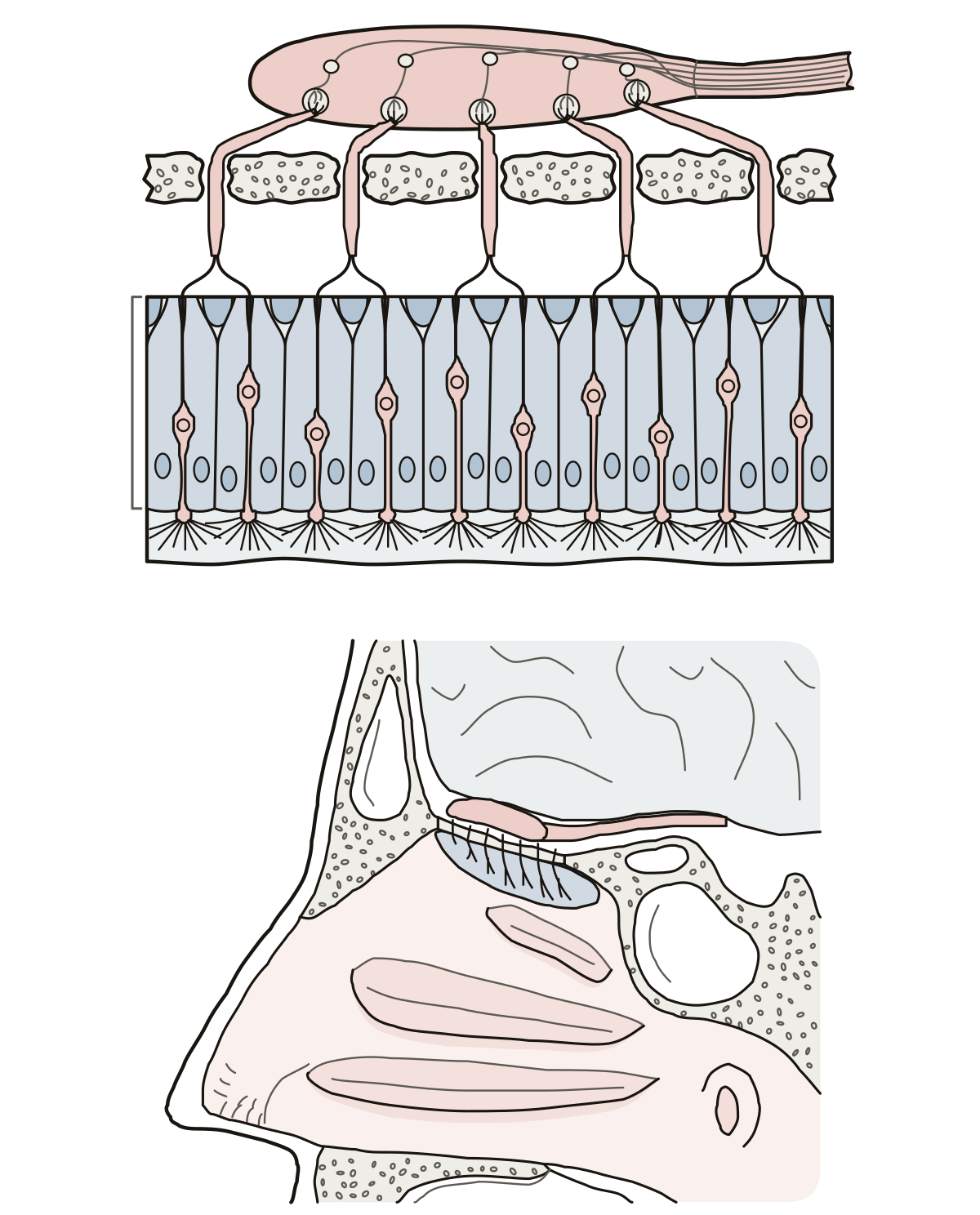 Olfactory System DiagramOlfactory epithelium, receptor neurons, supporting cells, cilia, mucus, olfactory nerve, cribriform plate, bulb and tract. Olfactory nerve (cranial nerve I)
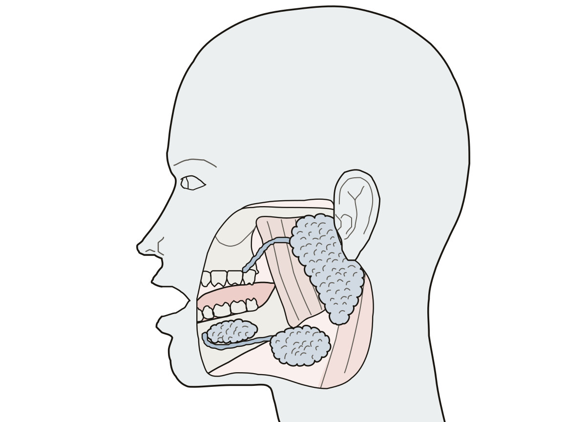 Salivary Glands DiagramParotid gland and duct, submandibular gland and duct, sublingual gland, tongue and mandible.
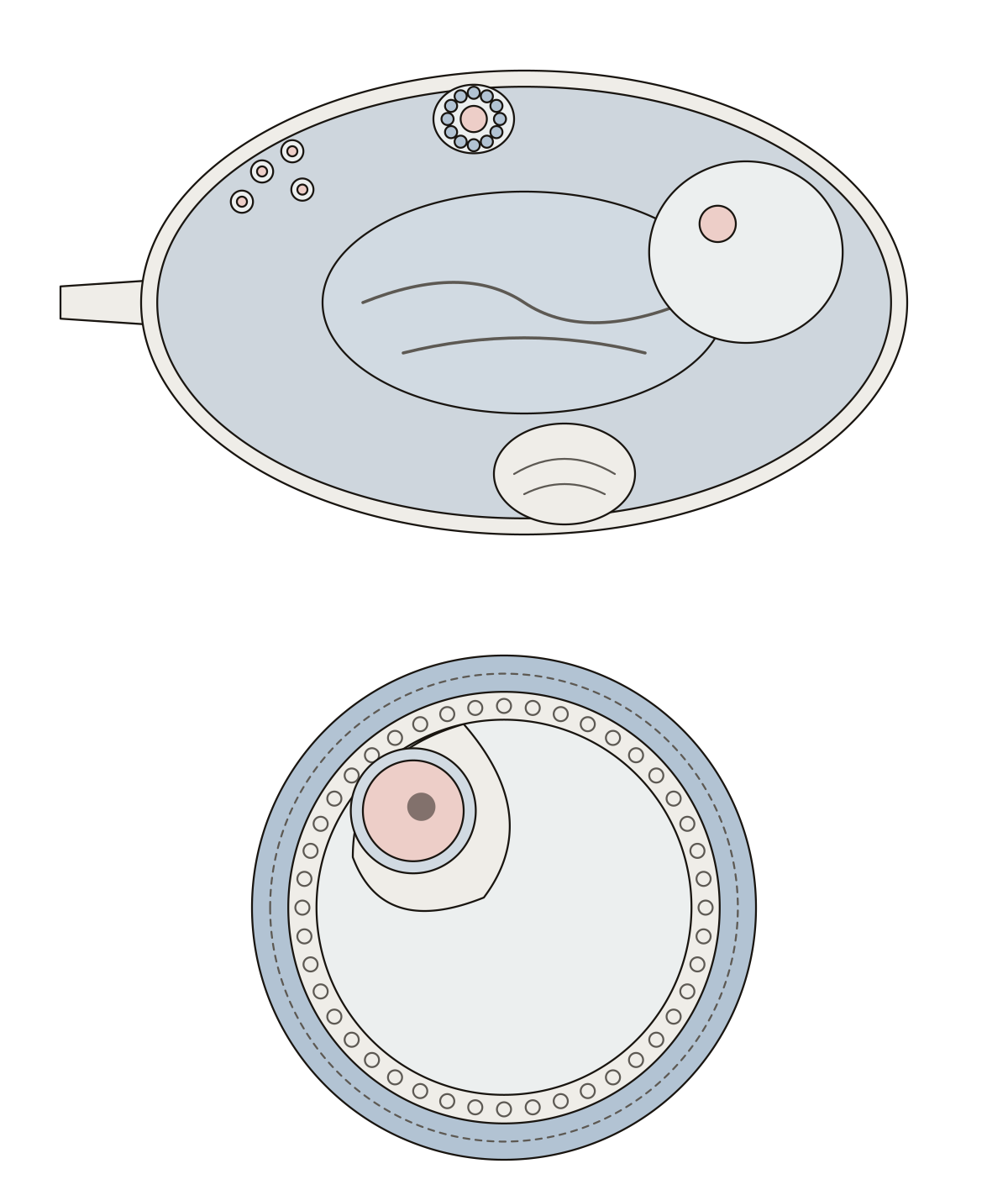 Ovary DiagramTunica albuginea, cortex, medulla, primordial, secondary and mature follicles, corpus luteum, oocyte, zona pellucida, granulosa, antrum, theca. Mature (tertiary) follicle
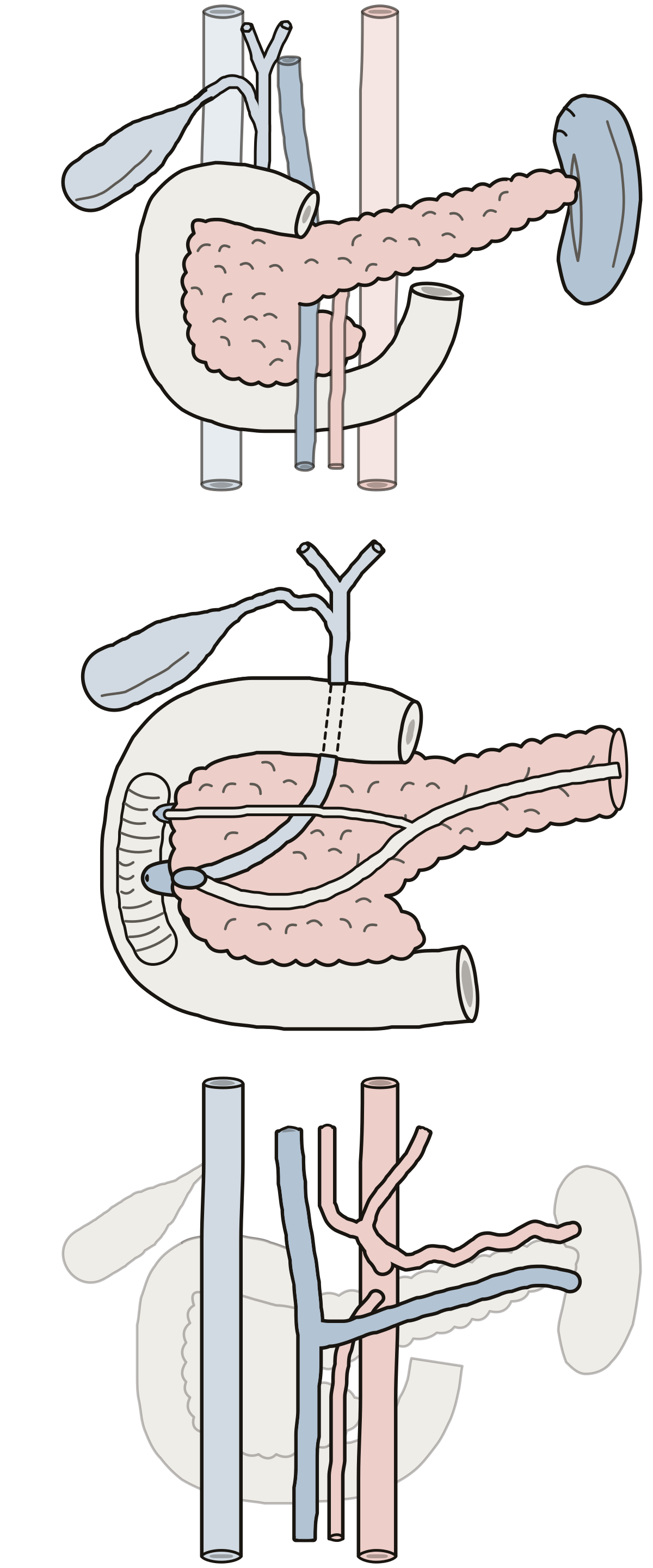 Upper Abdomen Organs DiagramThe four parts of the duodenum, head, body and tail of the pancreas, spleen, gallbladder, bile and pancreatic ducts, celiac trunk, portal vein.
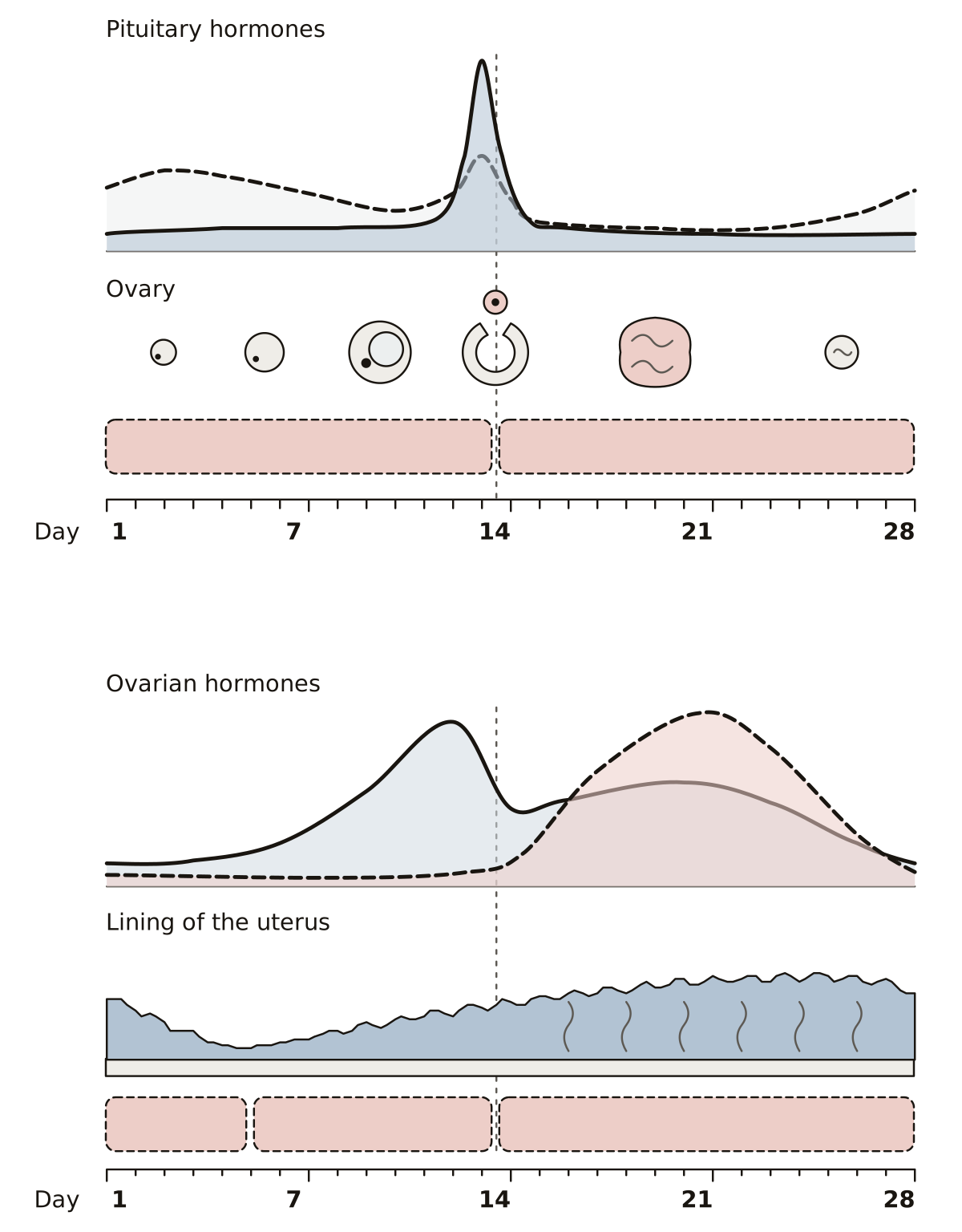 Uterine Cycle DiagramMenstrual, proliferative and secretory phases; follicular phase, ovulation and luteal phase; FSH, LH, estrogen, progesterone, corpus luteum and endometrium.
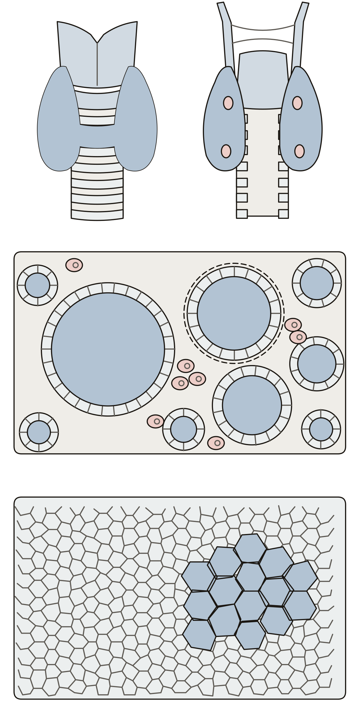 Thyroid Gland DiagramRight and left lobes, isthmus, parathyroid glands, thyroid follicle, colloid, follicular and parafollicular cells, chief and oxyphil cells.
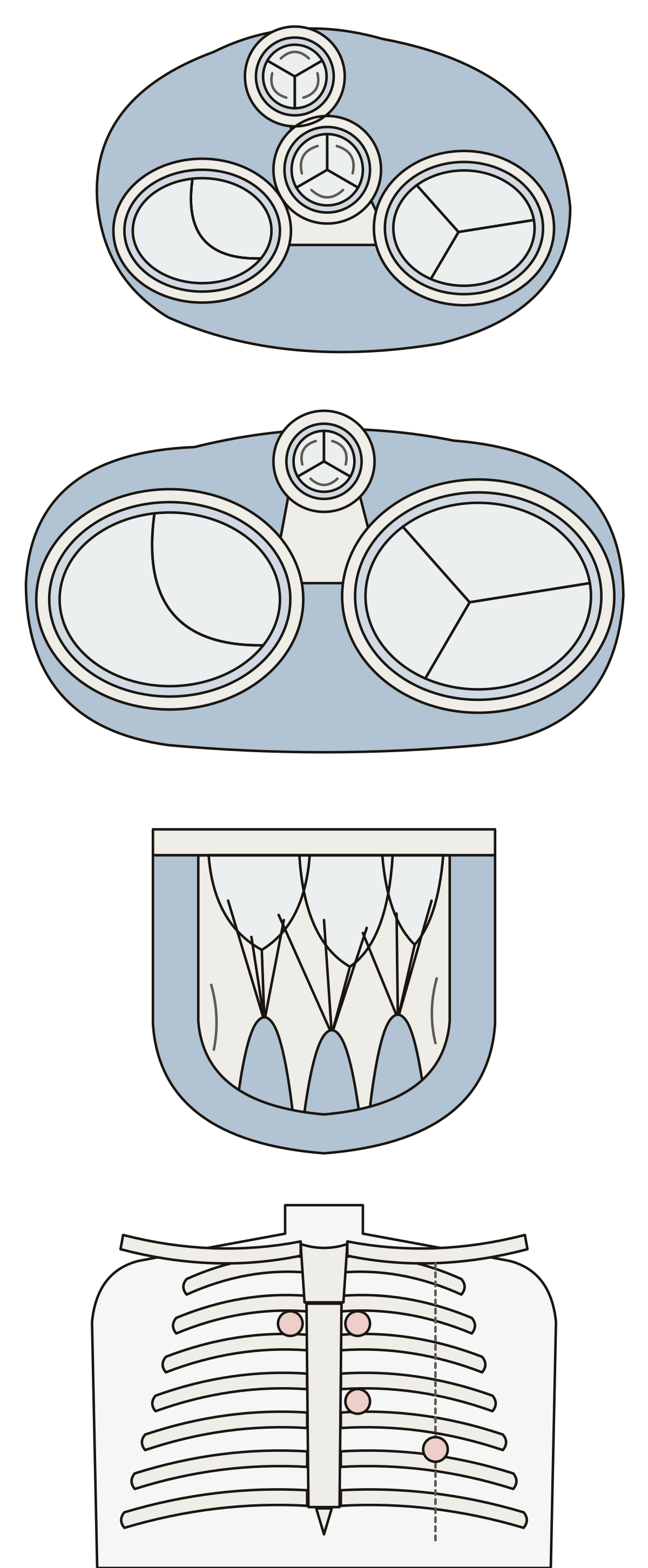 Heart Valves DiagramTricuspid, mitral, aortic and pulmonary valves, the cardiac skeleton, five named cusps, chordae tendineae, a papillary muscle and four listening areas.
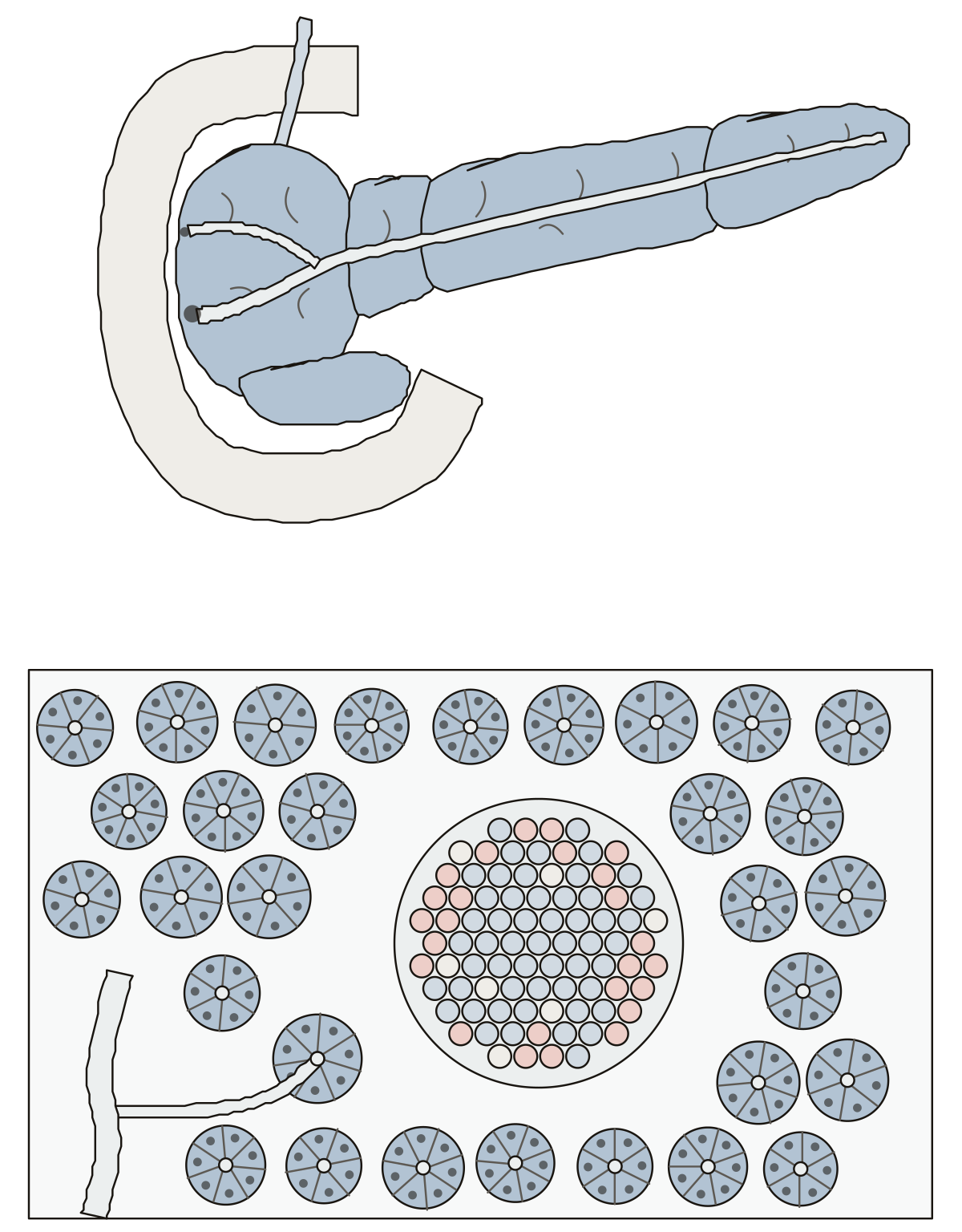 Pancreas DiagramHead, neck, body, tail, uncinate process, pancreatic and accessory ducts, duodenum, islet, alpha, beta and delta cells, acinus, intercalated duct.
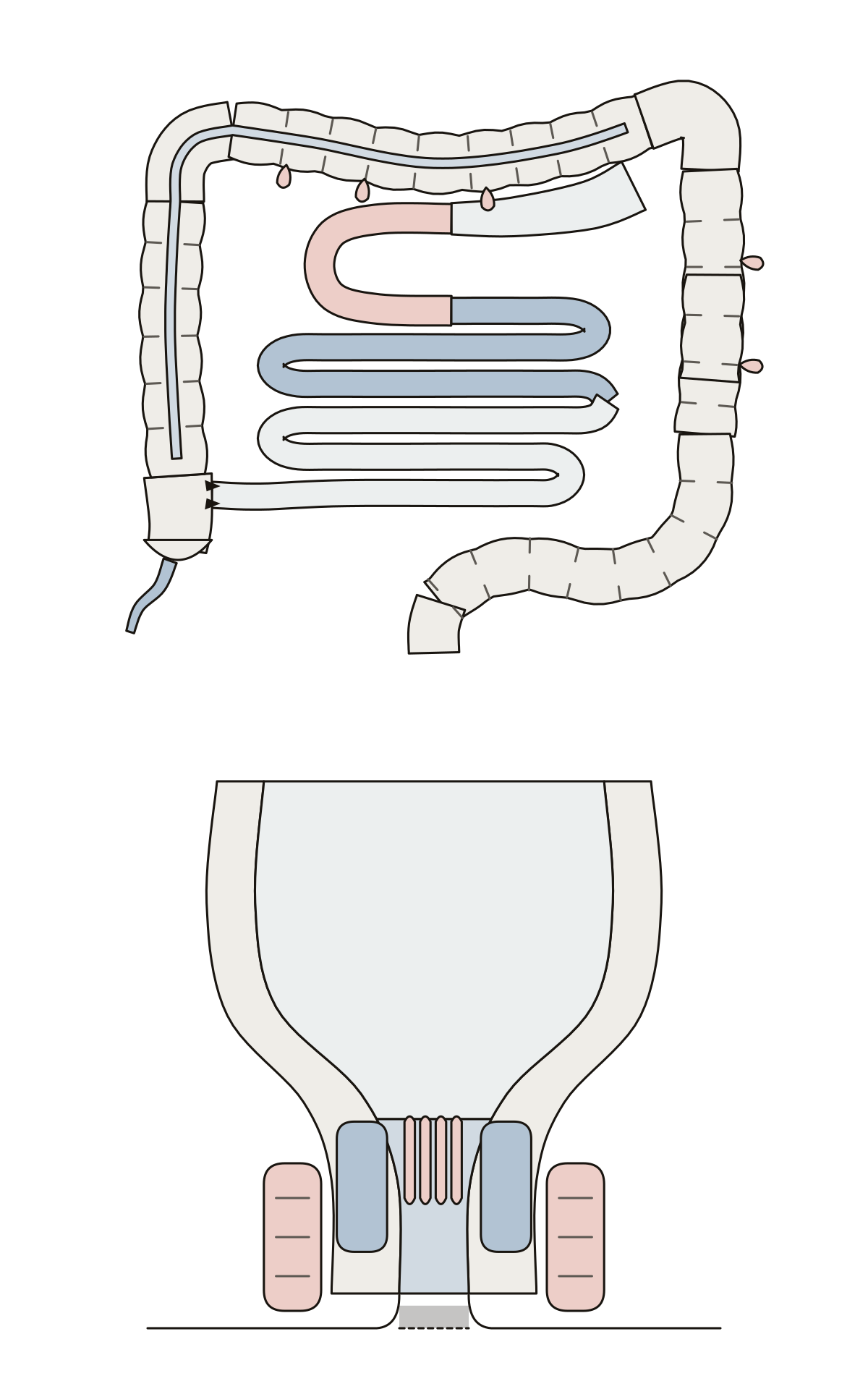 Small and Large Intestine DiagramThe small intestine inside the frame of the large intestine, from duodenum to sigmoid colon, and the rectum and anal canal cut through.
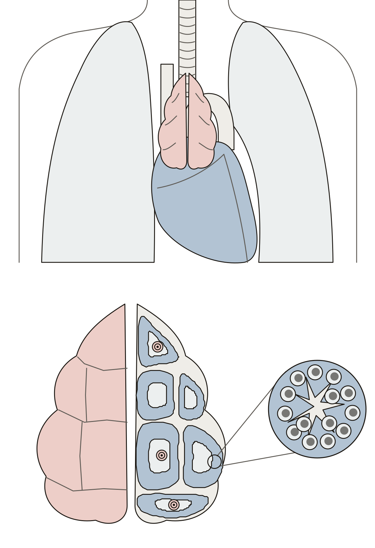 Thymus DiagramThymus, trachea, heart and lungs, then the capsule, lobules, cortex, medulla, thymic corpuscles and thymocytes of a lobe cut open.
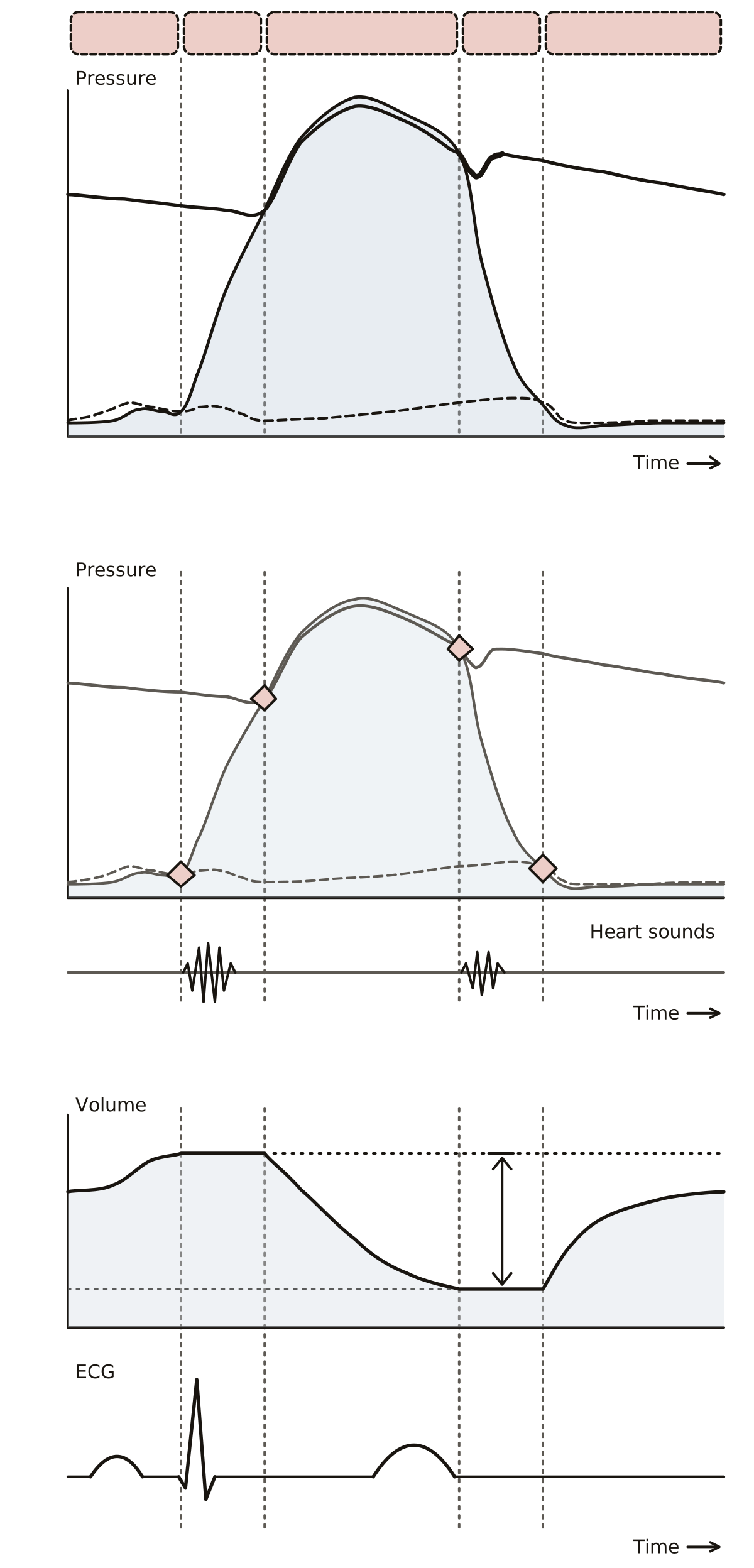 Cardiac Cycle QuizA schematic chart of one heartbeat: the five phases, aortic, ventricular and atrial pressure, the valve events, ventricular volume, the ECG waves and the two heart sounds.
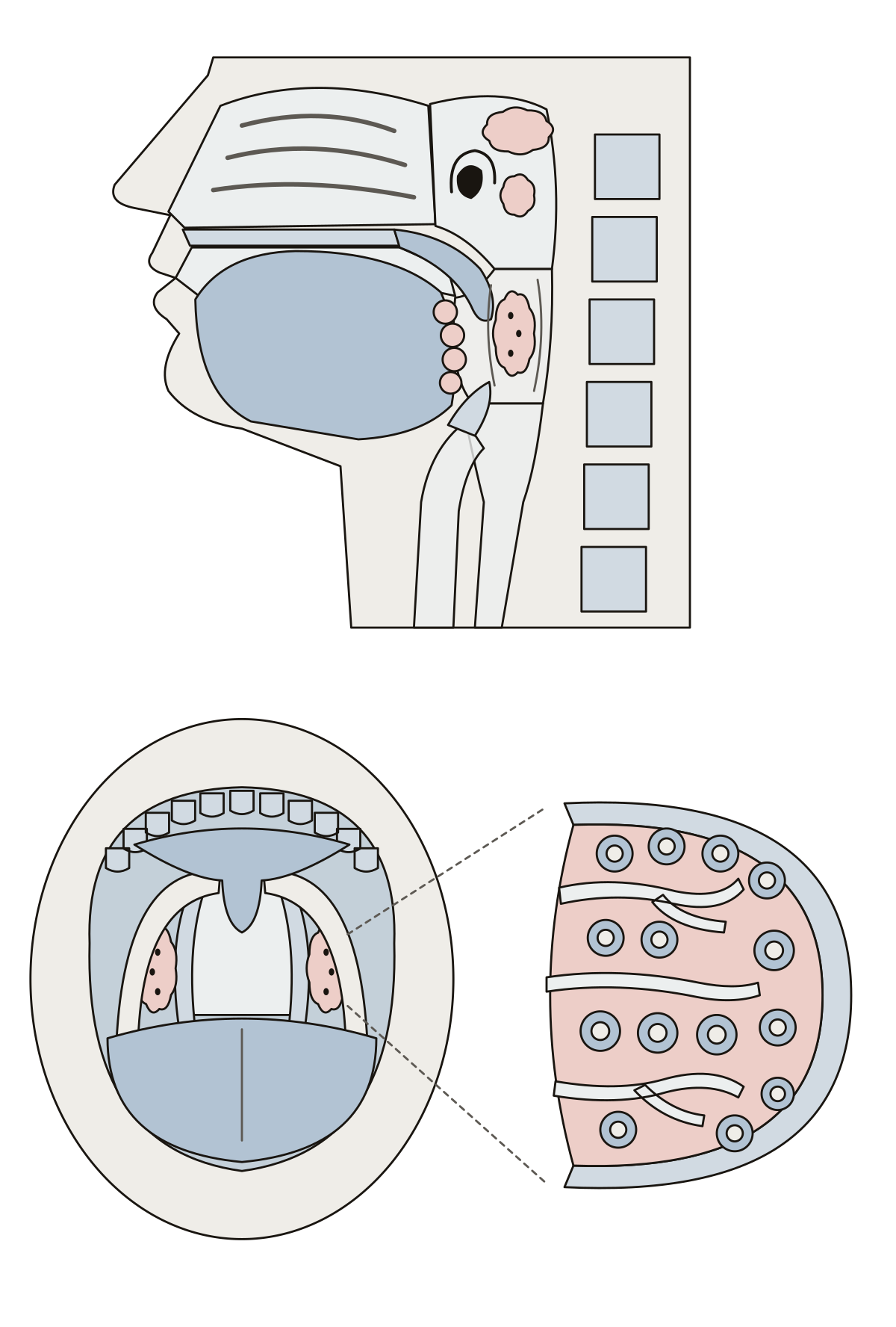 Tonsils DiagramThe pharyngeal, tubal, palatine and lingual tonsils in a midsagittal section of the throat, then the arches, crypts, lymphoid nodules and capsule of a palatine tonsil.
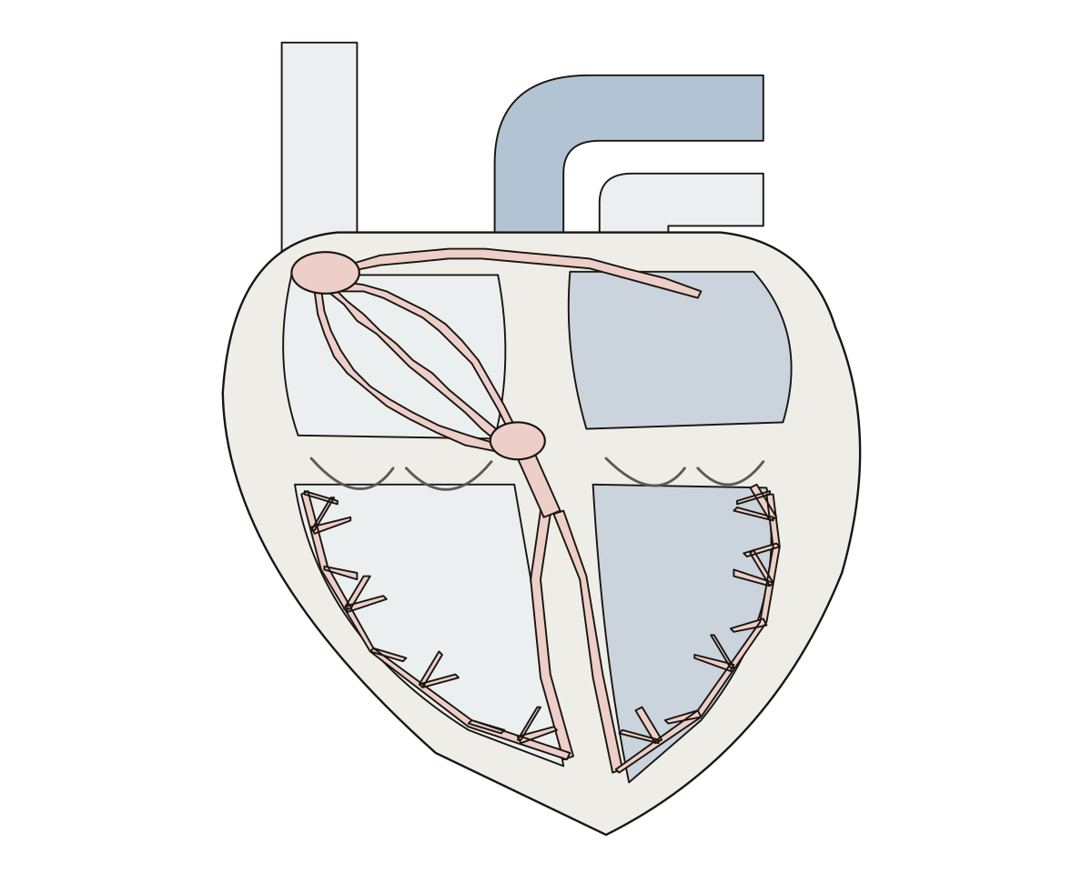 Electrical Conduction of the Heart DiagramThe sinoatrial node, internodal pathways, Bachmann’s bundle, atrioventricular node and bundle, bundle branches and Purkinje fibers on a heart cut open, with its four chambers.
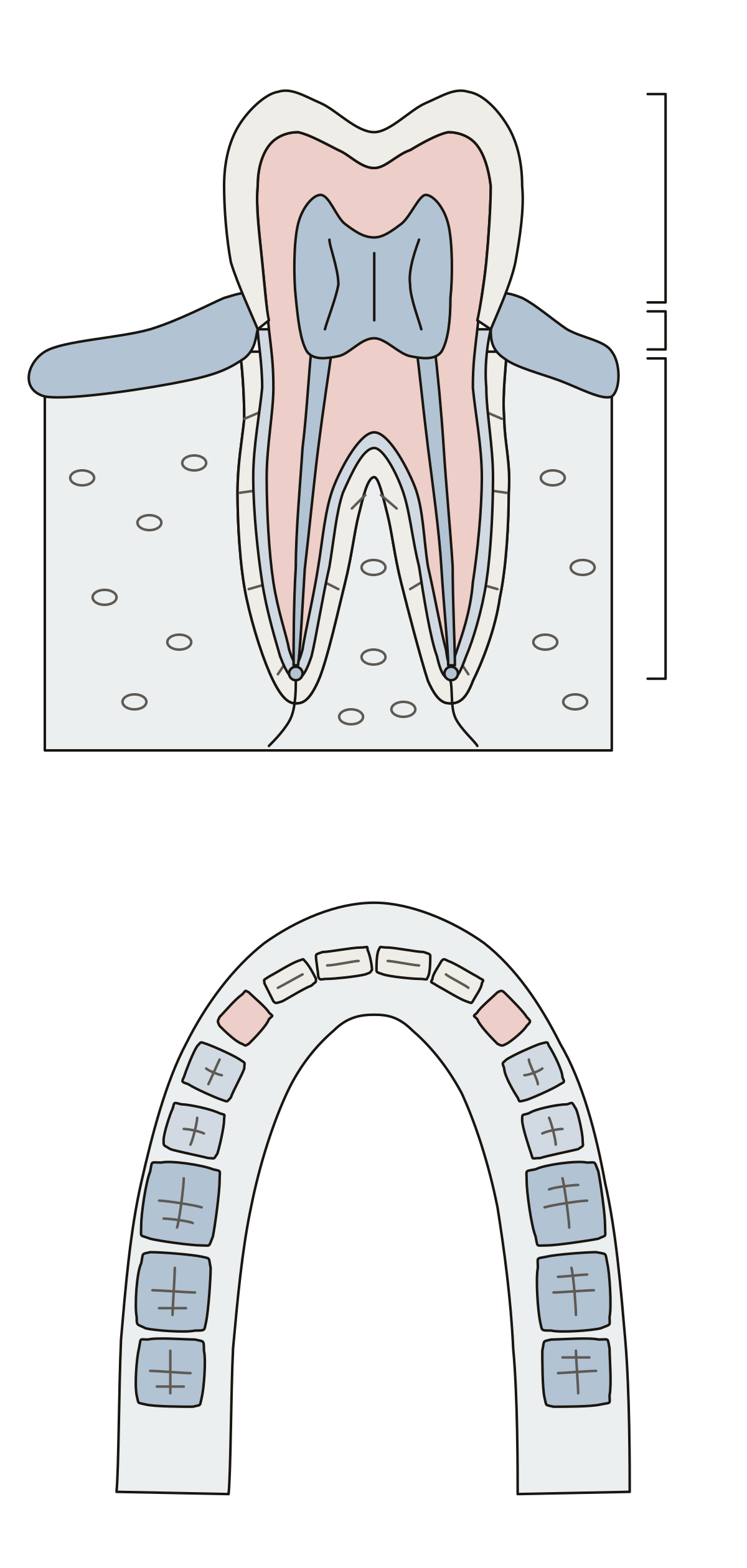 Parts of a Tooth and Their FunctionsA molar cut in half: crown, neck and root, enamel, dentin, pulp, cementum and the ligament that holds it in the jaw, each with its job, then the four types of teeth.
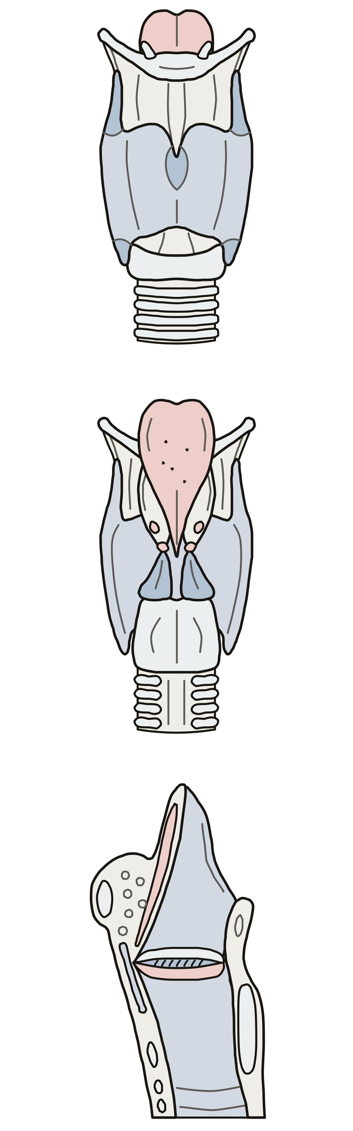 Larynx Anatomy QuizThe larynx in three views: ten parts from the front, the arytenoid, corniculate and cuneiform cartilages from behind, and the vocal and vestibular folds in a cut down the middle.
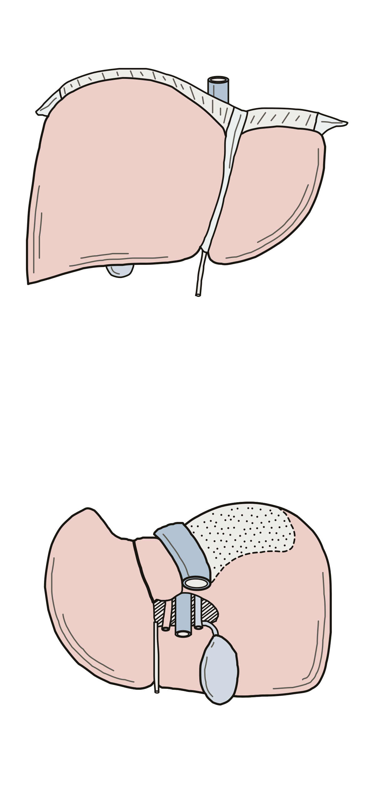 Liver Anatomy QuizThe liver in two views: two lobes and four ligaments from the front, then the underside with the caudate and quadrate lobes, the gallbladder and the vessels at the porta hepatis.
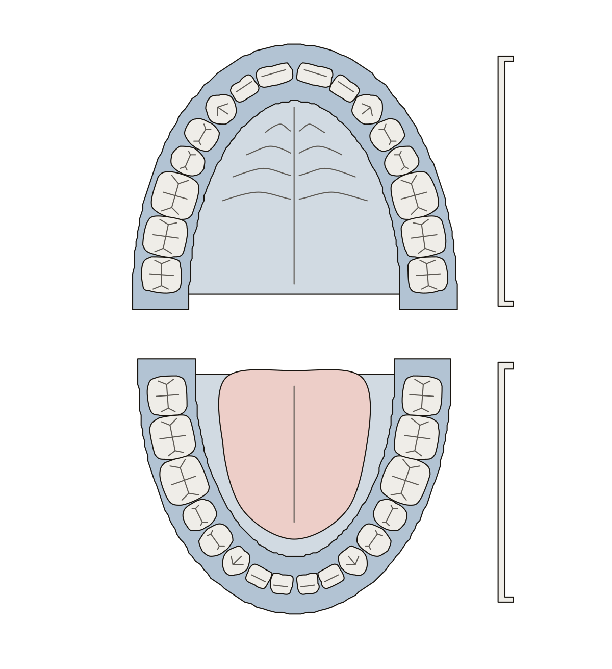 Teeth DiagramCentral incisor, lateral incisor, canine, first and second premolar, first, second and third molar, upper and lower dental arch.
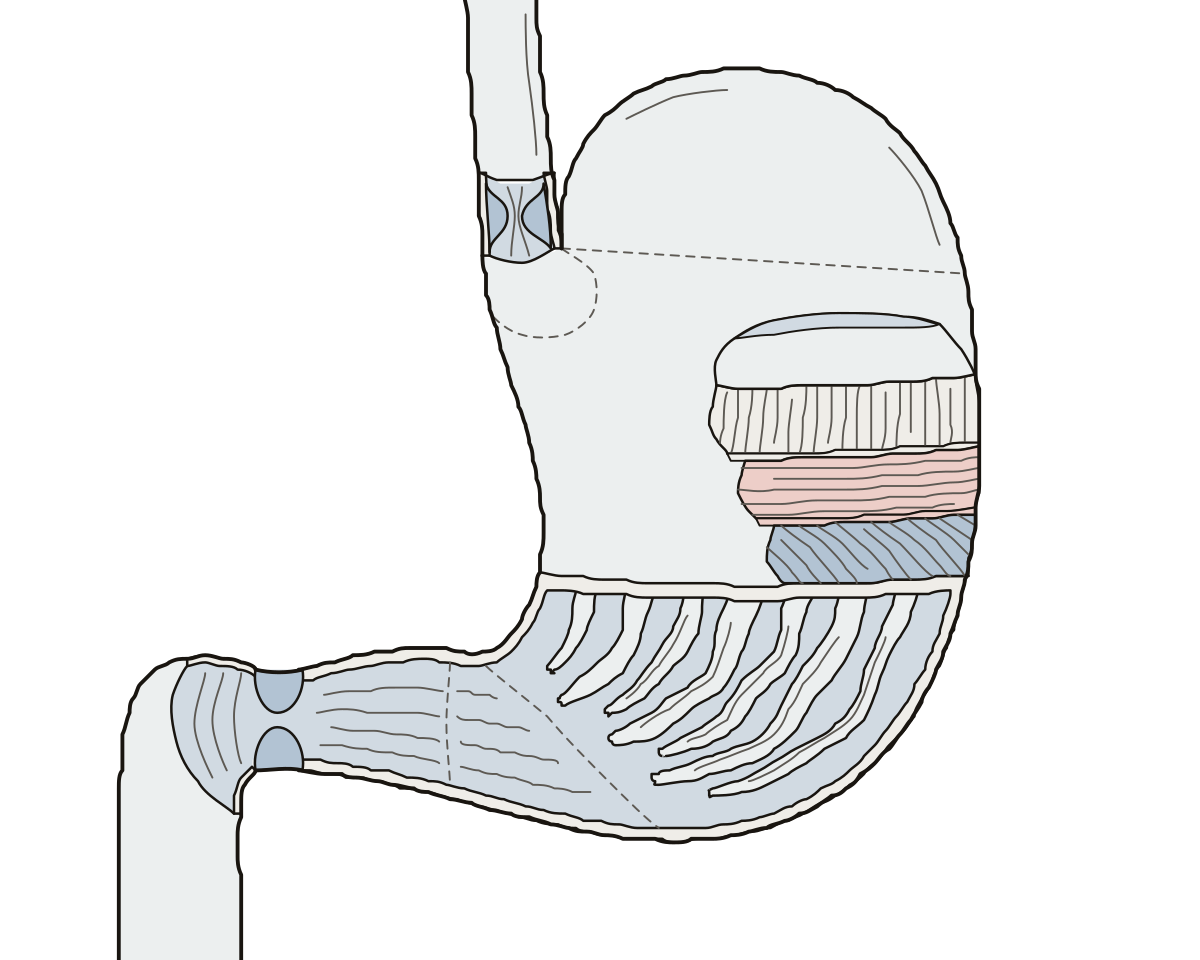 Stomach Anatomy QuizSixteen parts of the stomach on one drawing, from the esophagus to the duodenum: regions, sphincters, curvatures, rugae and three muscle layers.
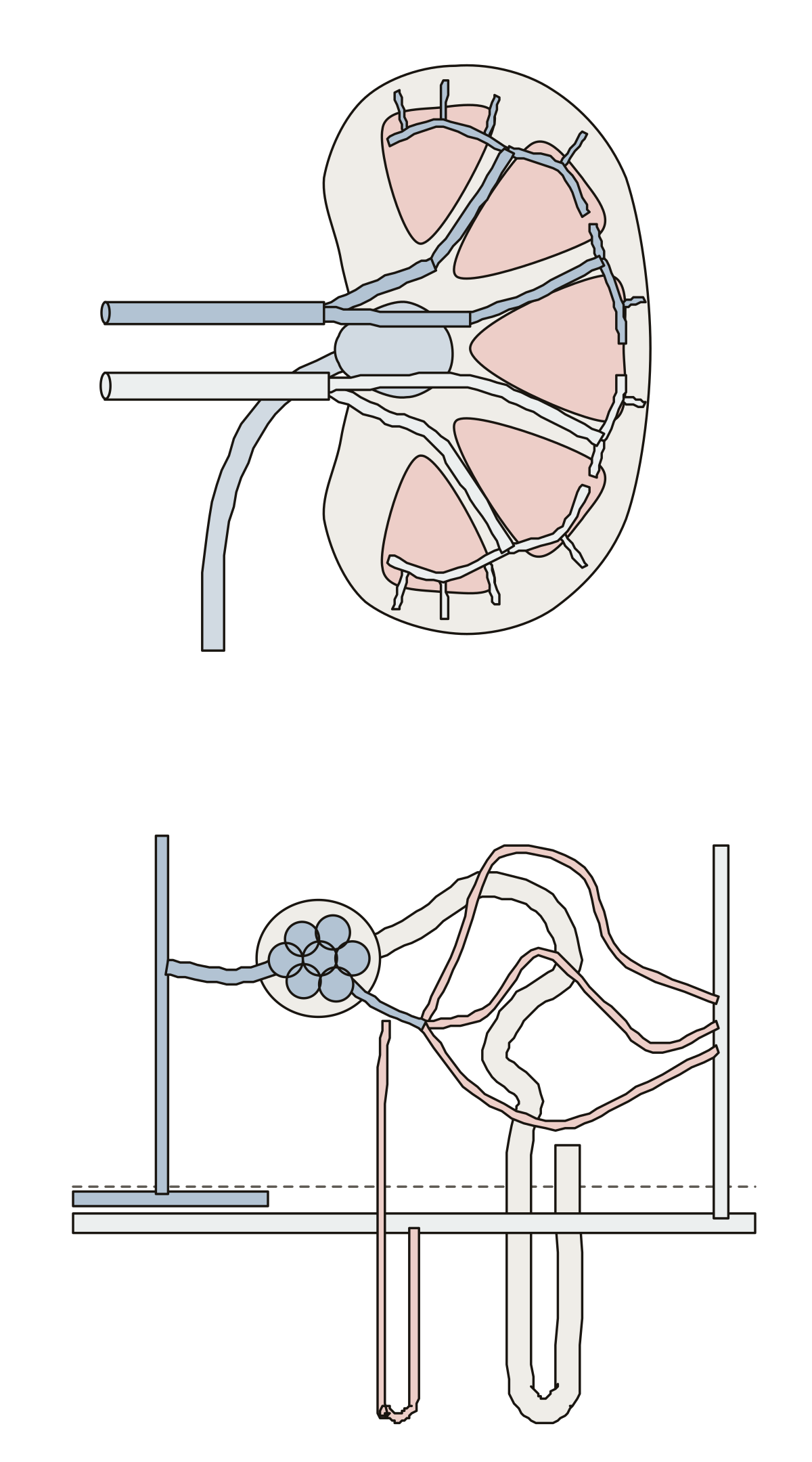 Kidney Blood Vessels DiagramRenal artery to renal vein: segmental, interlobar, arcuate and cortical radiate vessels, afferent and efferent arterioles, peritubular capillaries, vasa recta.
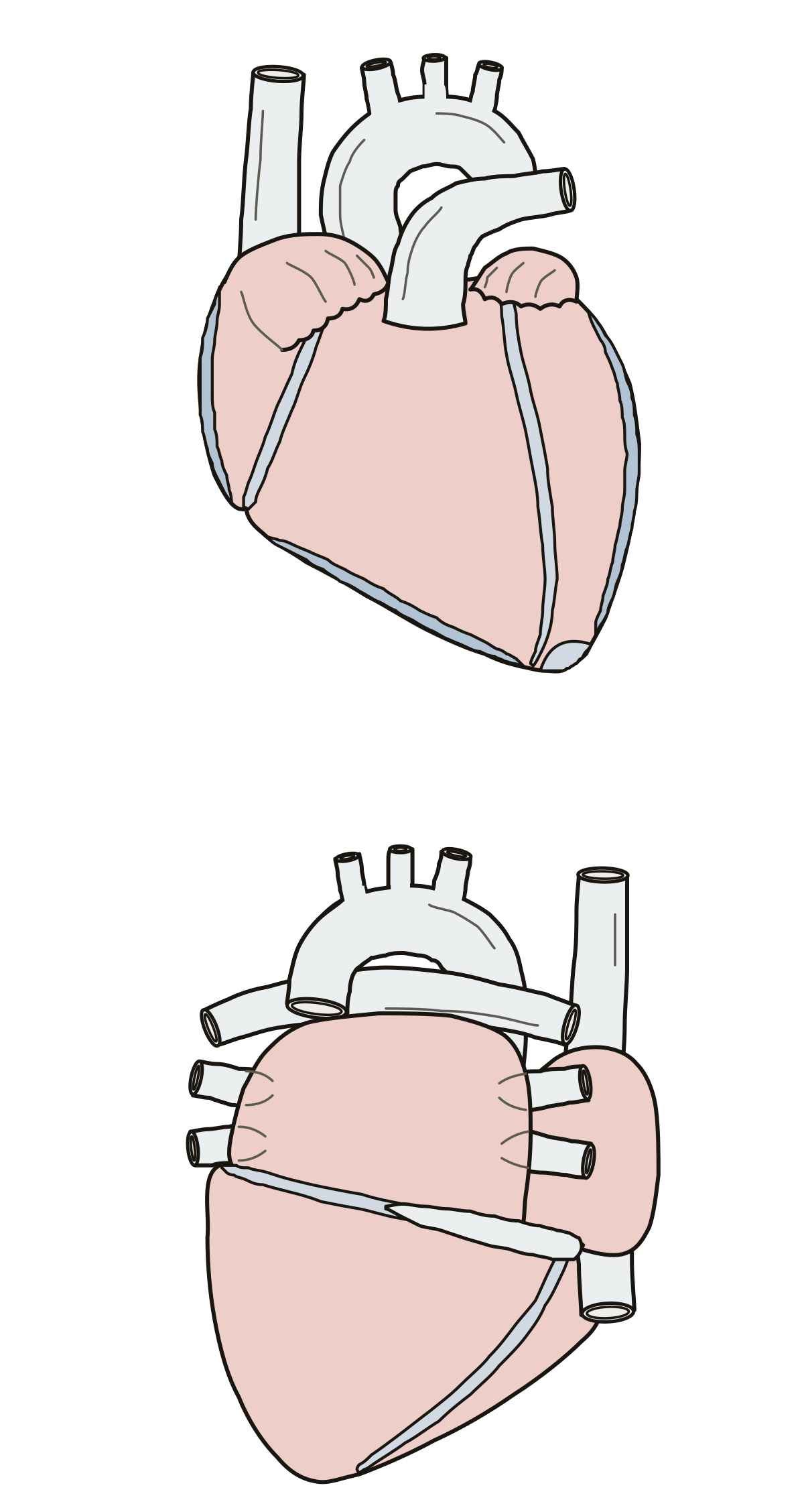 Anterior Surface of Heart Diagram, with the Back and BaseThe heart from the front and from behind: auricles, coronary and interventricular sulci, three borders, apex, base and diaphragmatic surface.
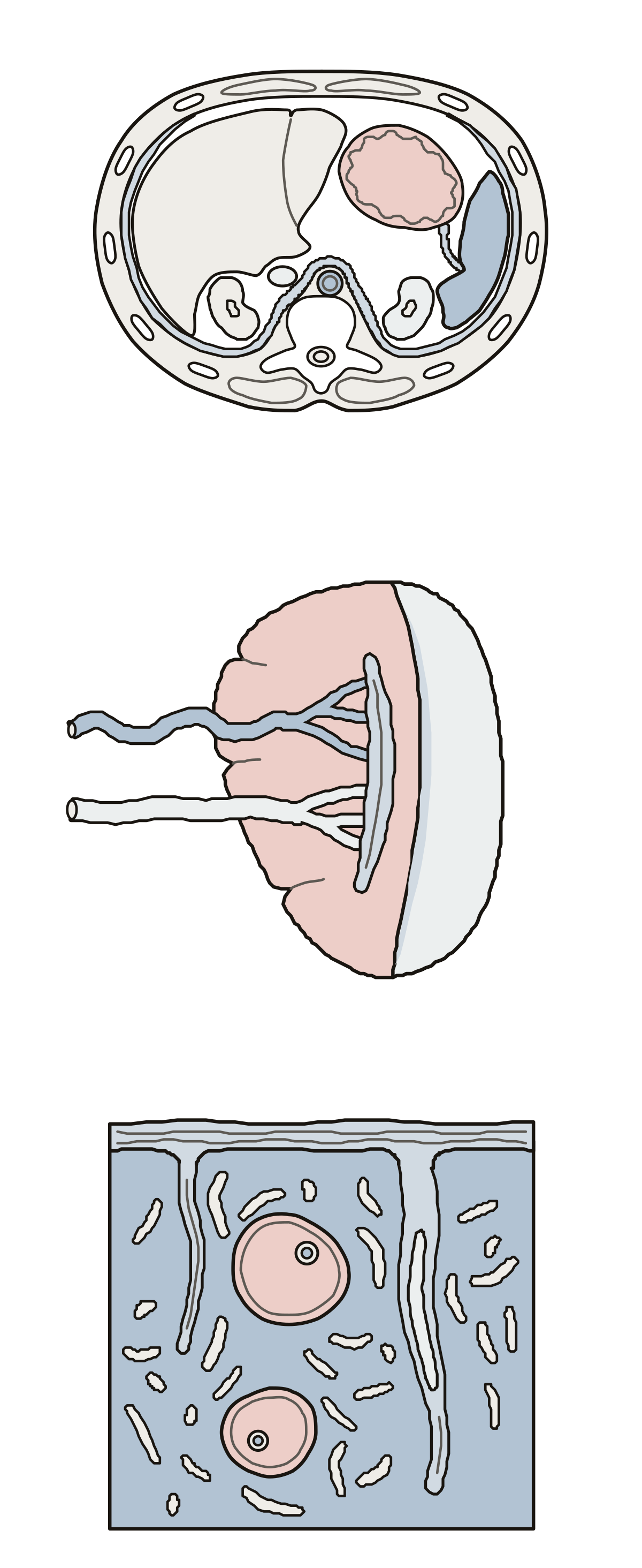 Spleen DiagramSpleen, stomach, left kidney and diaphragm in cross-section; hilum, gastric and renal surfaces; capsule, trabeculae, white pulp and red pulp.
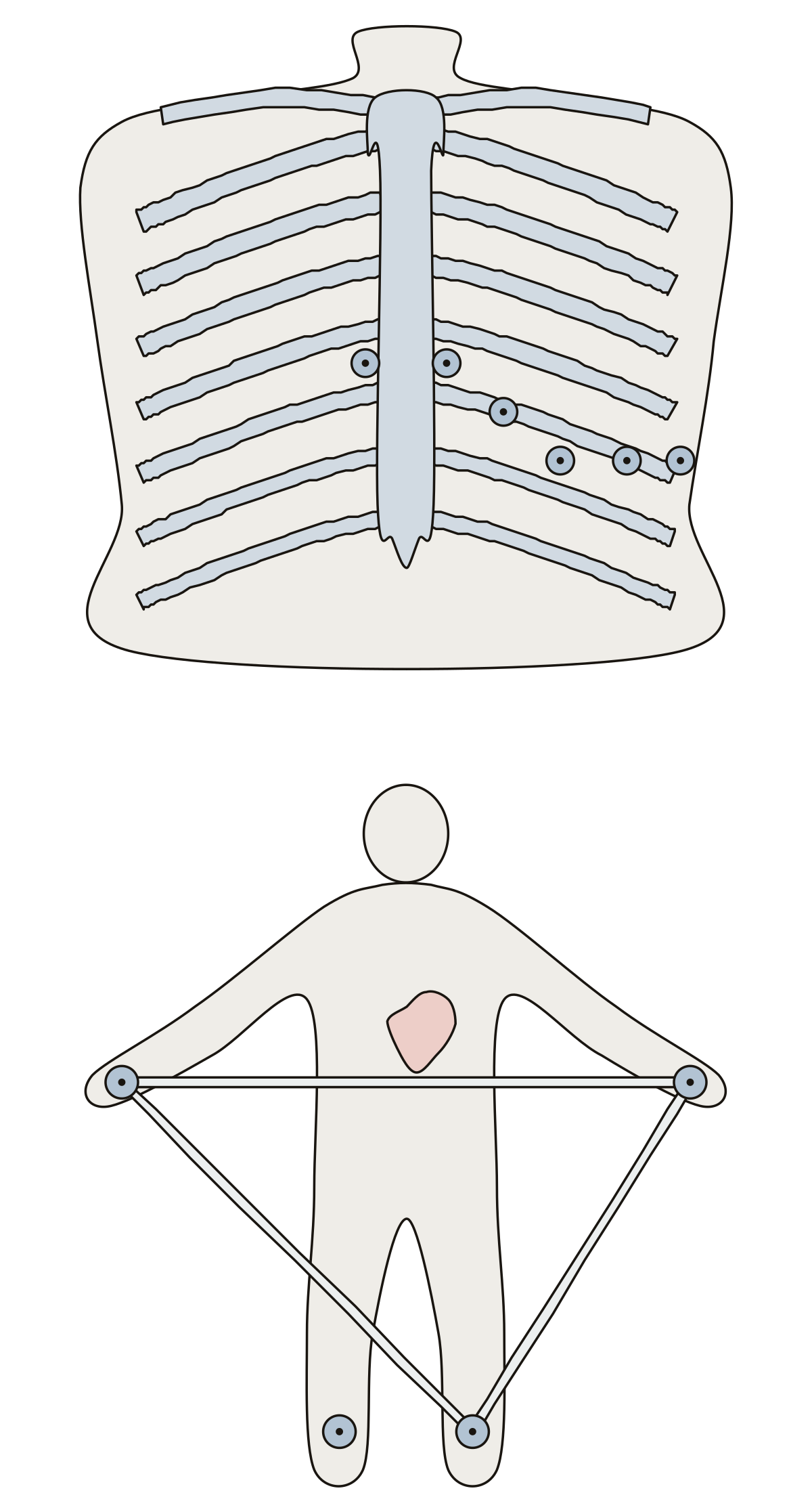 12 Lead Heart DiagramV1 to V6 on the chest, the right arm, left arm, right leg and left leg electrodes, and leads I, II and III.
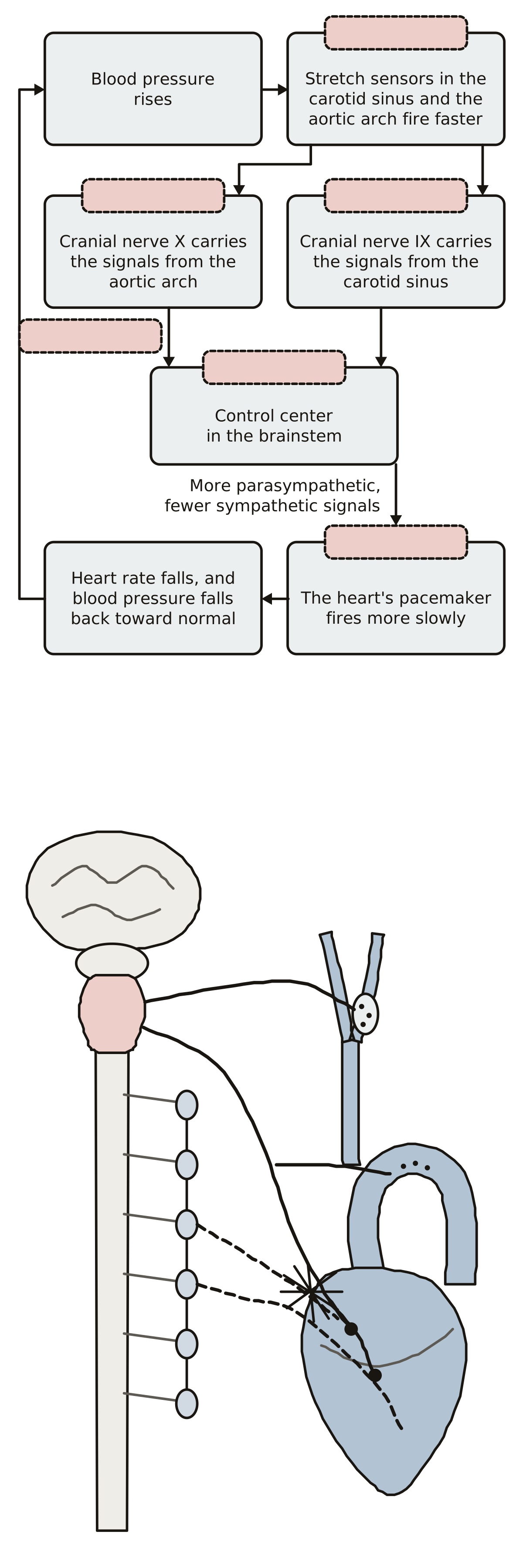 Heart Rate Feedback Loop DiagramThe baroreceptor reflex as a loop with six blank tags, then the carotid sinus, aortic arch, sympathetic trunk, cardiac plexus and the nerves between the brainstem and the heart.
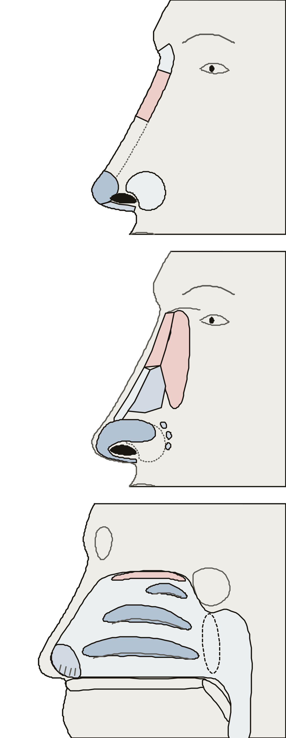 Diagram of the Nose and LabelsRoot, bridge, tip, ala, nostril and columella; the nasal bone and four kinds of cartilage; the vestibule, three conchae, olfactory region and choana.
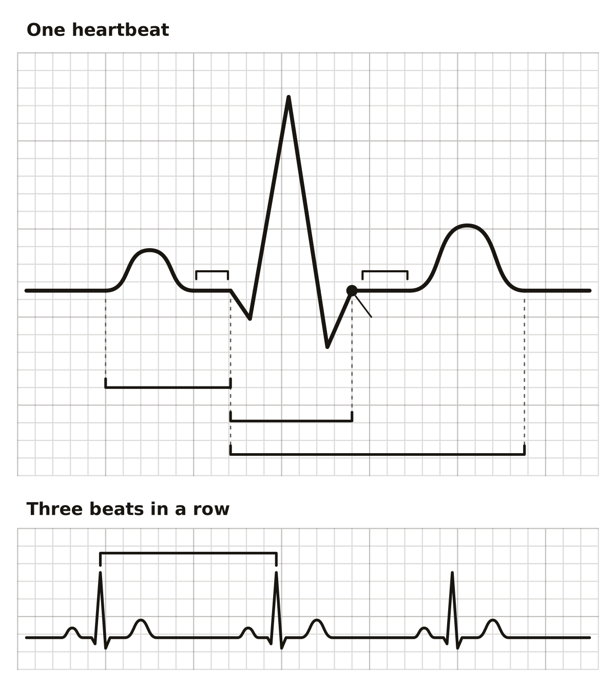 ECG Anatomy and Physiology QuizOne normal heartbeat as an ECG trace: the P, Q, R, S and T waves, the QRS complex, the PR and ST segments, the J point, and the PR, QT and R-R intervals.
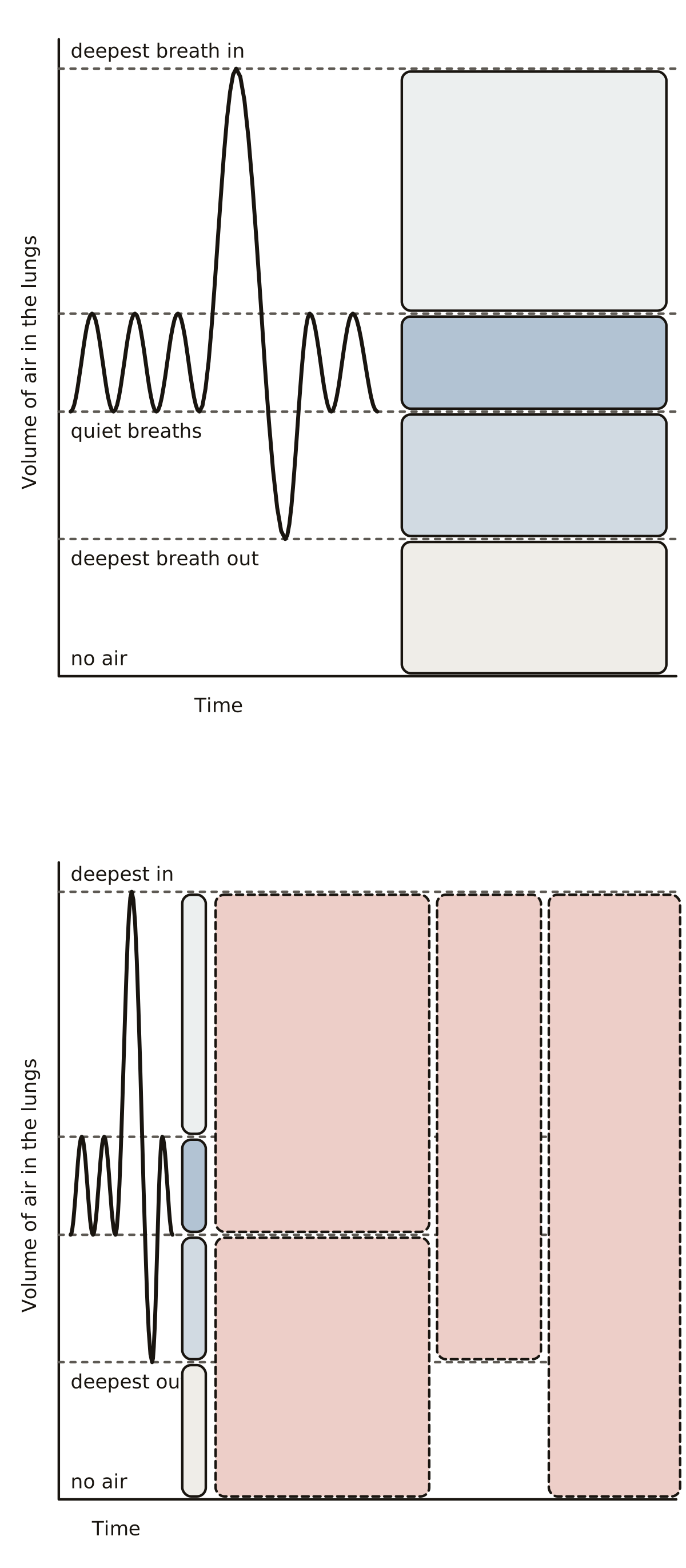 Lung Volumes and Capacities DiagramTidal volume, inspiratory and expiratory reserve volume and residual volume as blocks beside a spirometer trace, then inspiratory, functional residual, vital and total lung capacity as bars.
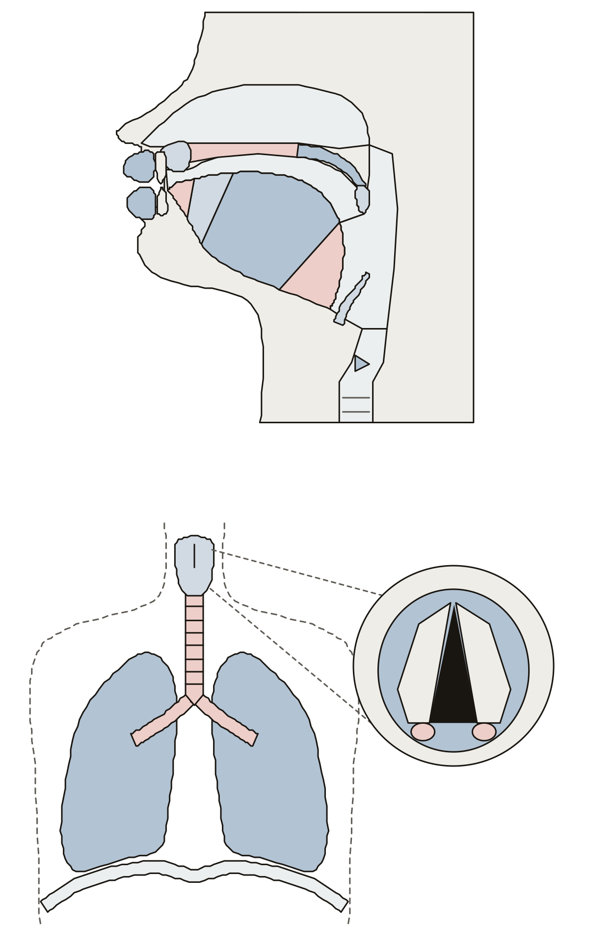 Speech Anatomy QuizLips, teeth, alveolar ridge, hard and soft palate, uvula, tongue tip, blade, body and root, the oral and nasal cavities, pharynx and epiglottis, then lungs, diaphragm, trachea, larynx, vocal folds and glottis.
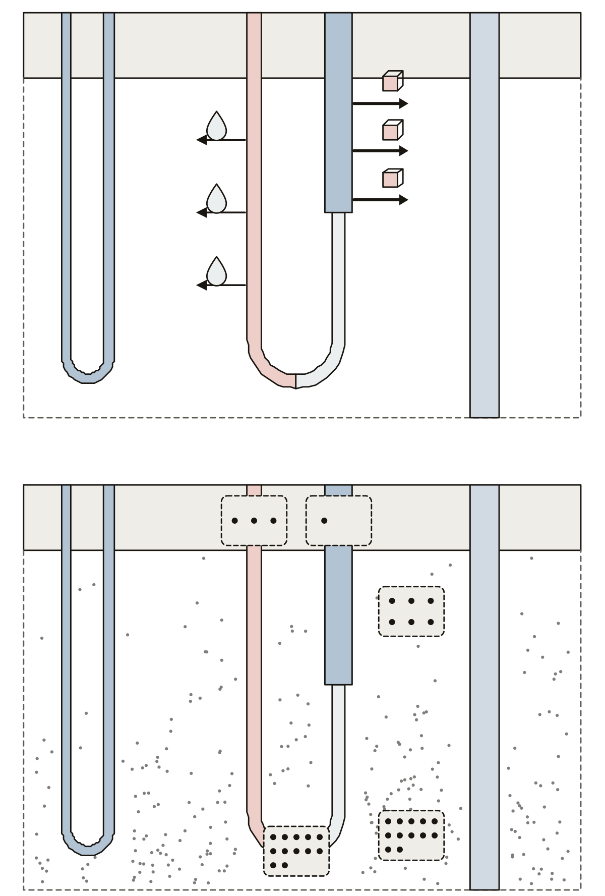 Countercurrent Multiplier DiagramDescending limb, thin and thick ascending limb, collecting duct, vasa recta, the water and salt that leave the loop, and the concentration of the fluid at five places.
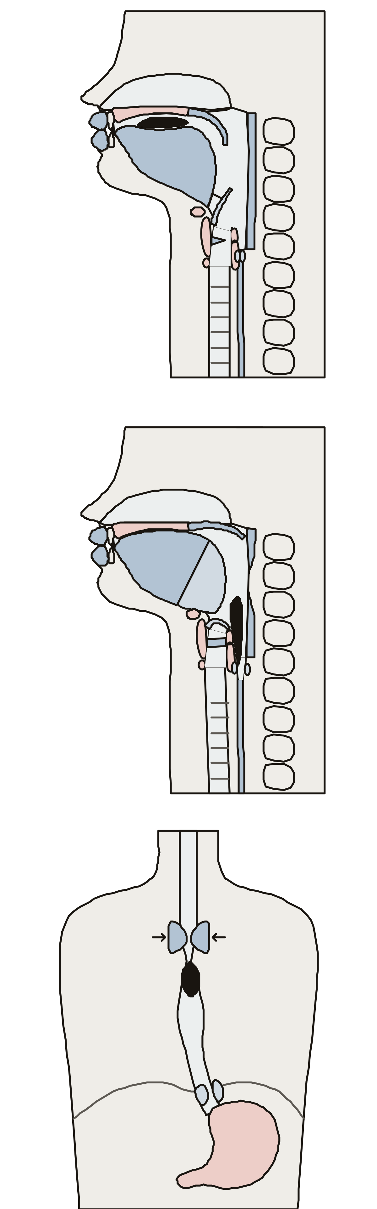 Swallowing Anatomy and Physiology QuizTongue, bolus, hard and soft palate and valleculae in the oral phase; epiglottis, larynx, hyoid bone, vocal folds, constrictors and upper sphincter in the pharyngeal phase; the peristaltic wave and lower sphincter in the esophagus.
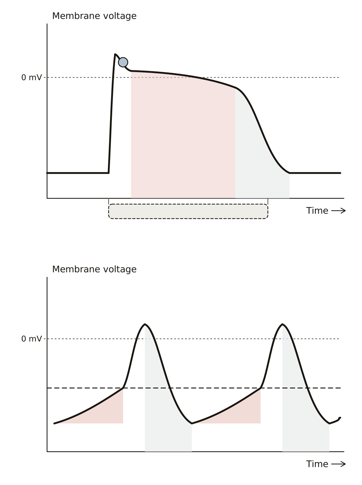 Heart Action Potential DiagramPhases 0 to 4, the plateau and the refractory period of a heart muscle cell, then the pacemaker potential, threshold, rise and fall of a sinoatrial node cell.
- Spleen Anatomy QuizA quiz from the first question: find the spleen and its neighbors, the hilum and surfaces of the inner face, and the pulp, capsule and trabeculae of the tissue.
- Blood Vessels of the Kidney Anatomy Labeling QuizA quiz from the first question: find the renal, segmental, interlobar, arcuate and cortical radiate vessels, then the arterioles and capillaries of one nephron.
- ECG Diagram UnlabeledA blank ECG trace of one normal heartbeat to print: waves, segments and intervals to name by hand, with a strip of three beats and an answer key.
- Spleen Diagram No LabelsThe spleen drawing with nothing written on it, to print and fill in: the organ in place beside the stomach and its inner face with the hilum, a word bank and an answer key.
- Speech Organs Diagram UnlabeledThe organs of speech in the head and throat with nothing written on them, to print and fill in: lips, teeth, palate, four parts of the tongue, three cavities and the epiglottis, with an answer key.
- The Human Liver Diagram UnlabeledThe liver from the front with no name on it, to print and fill in: two lobes, four ligaments and an answer key.
- Adrenal Gland Blank LabelingThe adrenal gland drawing with no name on it, to print and fill in: four lines at the gland on its kidney, six on the slice, and an answer key. Fasciculata · Reticularis · Capsule
Skin, hair and nails 6
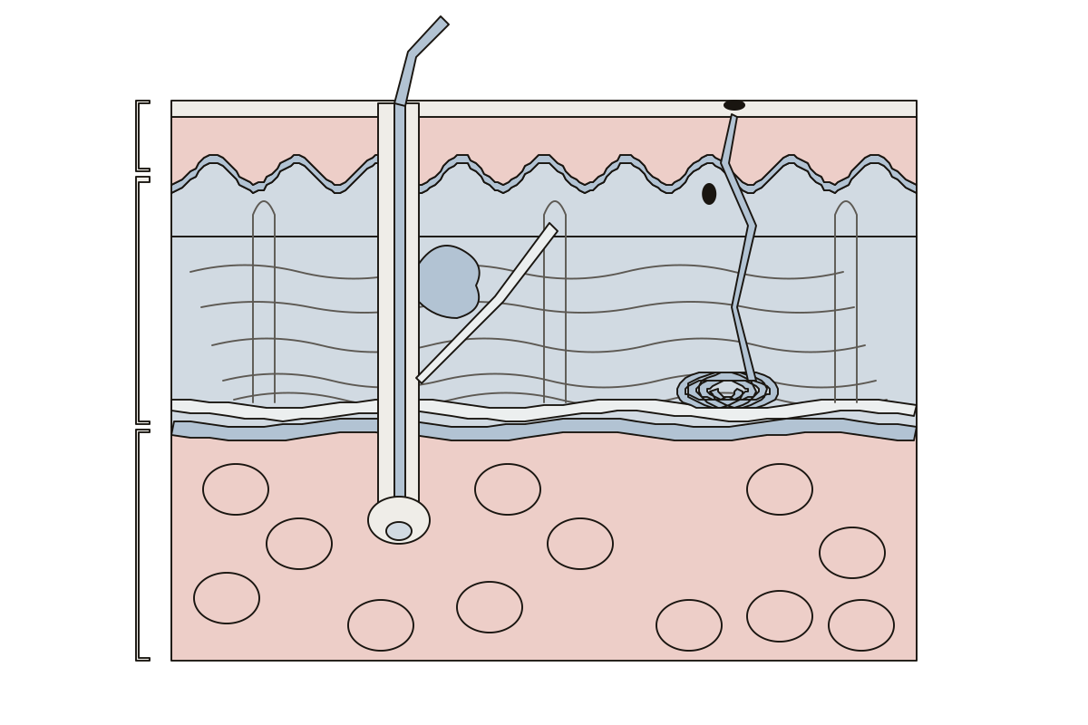 Skin Diagram Unlabeled, With a Labeled VersionOne block of skin in section: 17 parts to name, printable blank or labeled. Stratum basale · Hair follicle · Stratum corneum
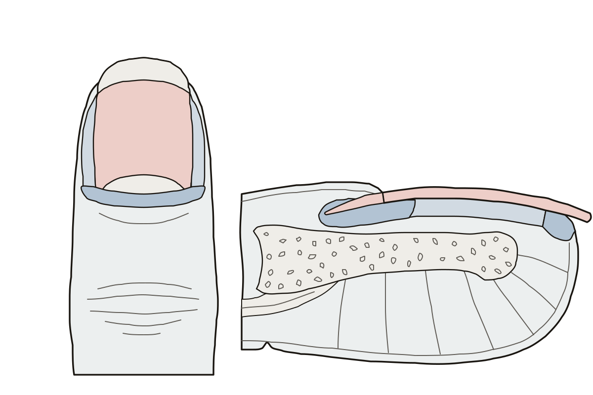 Nail Diagram LabeledA fingernail from above and cut lengthwise: 9 parts to name, labeled or blank. Eponychium · Lateral nail folds · Free edge
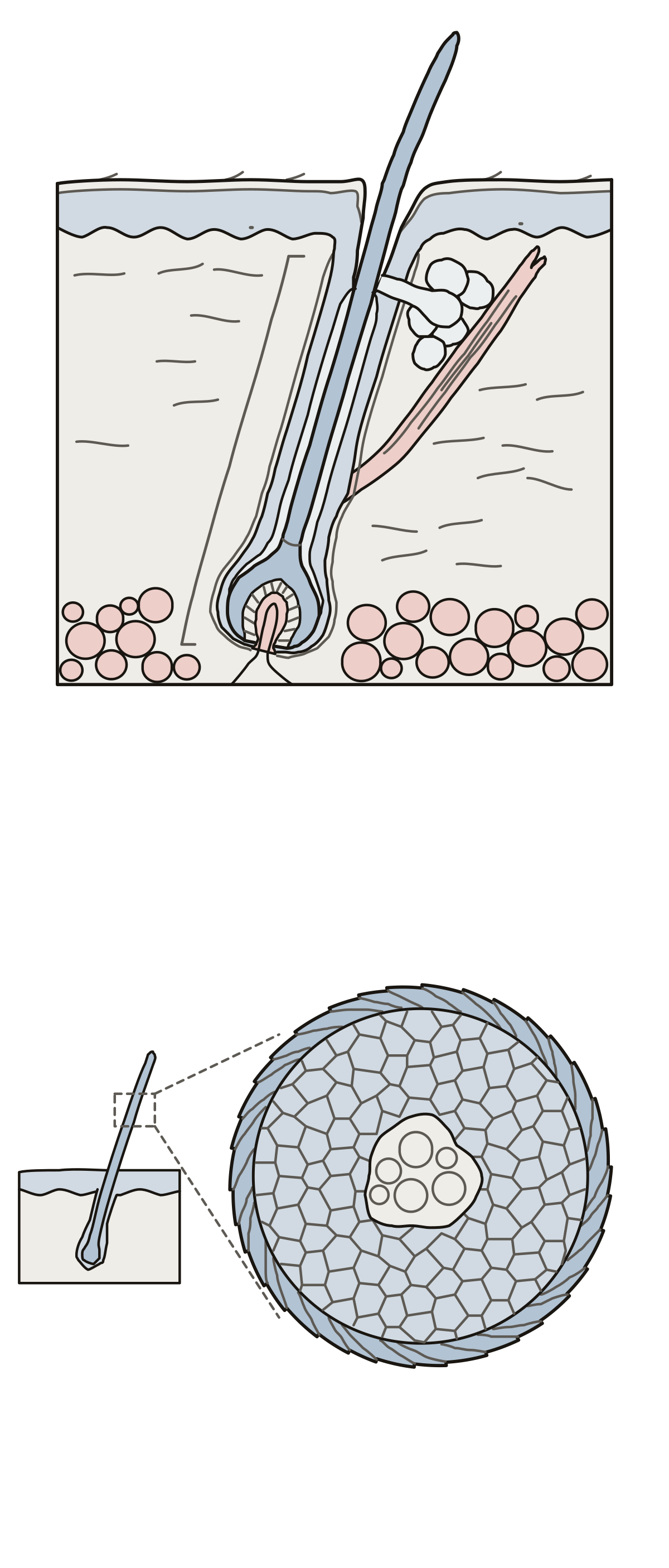 Hair Diagram LabeledA hair in its follicle and a strand cut across: 13 parts to name. Internal root sheath · Hair bulb · External root sheath
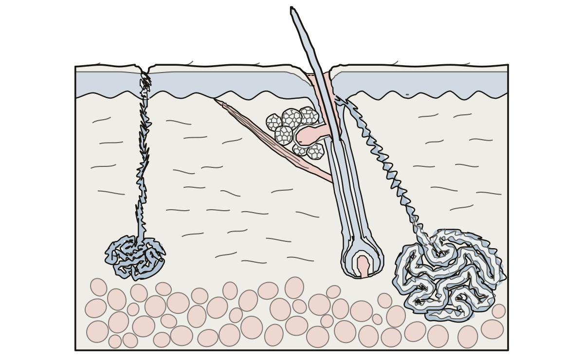 Sweat and Sebaceous Glands DiagramEccrine sweat gland, sweat duct and pore, apocrine sweat gland, sebaceous gland and sebum.
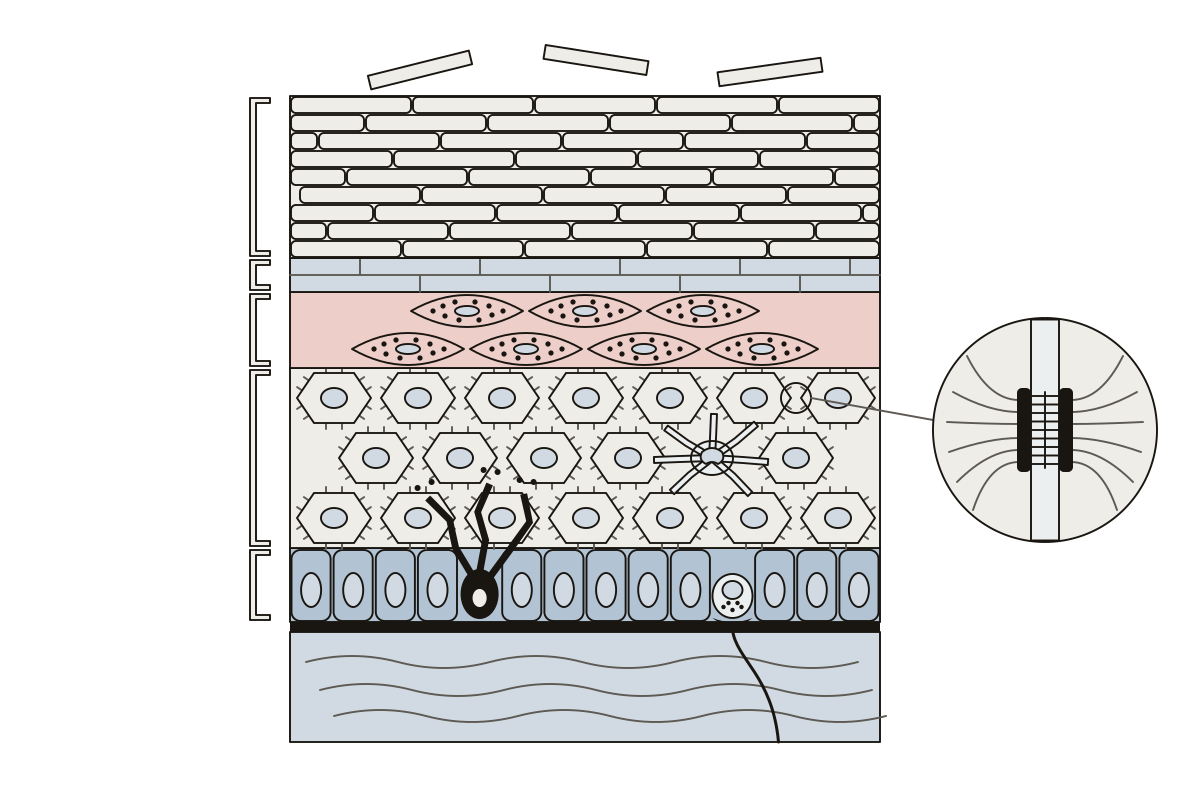 Epidermis Cell DiagramStratum basale, spinosum, granulosum, lucidum and corneum, keratinocyte, melanocyte, Langerhans cell, Merkel cell, desmosome, basement membrane and dermis.
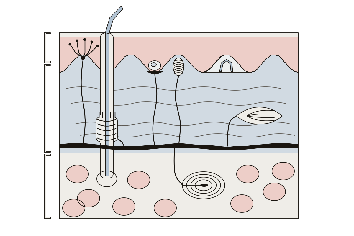 Skin Receptors DiagramFree nerve endings, Merkel cell and disc, Meissner corpuscle, Pacinian corpuscle, Ruffini corpuscle, hair follicle receptor, dermal papilla, epidermis, dermis and hypodermis.
The whole body 17
The whole body: a child figure with 20 everyday words, the regions, cavities, planes and directional terms of an anatomy course, and the feedback loops that keep it steady.
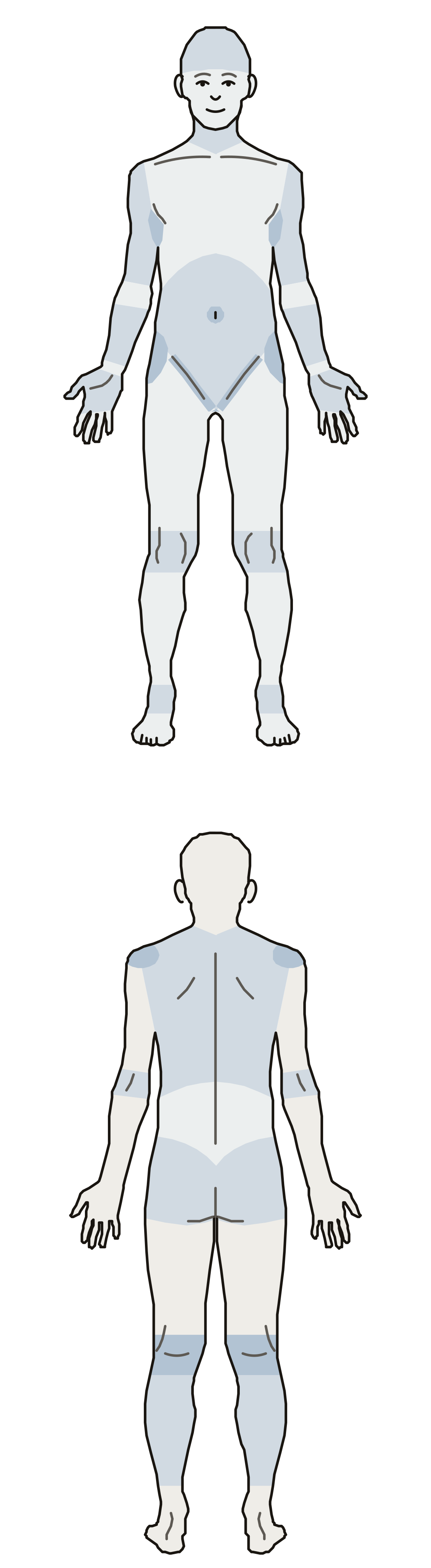 Labeling Body RegionsThe regional terms of anatomy on a body front and back: cranial to pedal, brachial to digital, dorsal, lumbar, gluteal, popliteal and sural. Crural · Digital · Abdominal
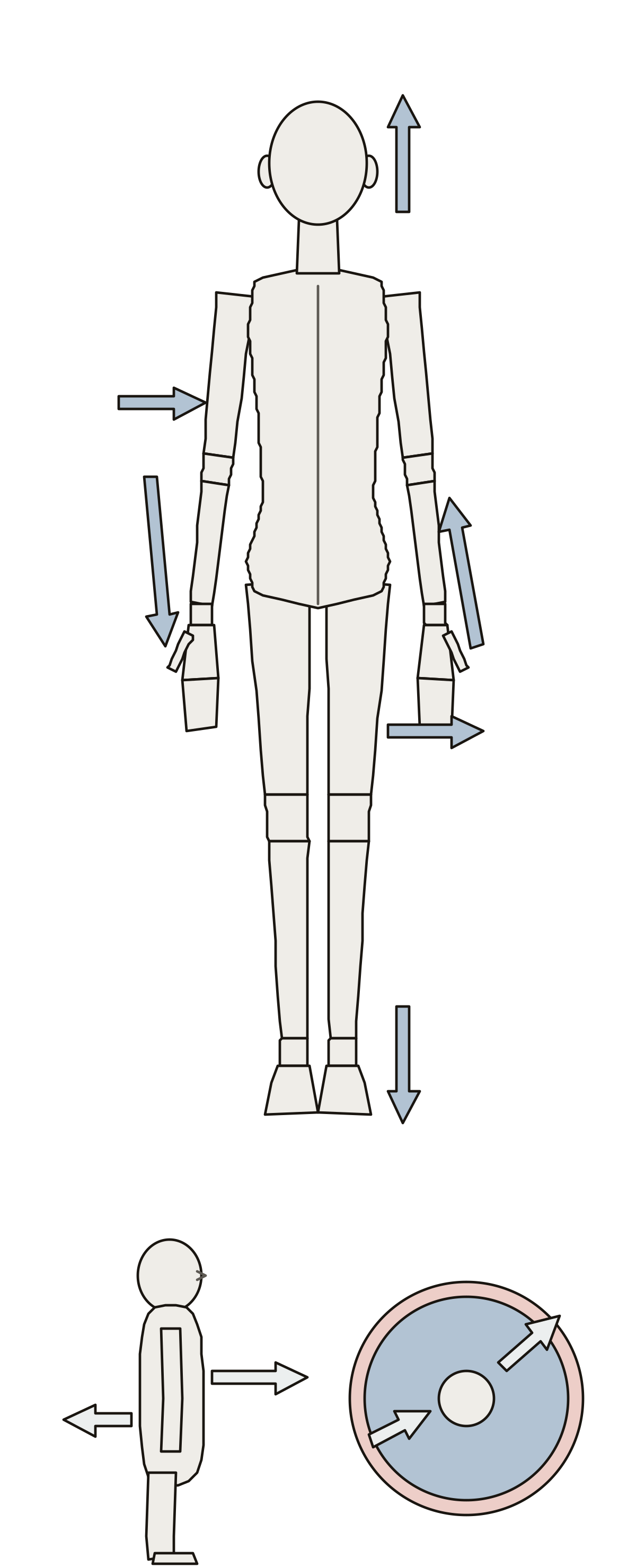 Directional Terms Anatomy QuizTen anatomical directional terms as arrows on the body: superior, inferior, medial, lateral, proximal, distal, anterior, posterior, superficial, deep. Proximal · Inferior · Distal
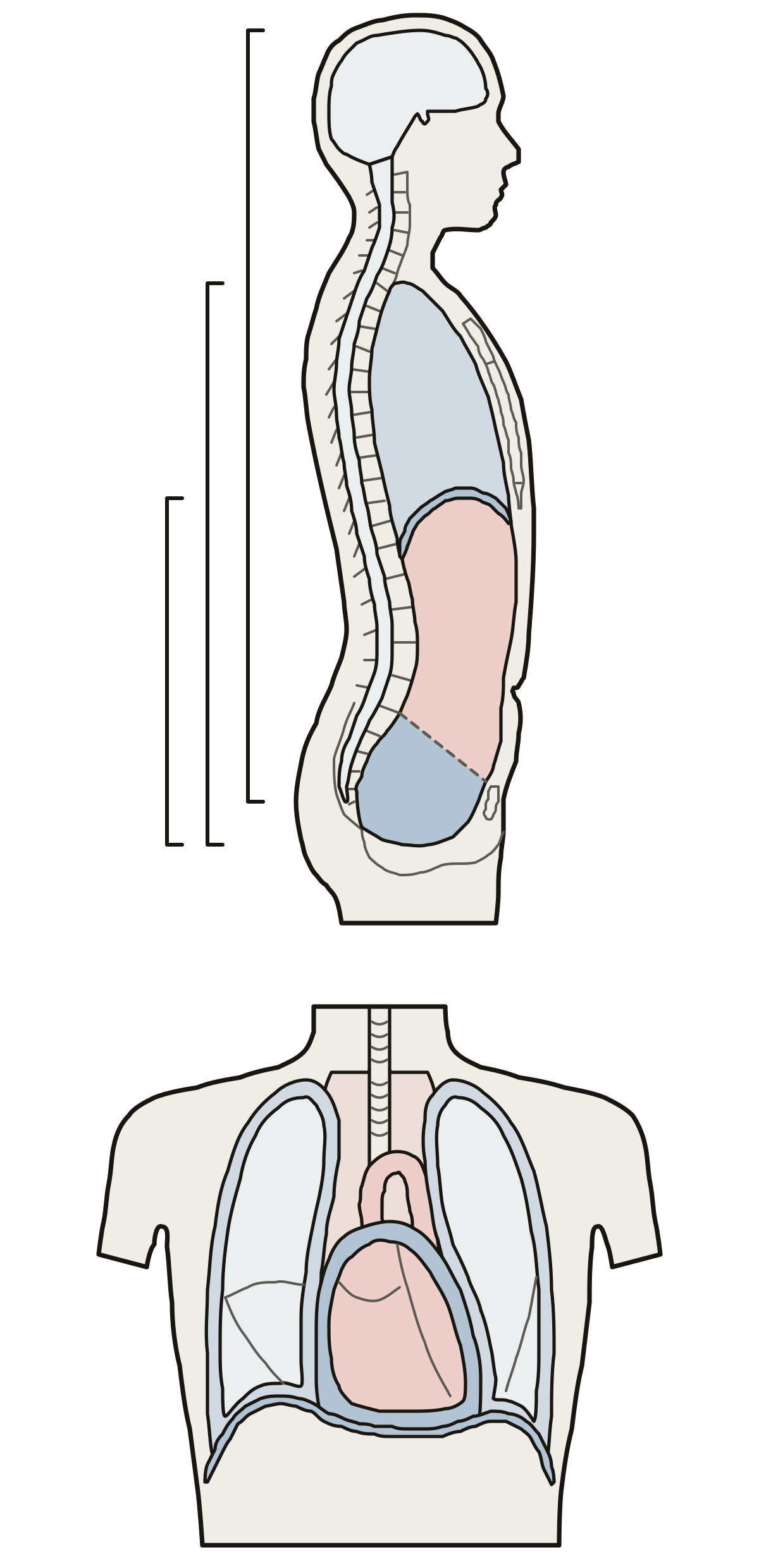 Body Cavities Unlabeled and LabeledThe body cavities: dorsal, cranial and spinal; ventral, thoracic, pleural, pericardial and mediastinum; the diaphragm; abdominopelvic, abdominal and pelvic. Dorsal · Ventral · Thoracic
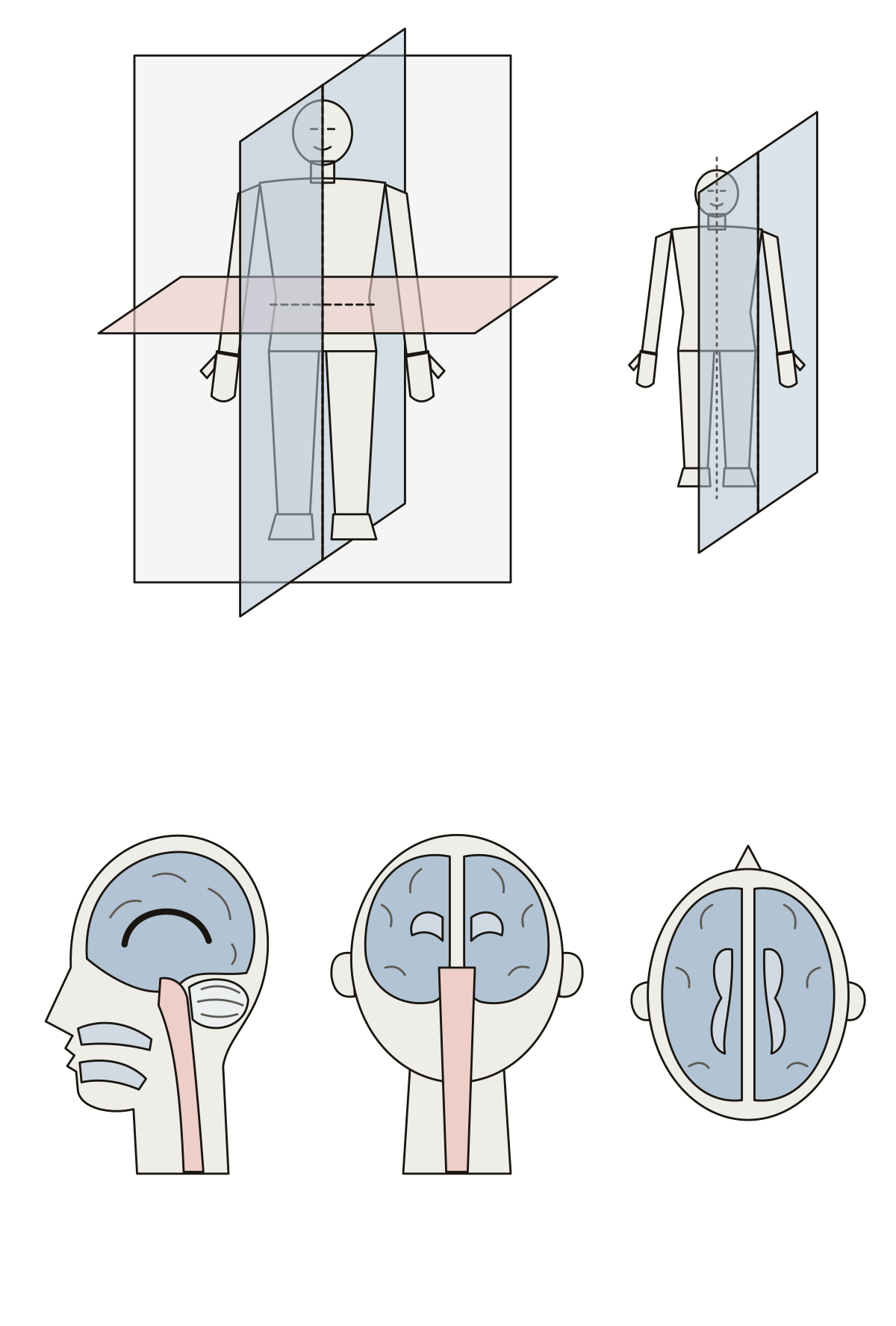 Label the Planes of the BodyThe median, sagittal, frontal and transverse planes on a figure in the anatomical position, then a sagittal, a frontal and a transverse section of the head.
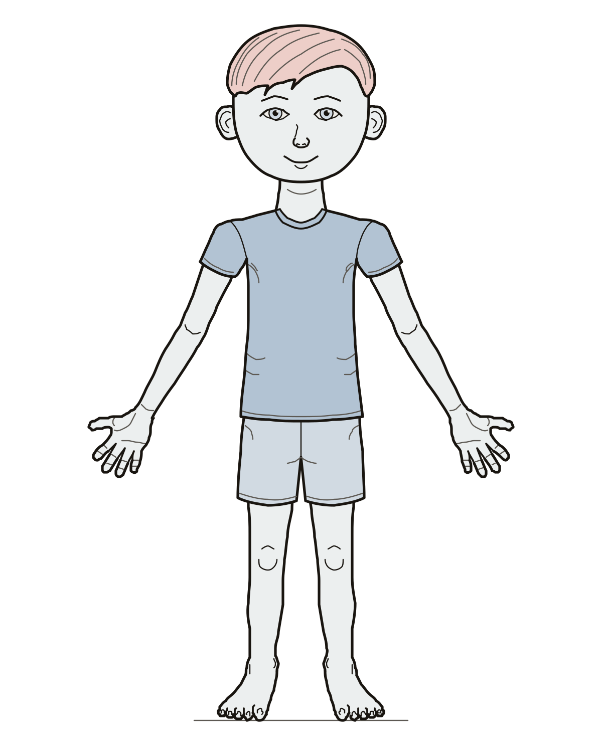 Labeling Parts of the BodyTwenty everyday body words on one child figure, head to toes: find or name each part, start with a first 10, and print the sheet blank or labeled.
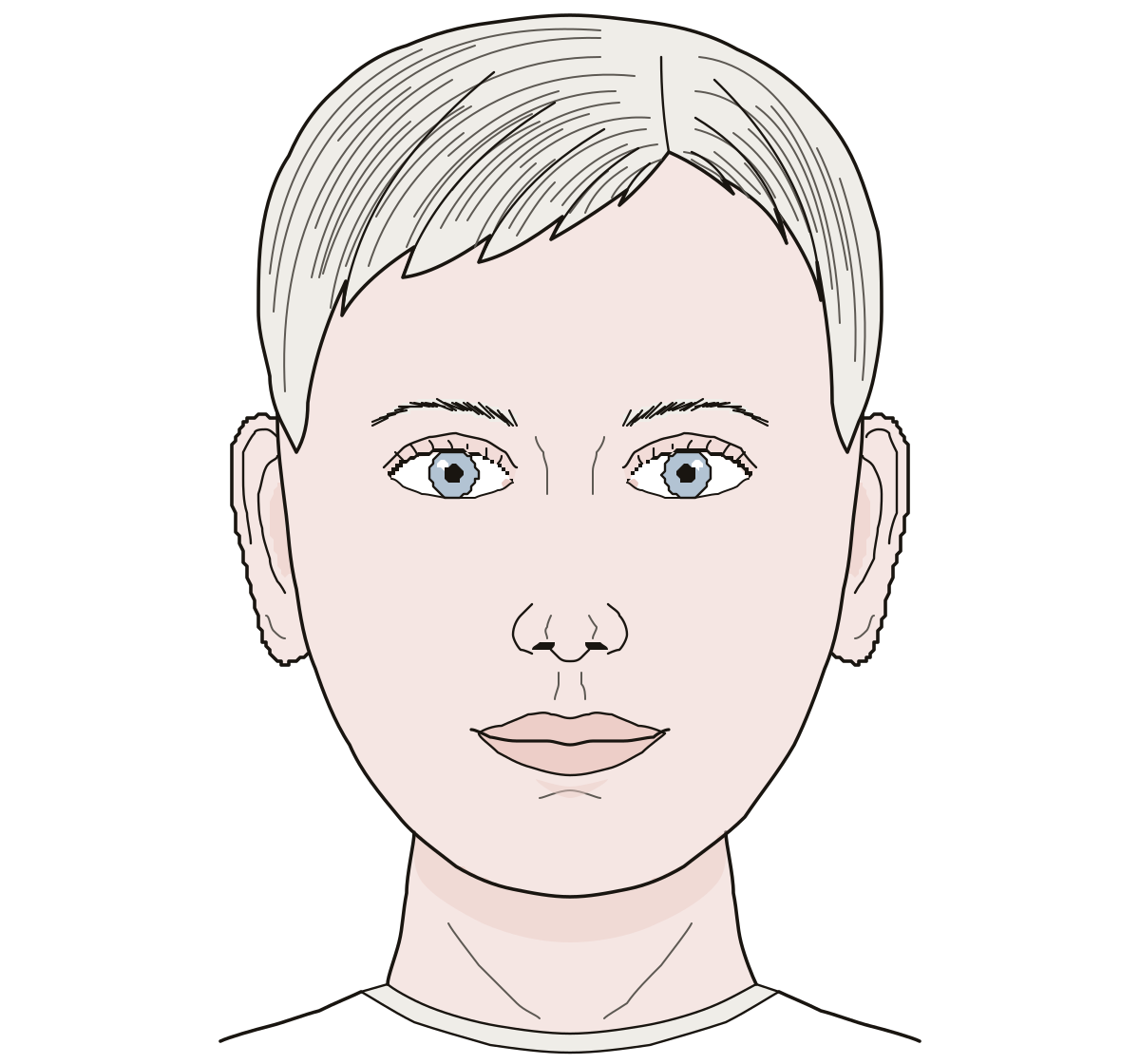 Parts of a FaceA face from the front: eyes, nose, ears, lips, cheeks and chin, then the forehead, temples, eyebrows, eyelids, eyelashes, nostrils and jaw. Jaw · Nostril · Forehead
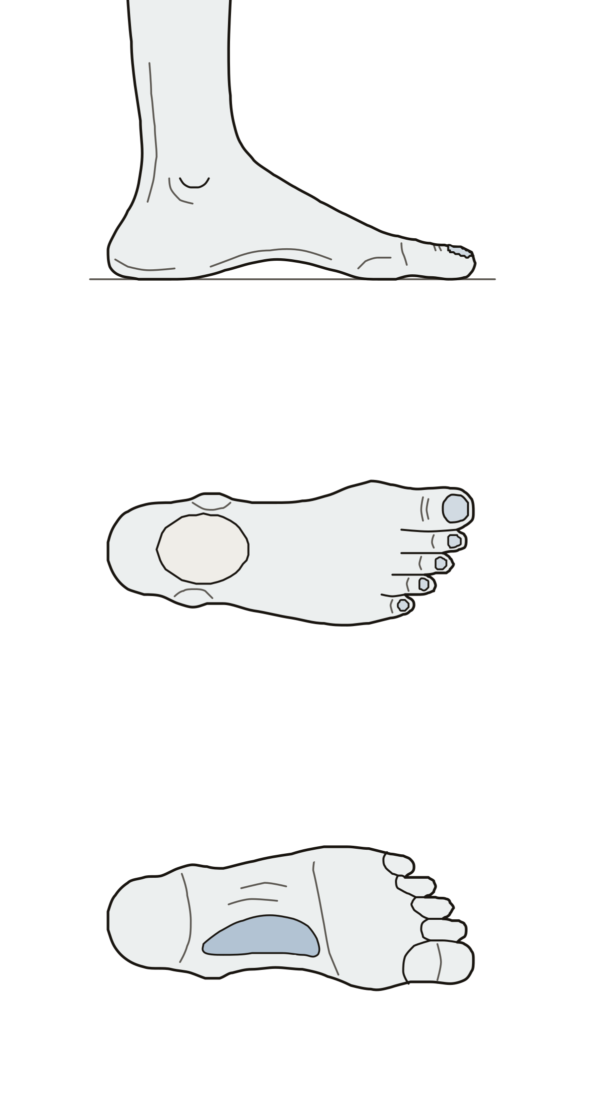 Parts of a FootSeventeen outside parts of one foot in three views: heel, arch, ball, sole, five toes, toenail, both ankle bumps and the Achilles tendon. Fourth toe · Toenail · Lateral malleolus
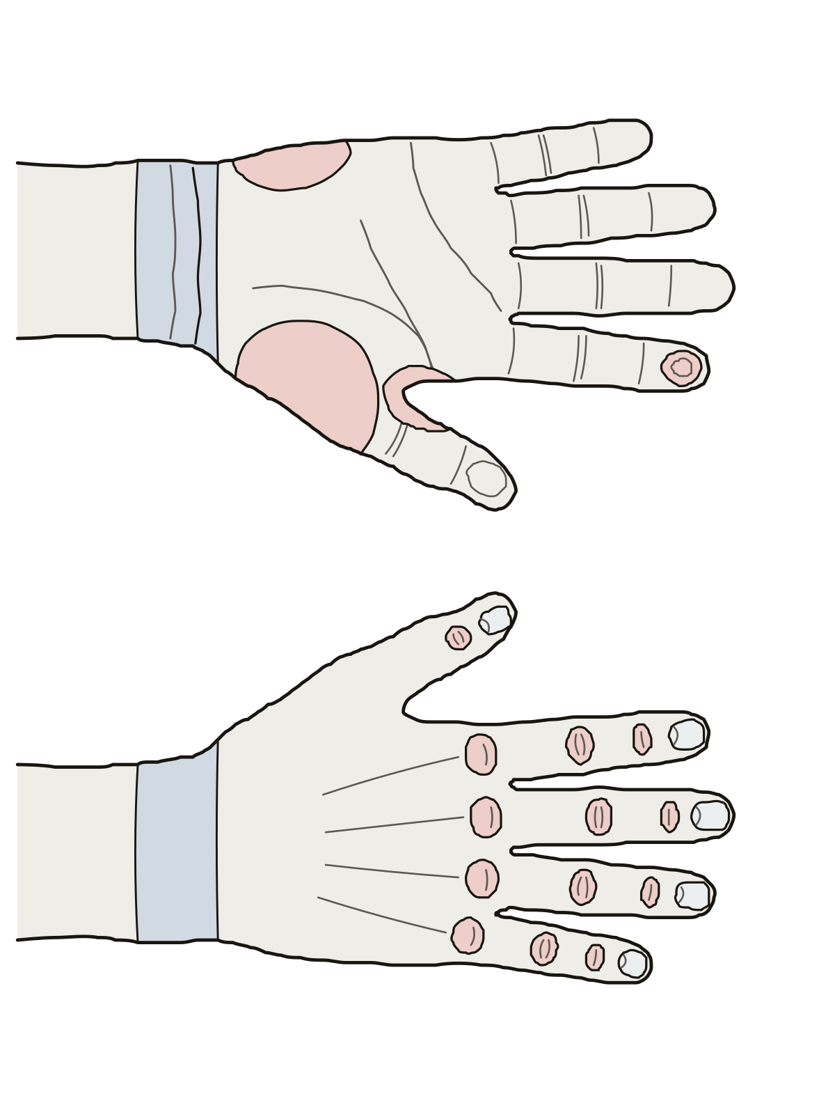 Hand DiagramThe outside of the hand: five fingers, palm, wrist, nails, the three rows of finger joints and the two muscle mounds of the palm. Palm (palmar surface) · Knuckles (metacarpophalangeal joints) · Middle finger (third digit)
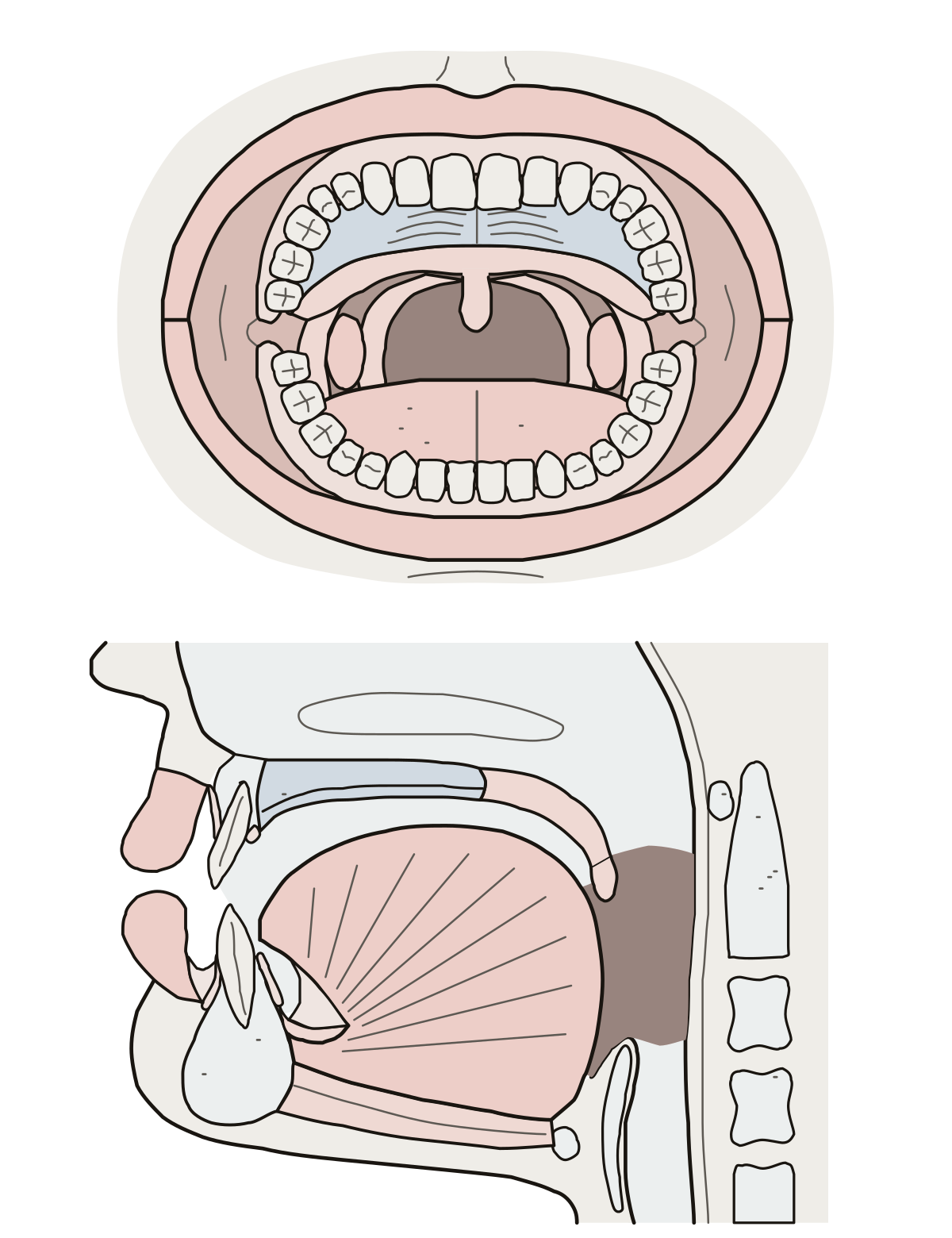 Parts of a MouthLips, teeth, gums, palate, uvula, tonsils and their folds, tongue, cheek, throat and the two frenula, in two views.
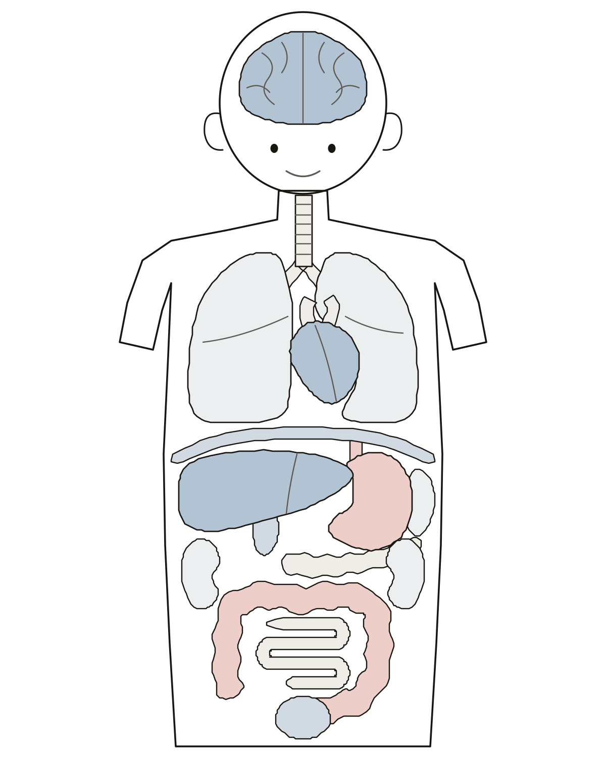 Label the Organs of the BodyA child's head and body with the organs drawn in: brain, lungs, heart, liver, stomach, kidneys, intestines and six more to name.
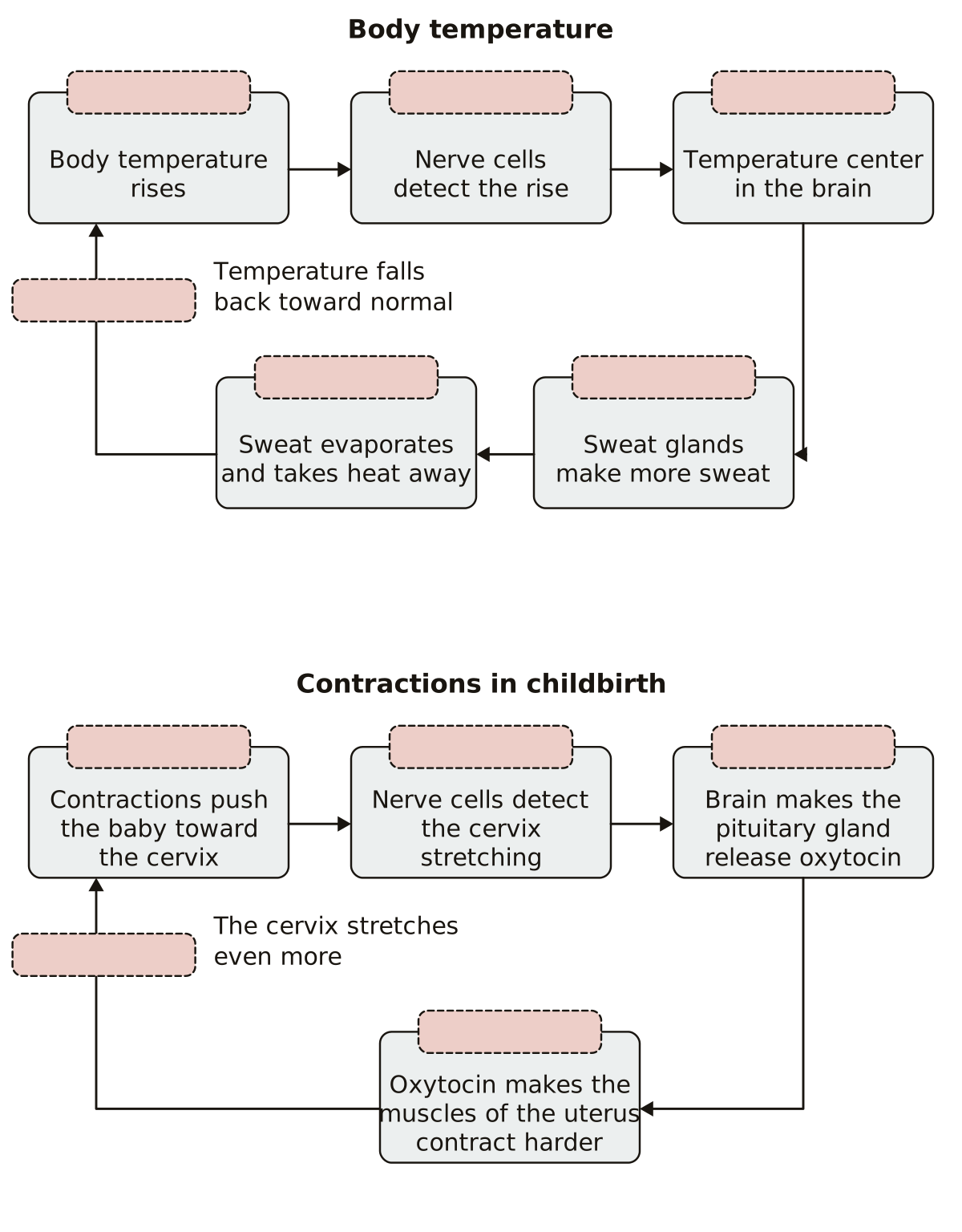 Feedback Loop DiagramStimulus, sensor, control center, effector and response on a body temperature loop and a childbirth loop, plus the kind of feedback each one shows.
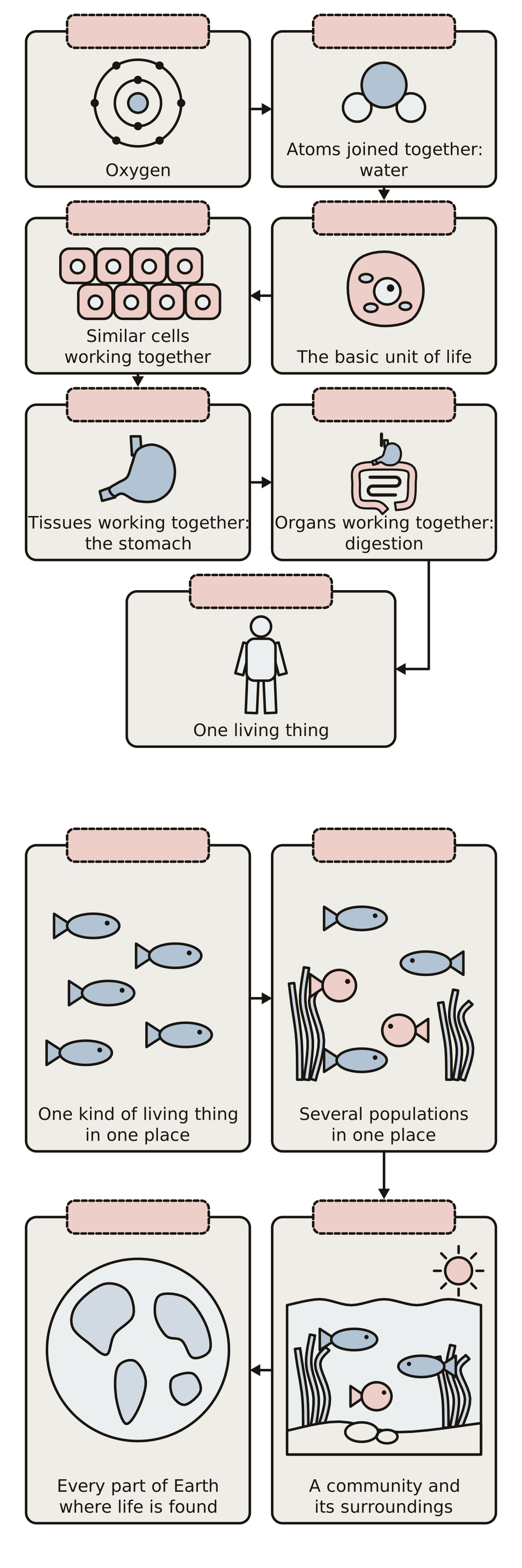 Cell Tissue Organ Organ System Organism DiagramAtom, molecule, cell, tissue, organ, organ system and organism on one chart, and population, community, ecosystem and biosphere on a second, each with a picture and a blank tag.
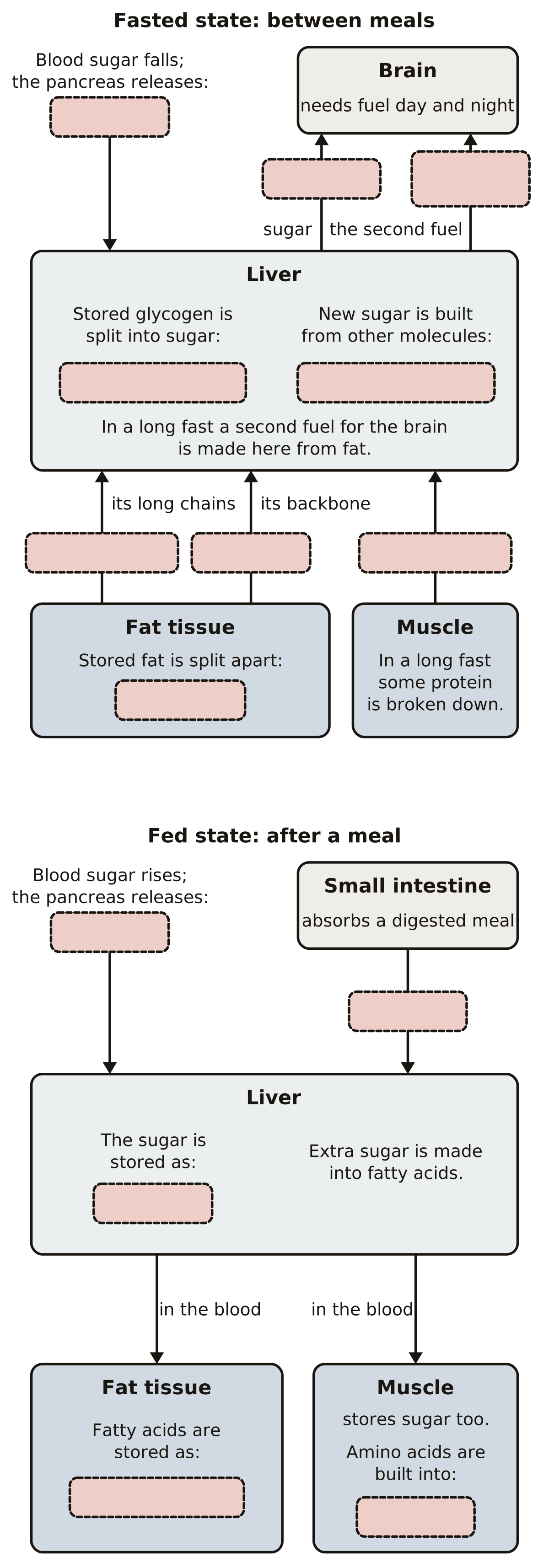 Fasted State Metabolism DiagramLiver, fat tissue, muscle and brain as boxes joined by arrows: glycogenolysis, gluconeogenesis, lipolysis, fatty acids, glycerol, ketone bodies and amino acids in the fasted state, then glycogen, triglycerides and protein in the fed state.
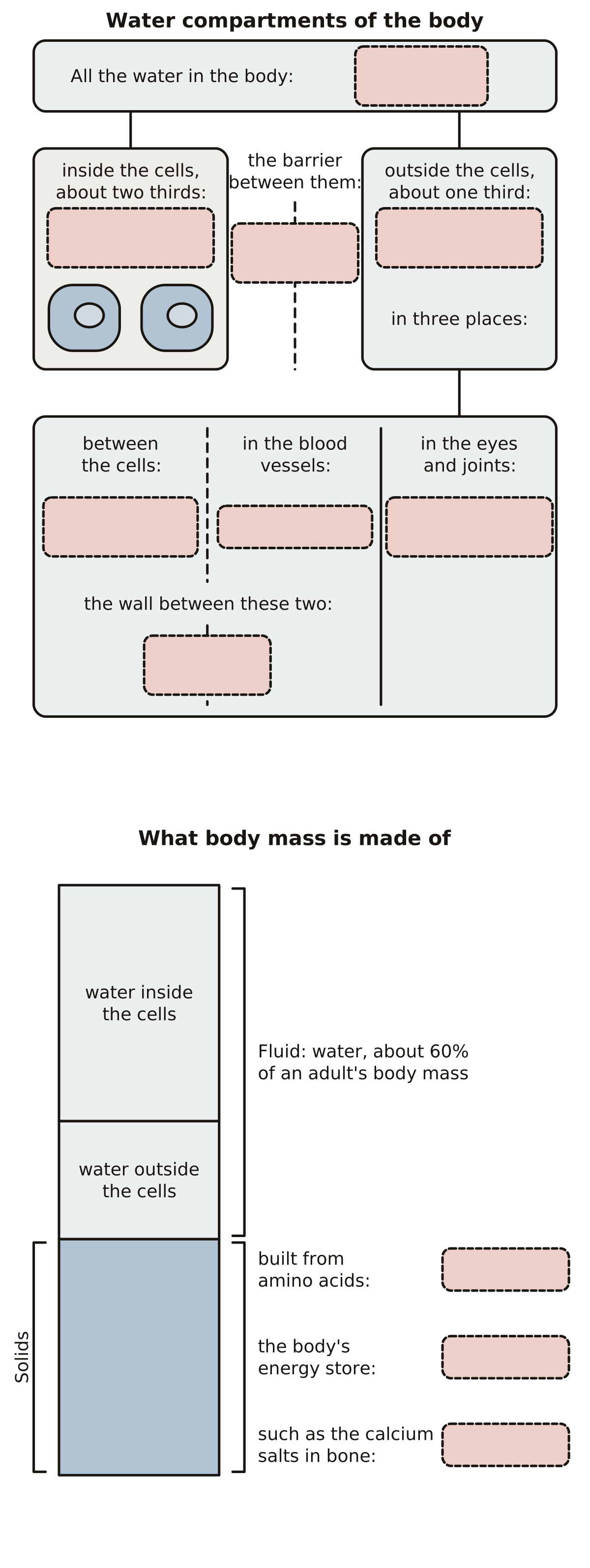 Body Fluid Compartments DiagramTotal body water, intracellular fluid, extracellular fluid, interstitial fluid, blood plasma and transcellular fluid, the cell membrane and the capillary wall between them, then protein, fat and minerals on a bar of body mass.
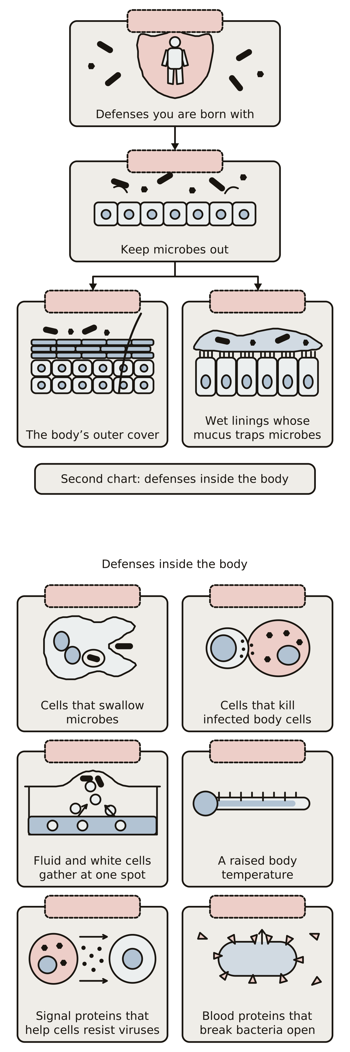 Innate Defenses DiagramInnate immunity, barriers, skin and mucous membranes on one chart; phagocytes, natural killer cells, inflammation, fever, interferons and complement on a second, each with a picture and a blank tag.
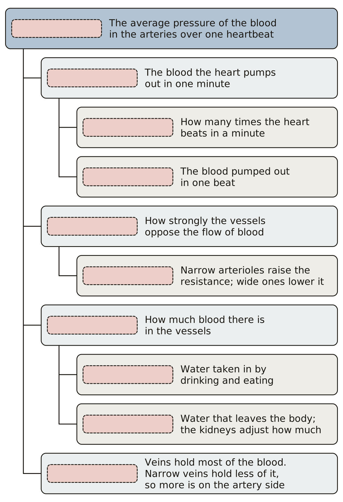 Mean Arterial Pressure DiagramA tree of ten boxes: mean arterial pressure, cardiac output with heart rate and stroke volume, peripheral resistance with the arterioles, blood volume with fluid intake and loss, and the veins.
- Cell to Organism Diagram Fill in the BlankThe chart from atom to organism with every name left off, to print and fill in: seven pictures, seven empty tags and an answer key.
Tissues under the microscope 29
Walls and tissues at the scale of a microscope slide, from a capillary cut across to 30 structures of connective tissue on 10 panels. One page draws a helper T cell meeting the cell that shows it an antigen. Others follow fat through one intestinal cell, a cut vessel through hemostasis and blood cells from one stem cell, and chart the four blood types.
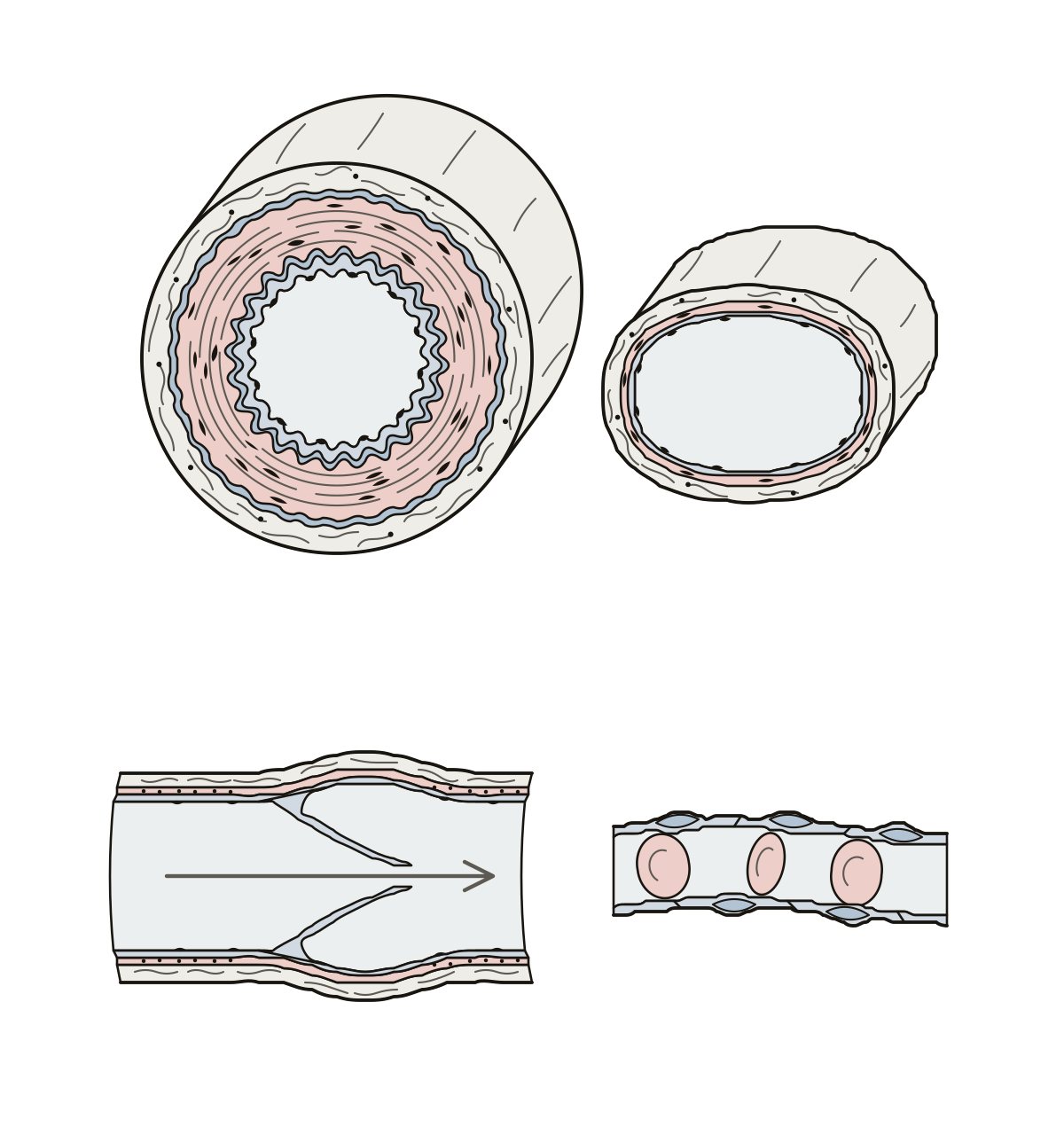 Blood Vessels DiagramBlood vessels: 10 numbered parts, from the three tunics, elastic membranes and lumen of an artery to a vein, a valve, a capillary and its endothelium. Lumen · Vein · Internal elastic membrane
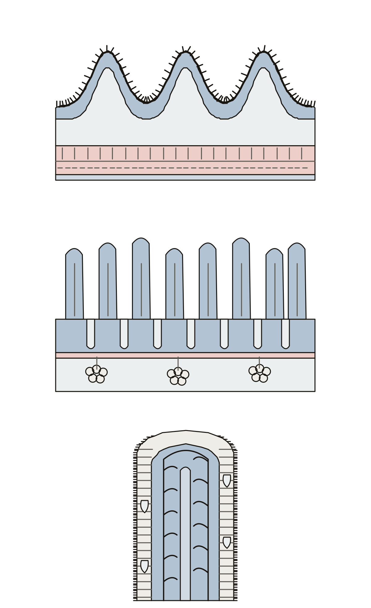 Small Intestine Wall DiagramCircular folds and the four layers, villi, intestinal and duodenal glands, and a villus with its cells, lacteal and capillaries. Circular folds (plicae circulares)
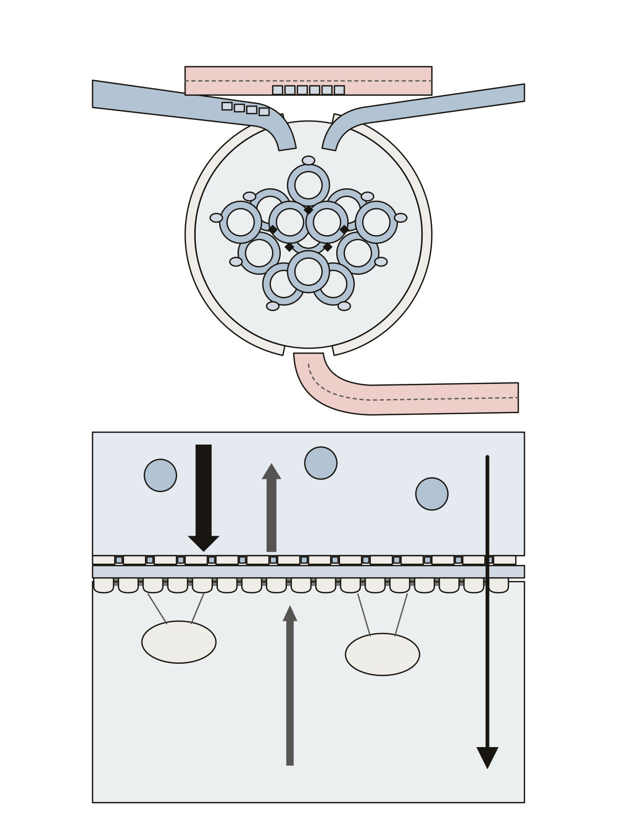 Renal Corpuscle DiagramGlomerulus, capsule, podocytes, arterioles, JG cells, macula densa, the filtration barrier and the four pressures.
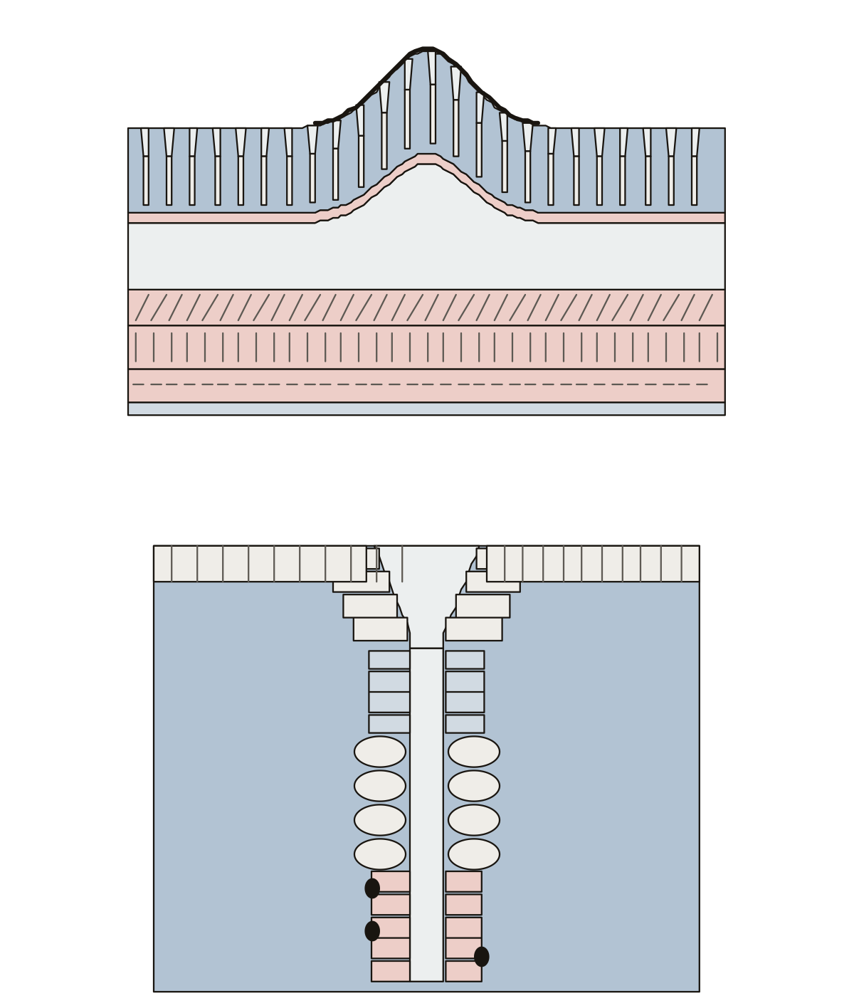 Stomach Wall DiagramRugae, gastric pits and glands, mucosa to serosa, three muscle layers, and parietal, chief and neck cells.
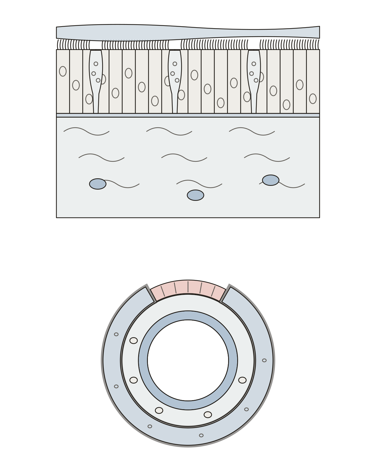 Mucous Membrane DiagramMucus, cilia, epithelium, goblet cells, basement membrane, lamina propria, and the trachea's mucosa, submucosa, cartilage and muscle. Hyaline cartilage (C-ring)
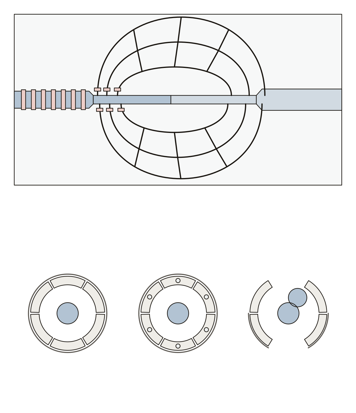 Capillary Bed DiagramArteriole, metarteriole, precapillary sphincters, capillaries, thoroughfare channel, venule, and continuous, fenestrated and sinusoid capillaries.
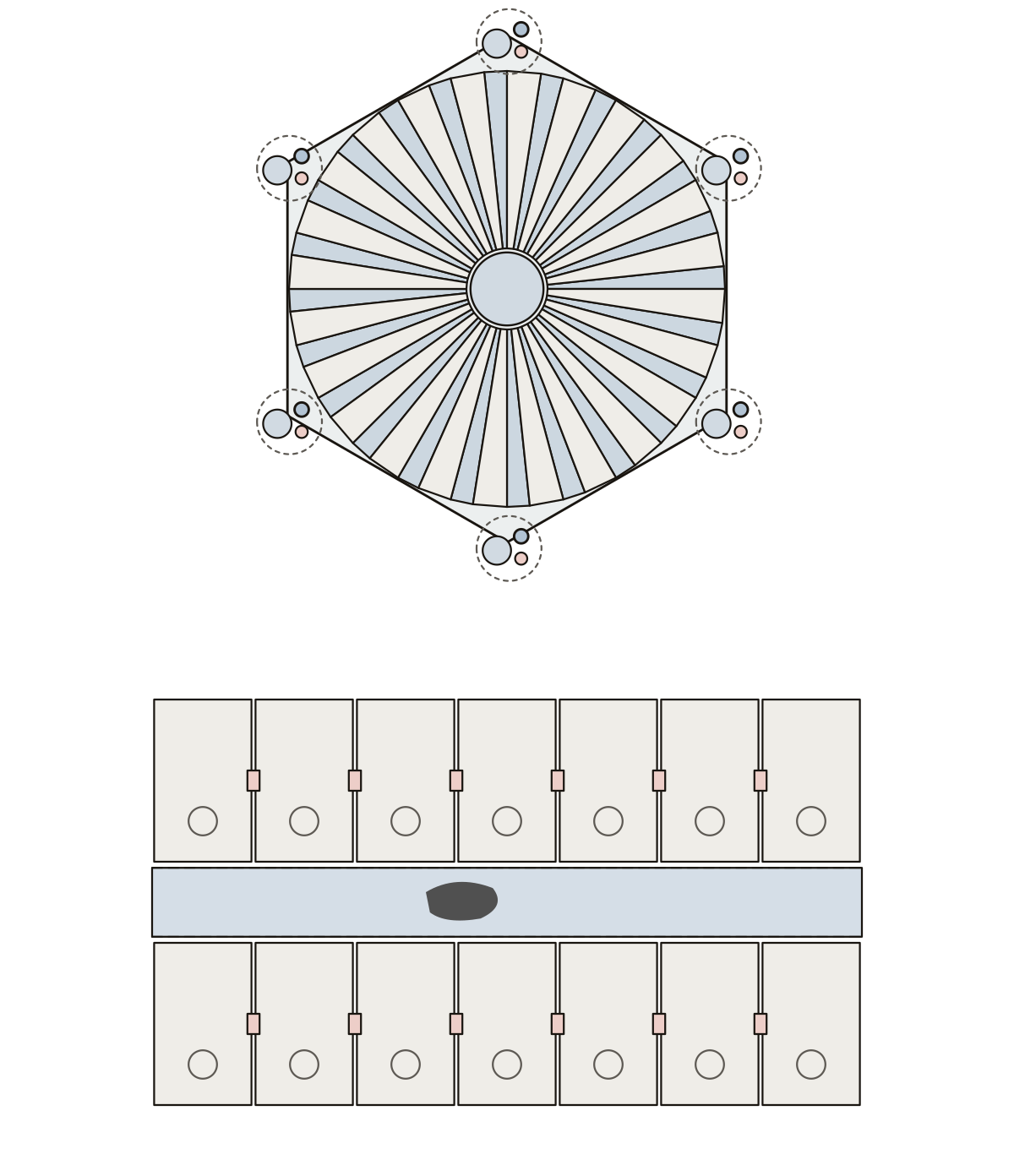 Liver Lobule DiagramHepatic lobule, central vein, hepatocytes, sinusoids, portal triad (vein, artery, bile duct), bile canaliculi, Kupffer cells.
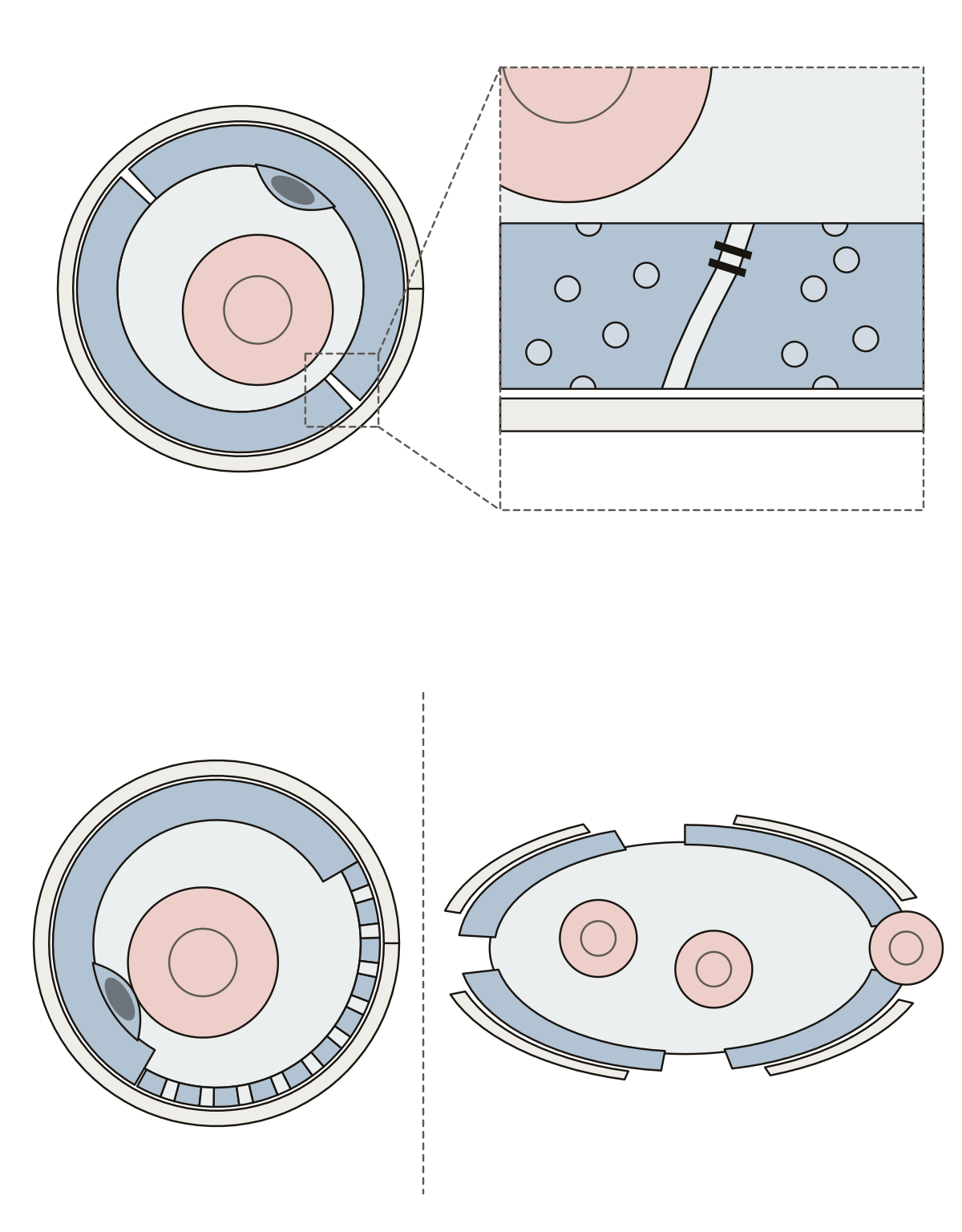 Capillary DiagramLumen, endothelial cell, basement membrane, intercellular cleft, tight junction, transport vesicles, fenestrations and the gaps of a sinusoid.
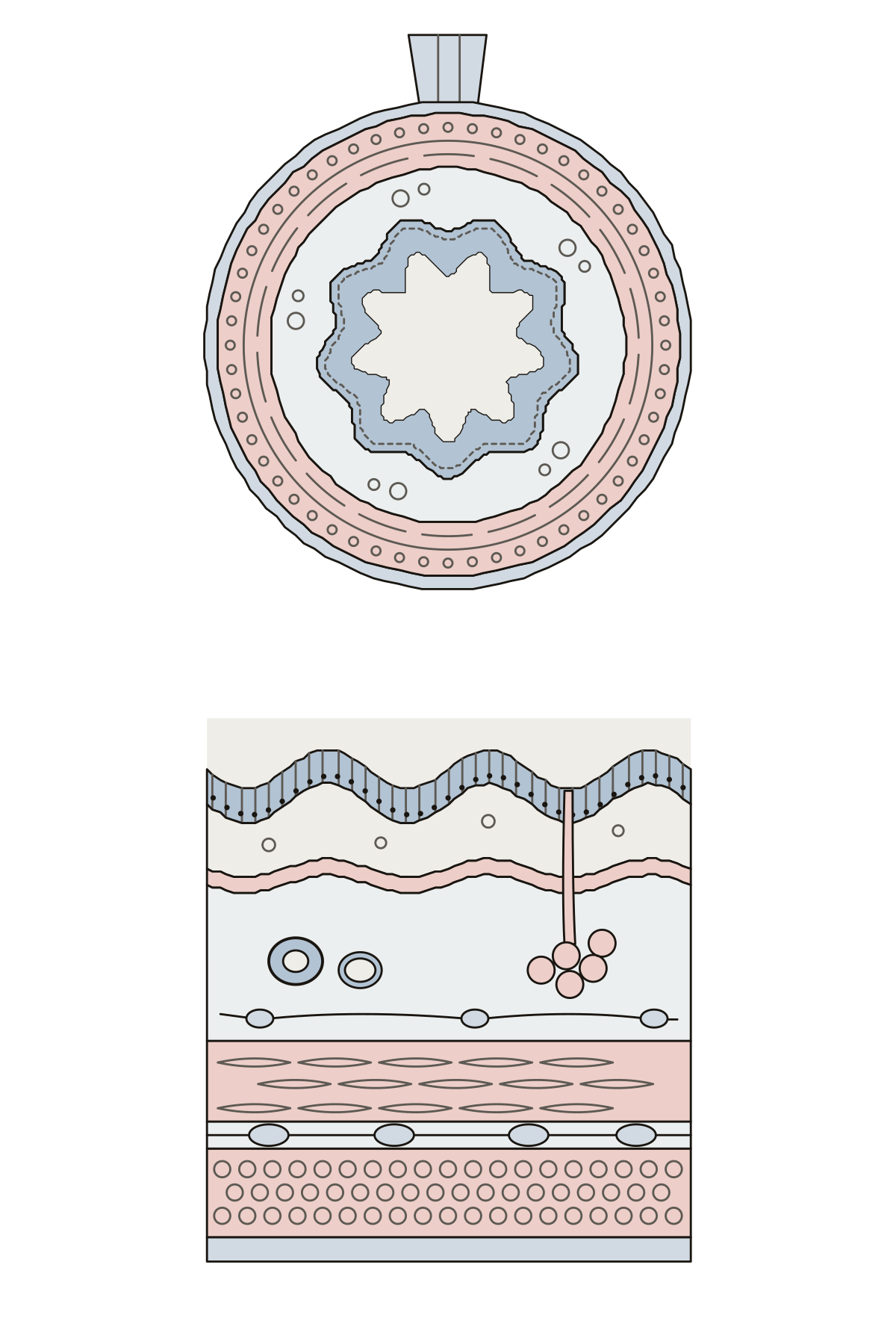 Digestive Tract Tissue Layers DiagramThe four layers of the digestive tract wall, from the lumen out to the serosa, and the epithelium, muscle layers and nerve plexuses inside them.
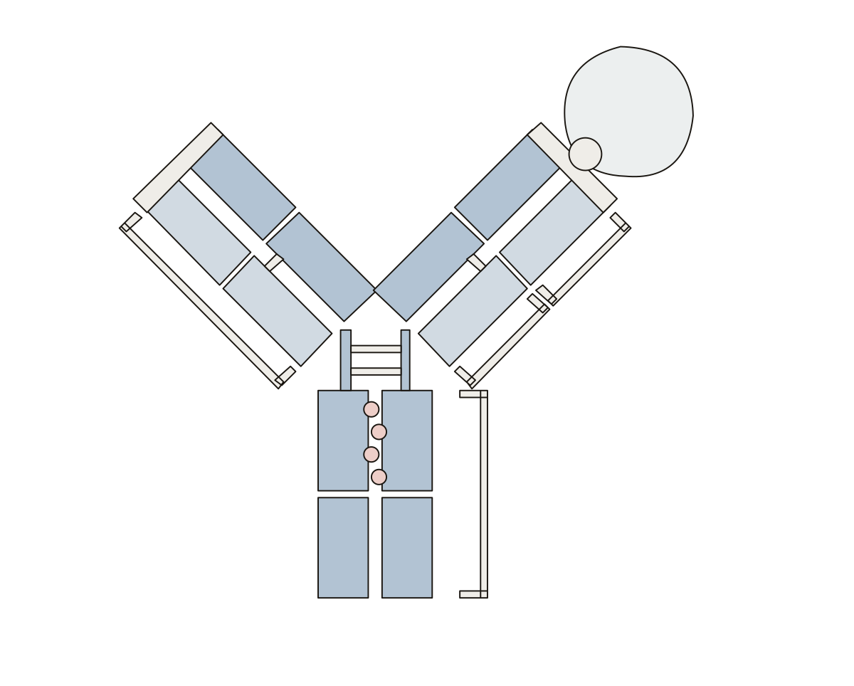 Antibody Diagram LabeledOne Y-shaped antibody holding an antigen: 12 parts to name. Carbohydrate · Heavy chain · Variable region
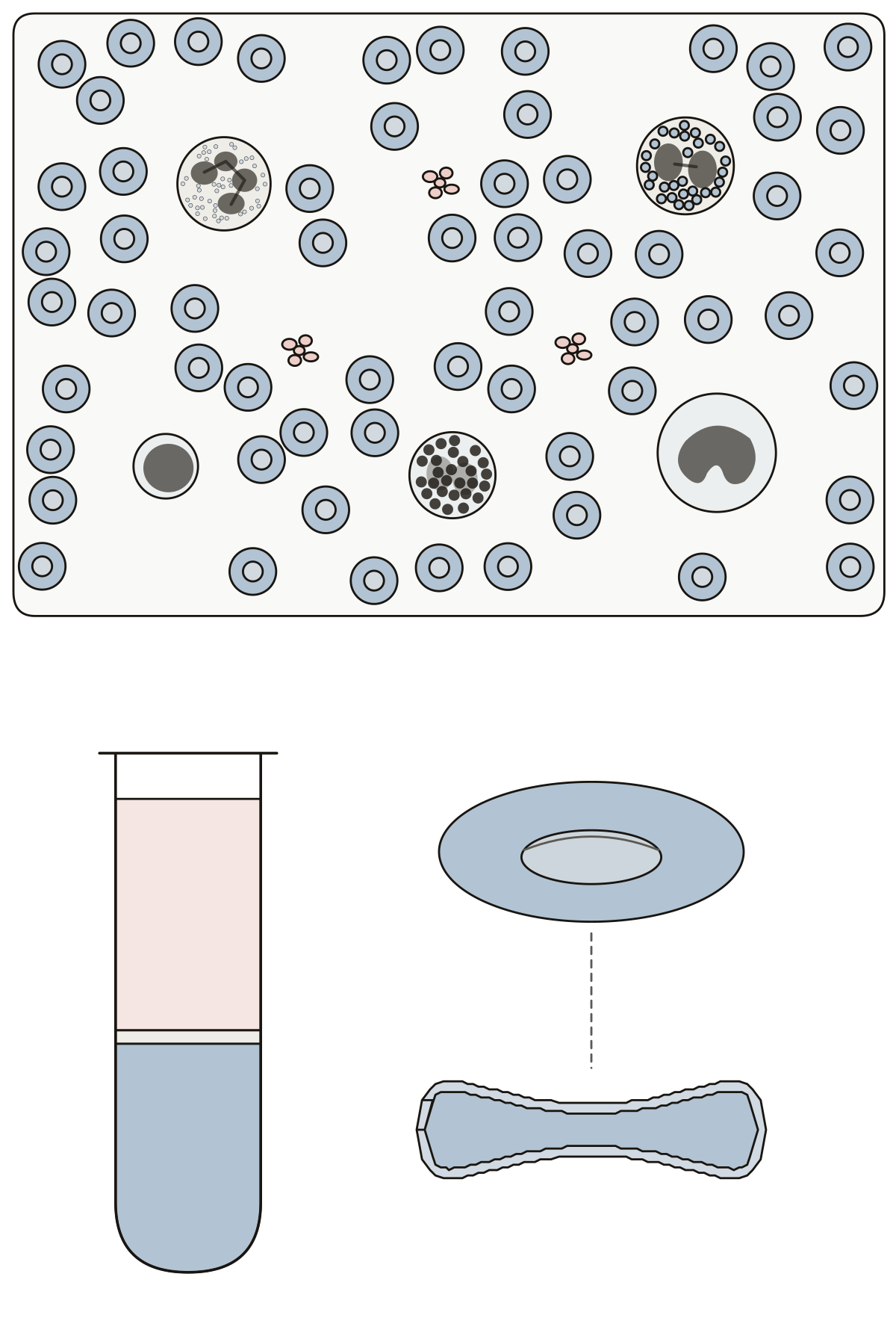 Formed Elements of Blood DiagramErythrocytes, platelets, neutrophil, eosinophil, basophil, lymphocyte and monocyte on a drawn blood smear, with plasma in a spun tube and a red blood cell cut in half.
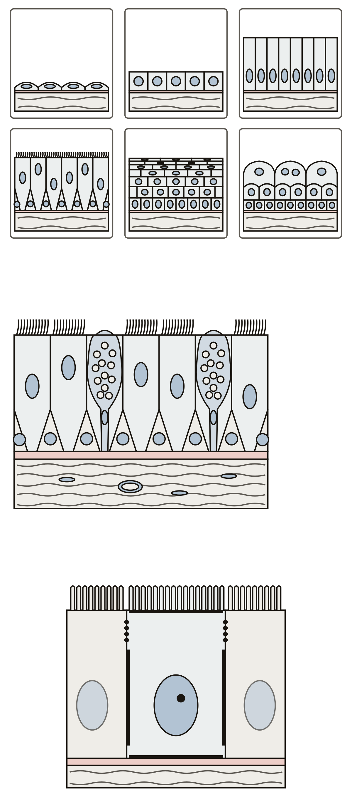 Epithelial Tissue Diagram LabeledSimple squamous, cuboidal and columnar, pseudostratified, stratified squamous and transitional epithelium, then cilia, goblet cells, the basement membrane, microvilli and tight junctions.
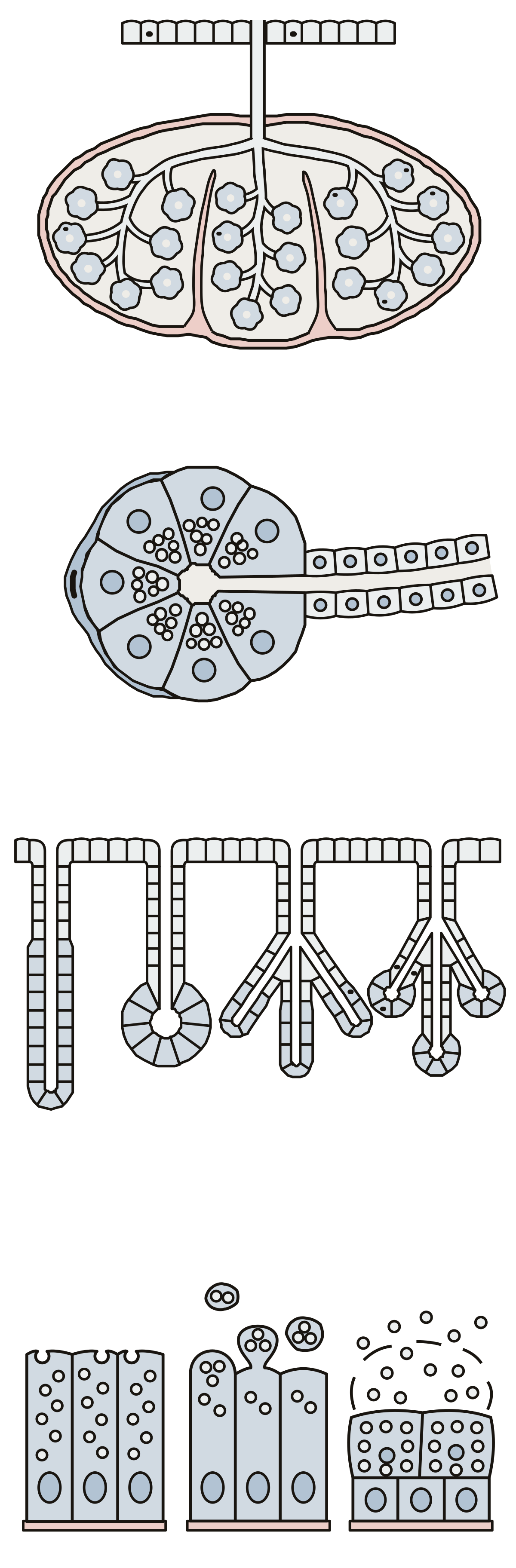 Exocrine Gland DiagramCapsule, septa, lobules, duct and acini; the secretory cells, lumen and myoepithelial cell of one acinus; simple, compound, tubular and alveolar glands; merocrine, apocrine and holocrine secretion.
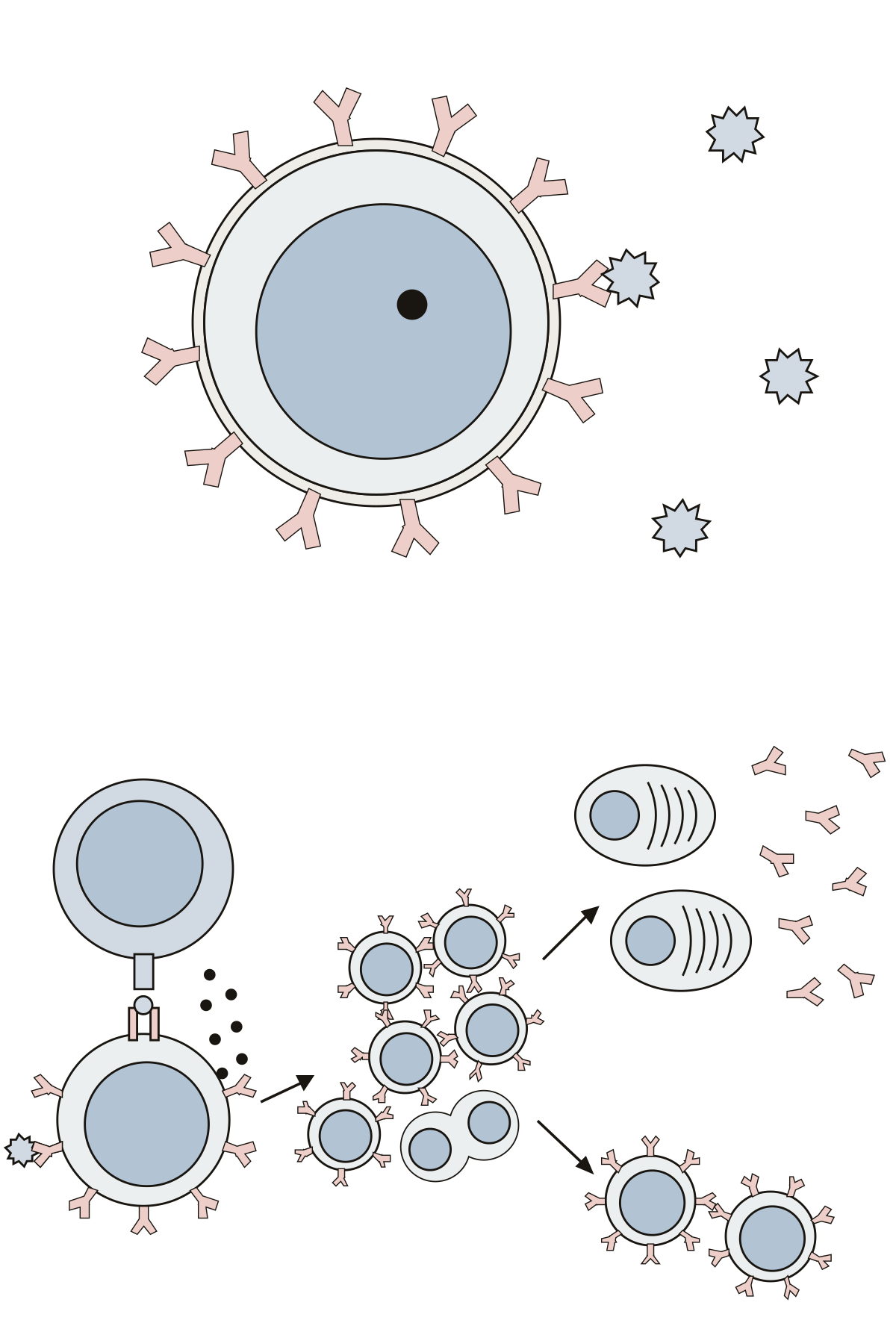 B Cell DiagramThe B cell receptor, an antigen, the nucleus and membrane of one B cell, then MHC class II, the helper T cell, cytokines, clonal expansion, plasma cells, antibodies and memory B cells.
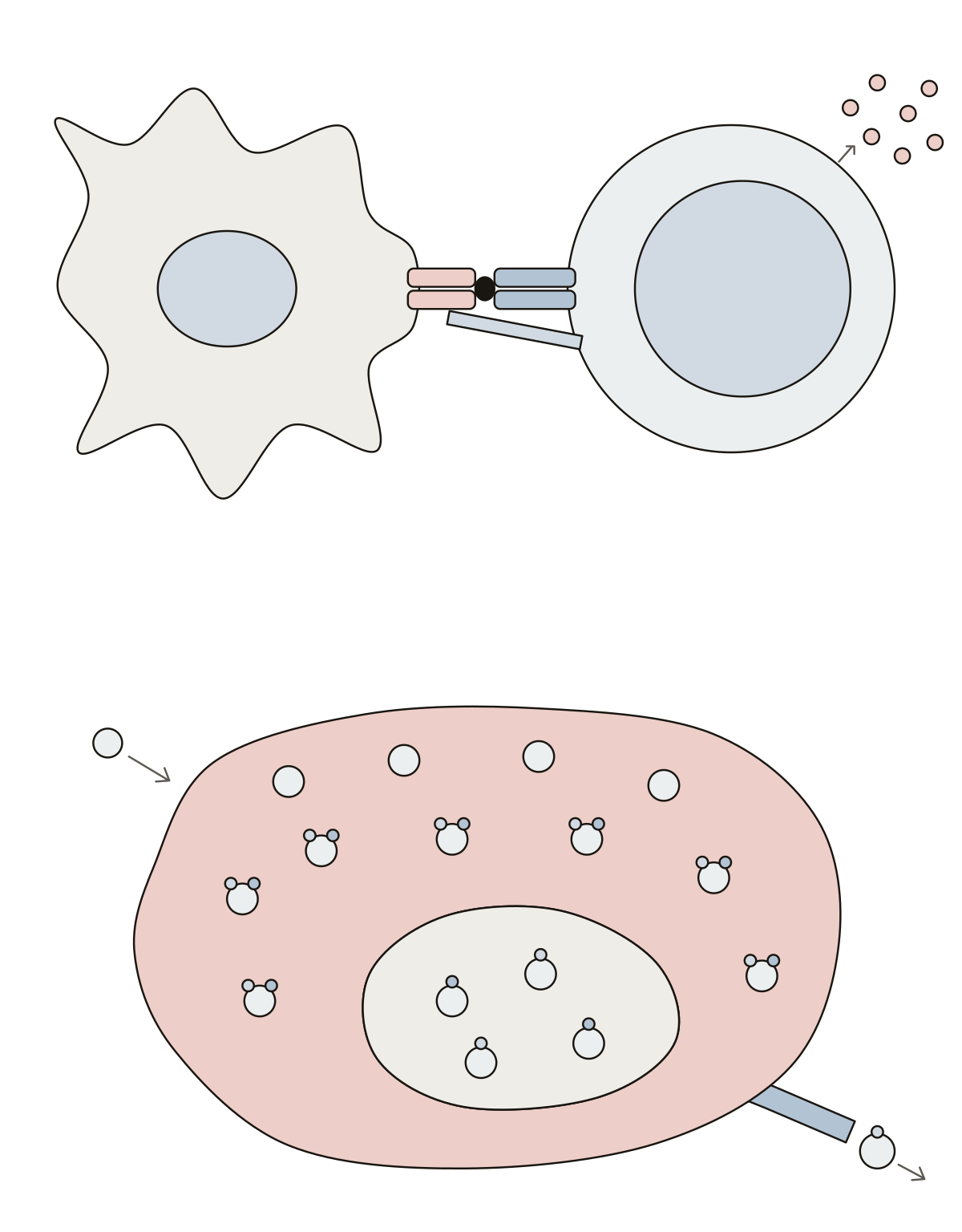 T Cell DiagramHelper T cell, T cell receptor, CD4, MHC class II molecule, antigen, cytokines, thymus cortex and medulla, double-negative and double-positive thymocytes, naive T cell.
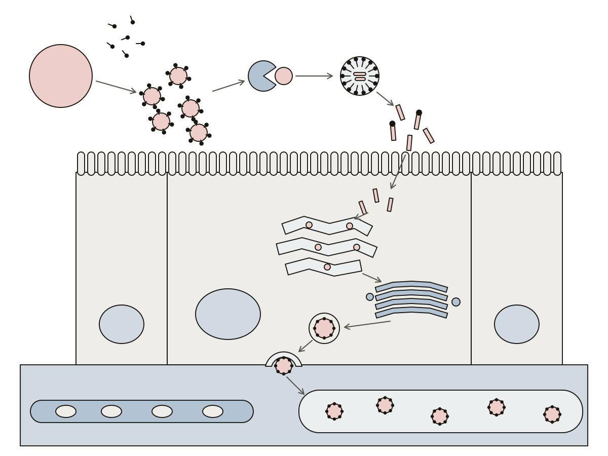 Lipid Absorption DiagramFat droplet, bile salts, emulsion droplets, pancreatic lipase, micelle, fatty acids and monoglycerides, brush border, enterocyte, smooth ER, Golgi apparatus, chylomicron, exocytosis, lacteal and blood capillary.
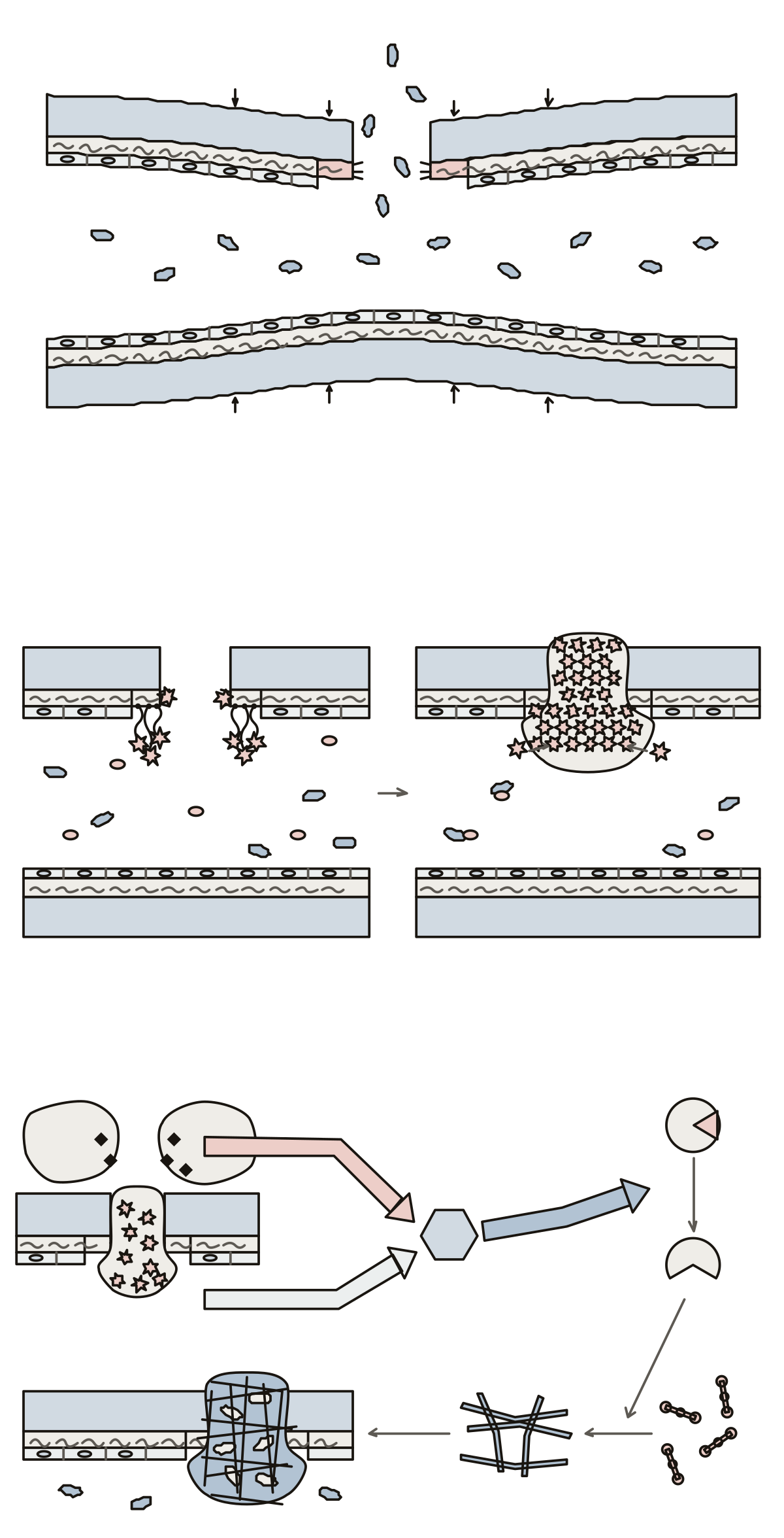 Hemostasis DiagramEndothelium, collagen fibers, vascular spasm, platelet, von Willebrand factor, platelet adhesion, platelet plug, tissue factor, extrinsic, intrinsic and common pathways, factor X, prothrombin, thrombin, fibrinogen, fibrin threads and blood clot.
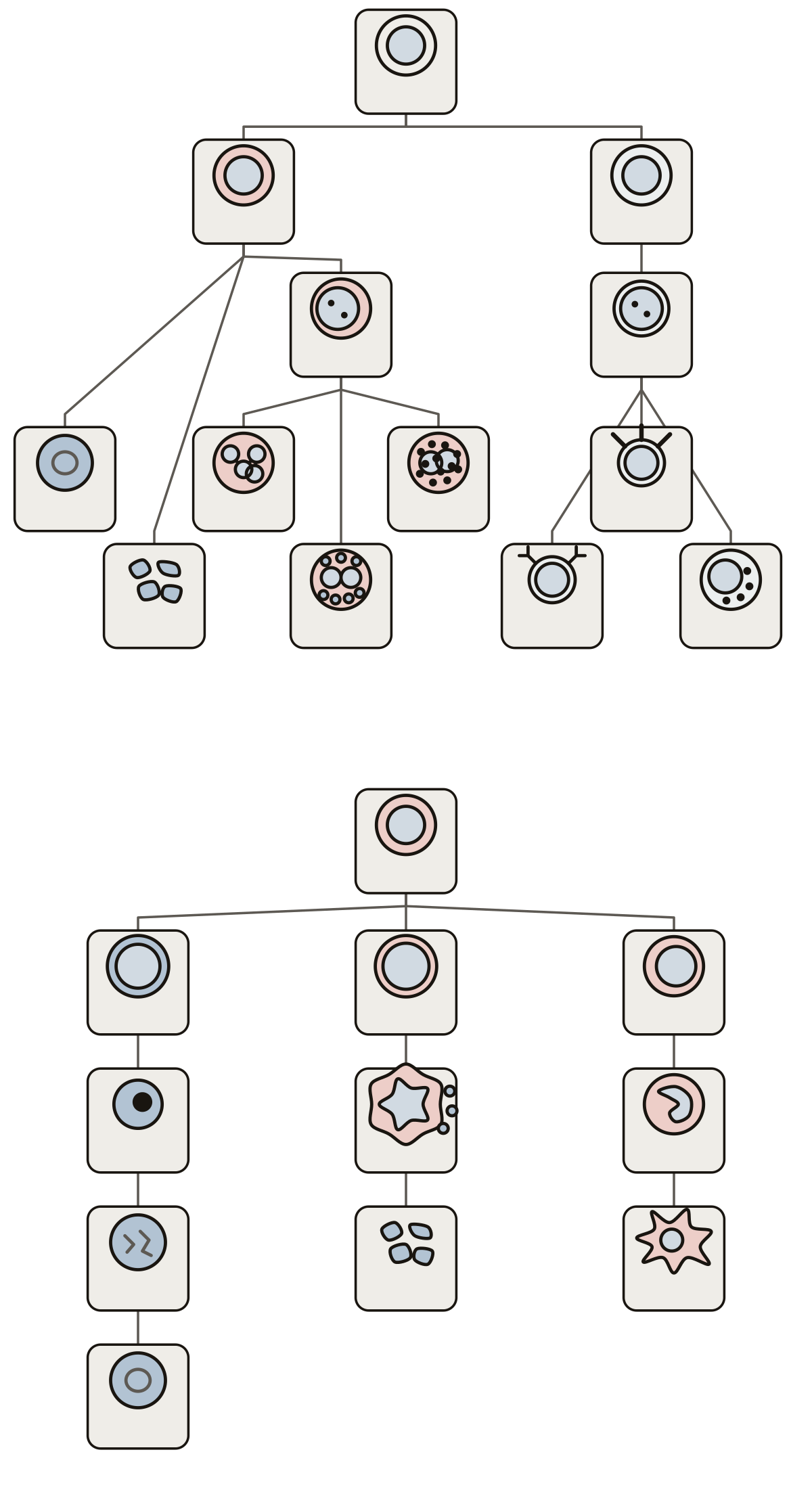 Blood Cell Formation DiagramBlood stem cell, myeloid and lymphoid stem cells, myeloblast, lymphoblast and the finished cells; then the stages of the red cell, platelet and monocyte lines.
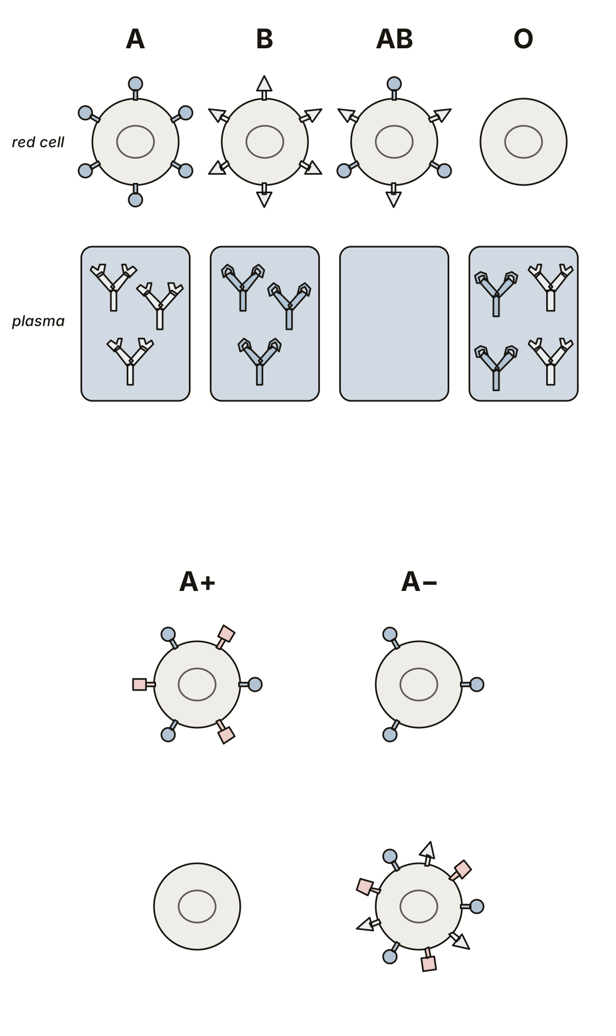 Blood Types DiagramThe four ABO types with their antigens and antibodies, then the D antigen, O negative and AB positive.
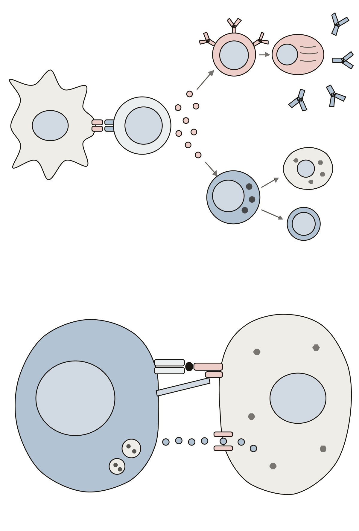 Cell Mediated Immune Response DiagramAntigen-presenting cell, helper and cytotoxic T cells, B cell, plasma cell and antibodies in one drawing; T cell receptor, MHC class I, CD8, perforin and granzymes in a second.
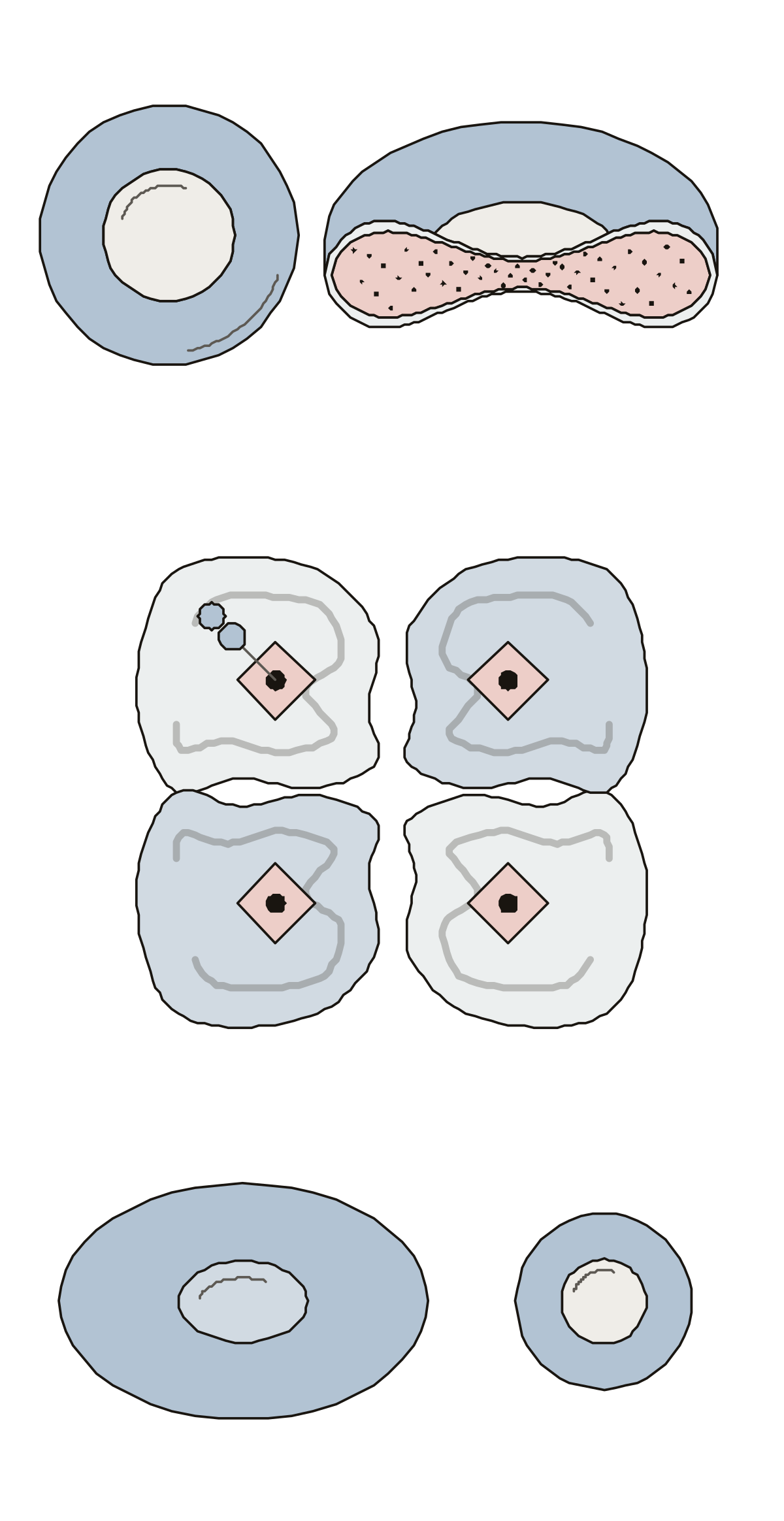 Diagram of a Human Red Blood Cell, Cut in HalfRim, center, cell membrane and hemoglobin on a cut cell; the alpha and beta chains, heme, iron and oxygen of one hemoglobin molecule; a frog cell with its nucleus.
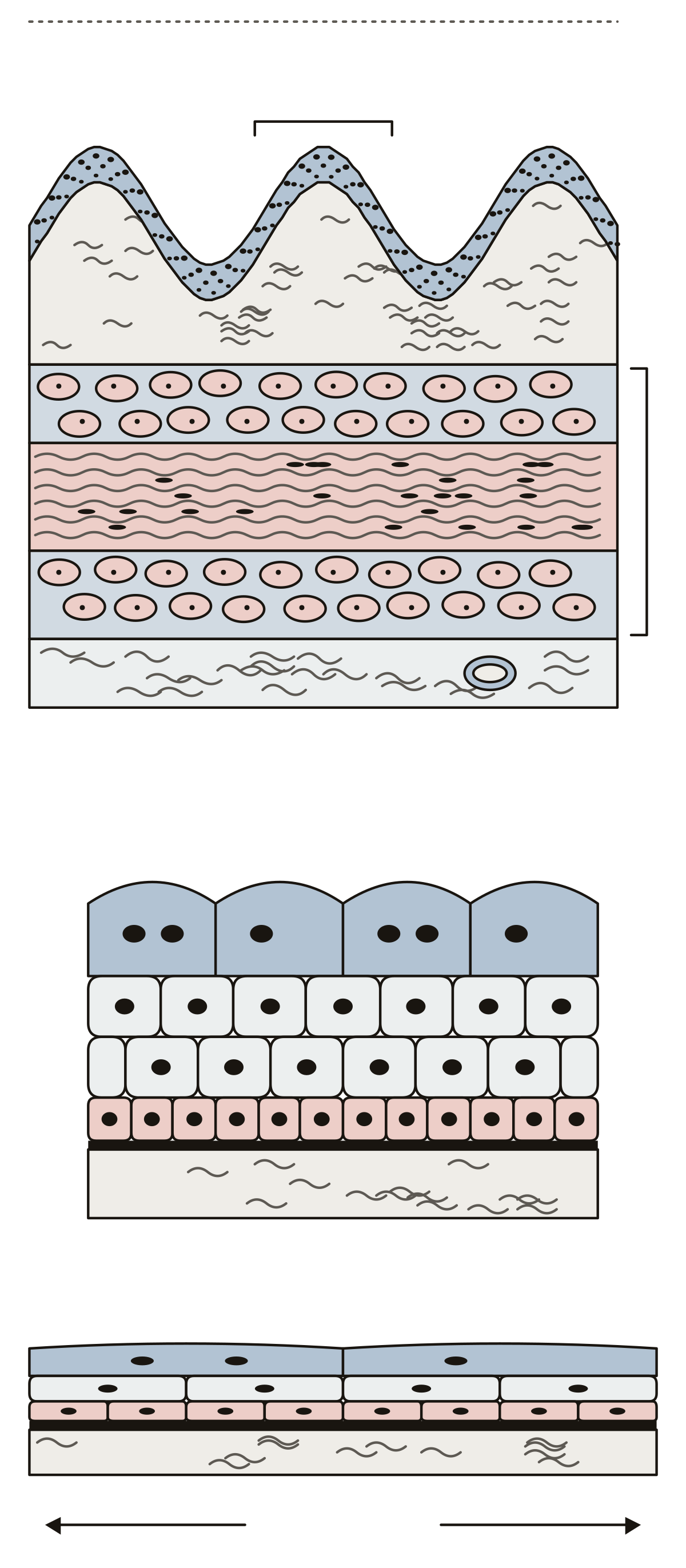 Bladder Slide LabeledTransitional epithelium, lamina propria, a ruga, the inner longitudinal, middle circular and outer longitudinal layers of the detrusor muscle and the adventitia, then umbrella, intermediate and basal cells on the basement membrane.
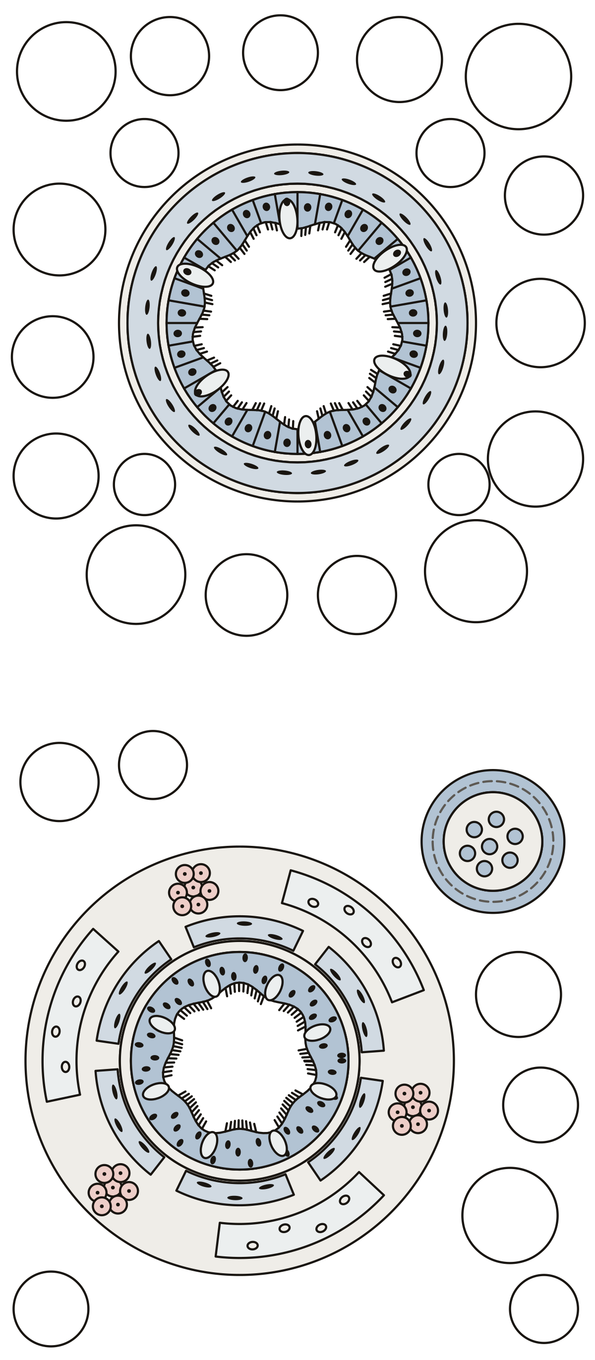 Bronchiole Histology LabeledSimple cuboidal epithelium, club cells, smooth muscle and alveoli around a bronchiole; then pseudostratified epithelium, goblet cells, cartilage plates, mucous glands and a pulmonary artery branch at a bronchus.
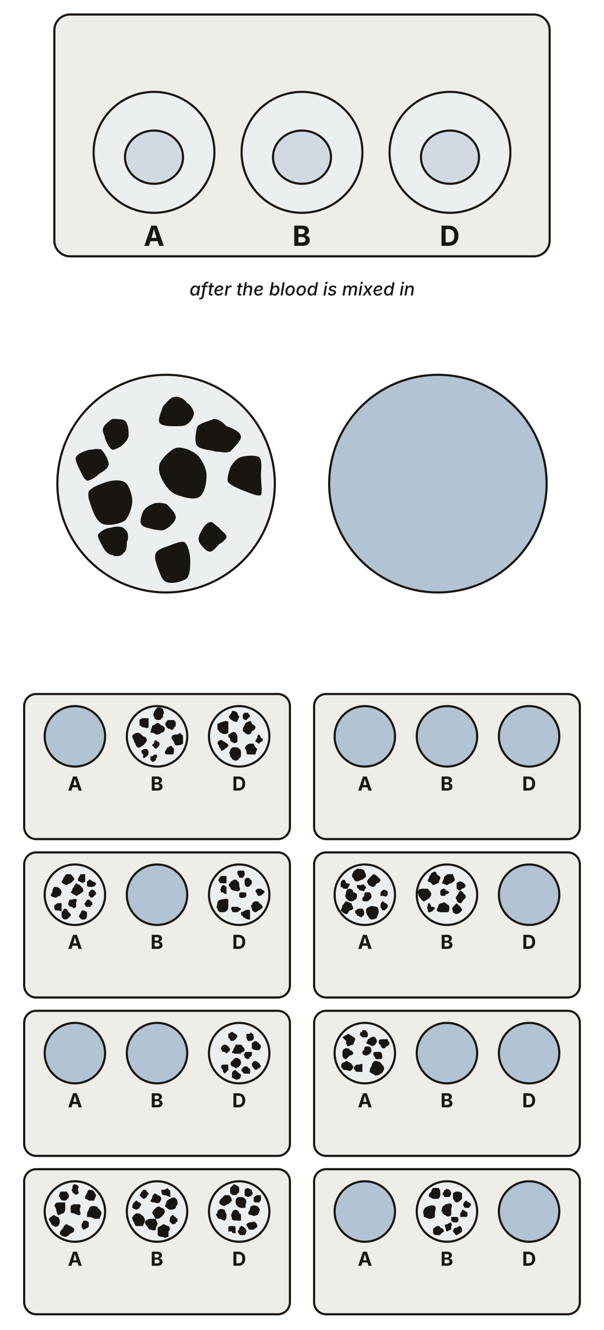 Blood Typing CardsThe anti-A, anti-B and anti-D wells of a typing card, a clumped and a smooth well, then eight cards to read, one for each blood type.
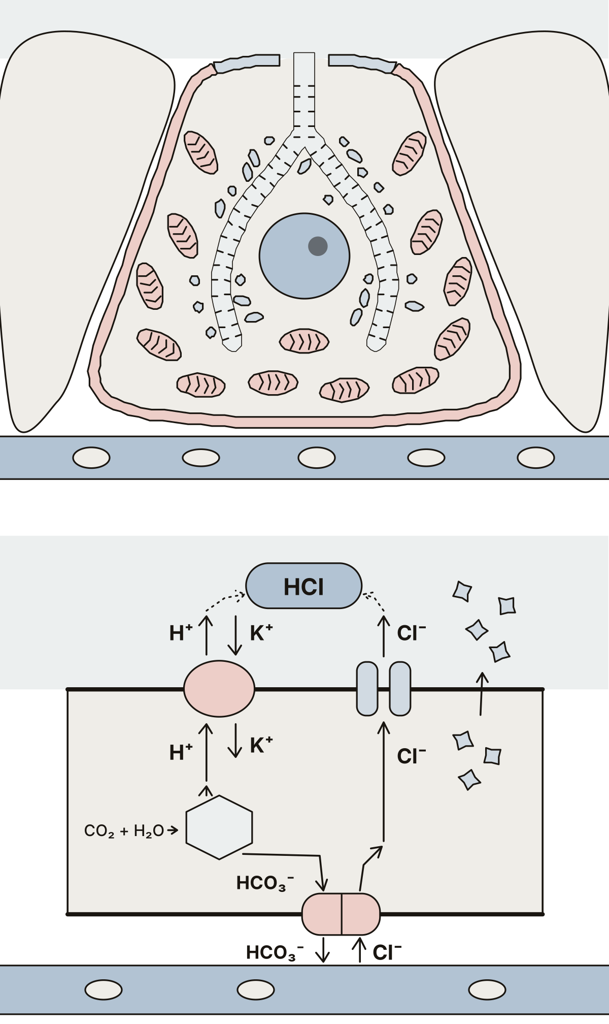 Parietal Cell DiagramOne parietal cell with its canaliculus, tubulovesicles, mitochondria, nucleus and two membrane faces, then the proton pump, chloride channel, carbonic anhydrase and exchanger that make hydrochloric acid, and intrinsic factor.
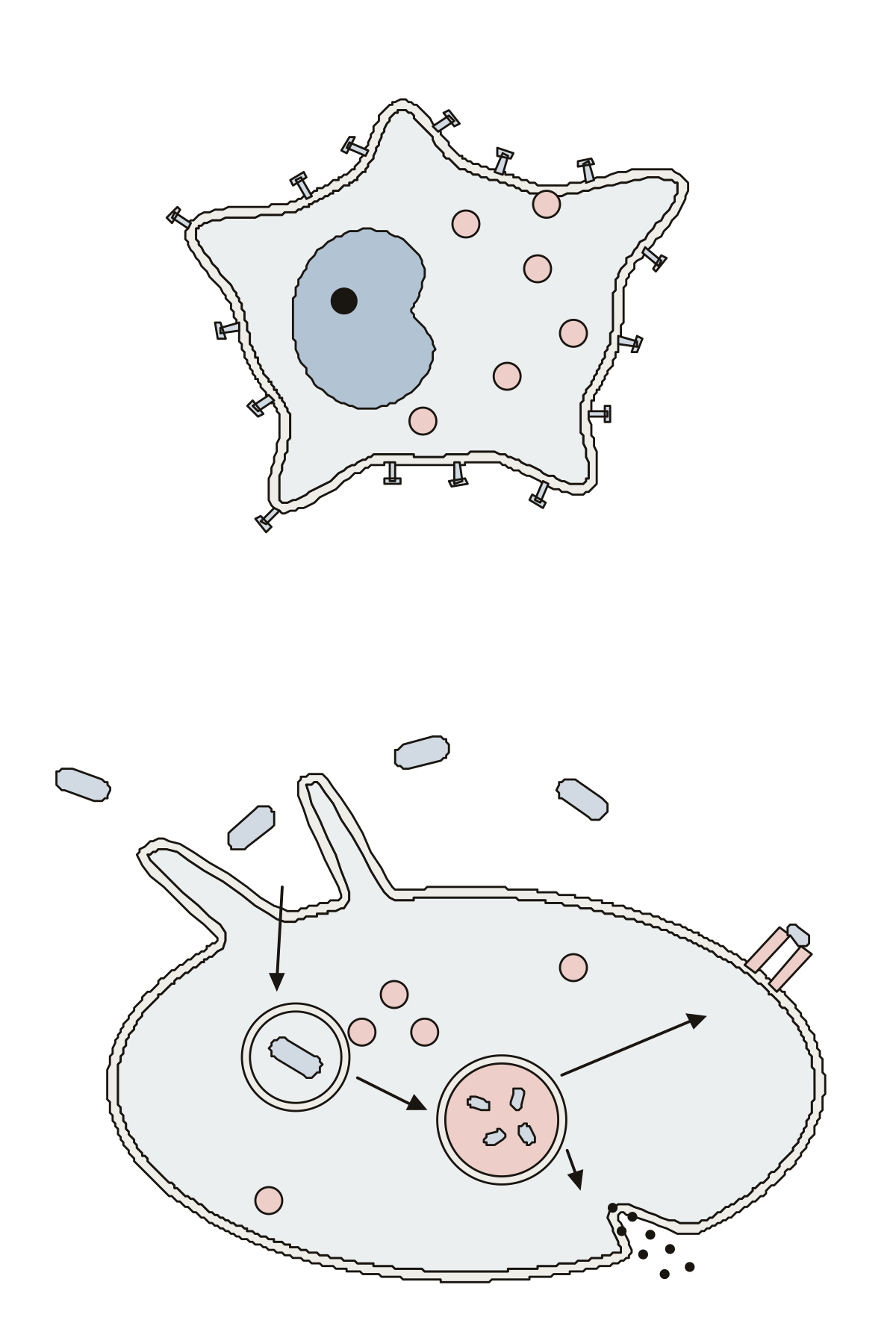 Macrophage Cell DiagramNucleus, pseudopod, lysosome, cell membrane and receptor on one macrophage, then the bacterium, phagosome, phagolysosome, exocytosis and the MHC class II molecule.
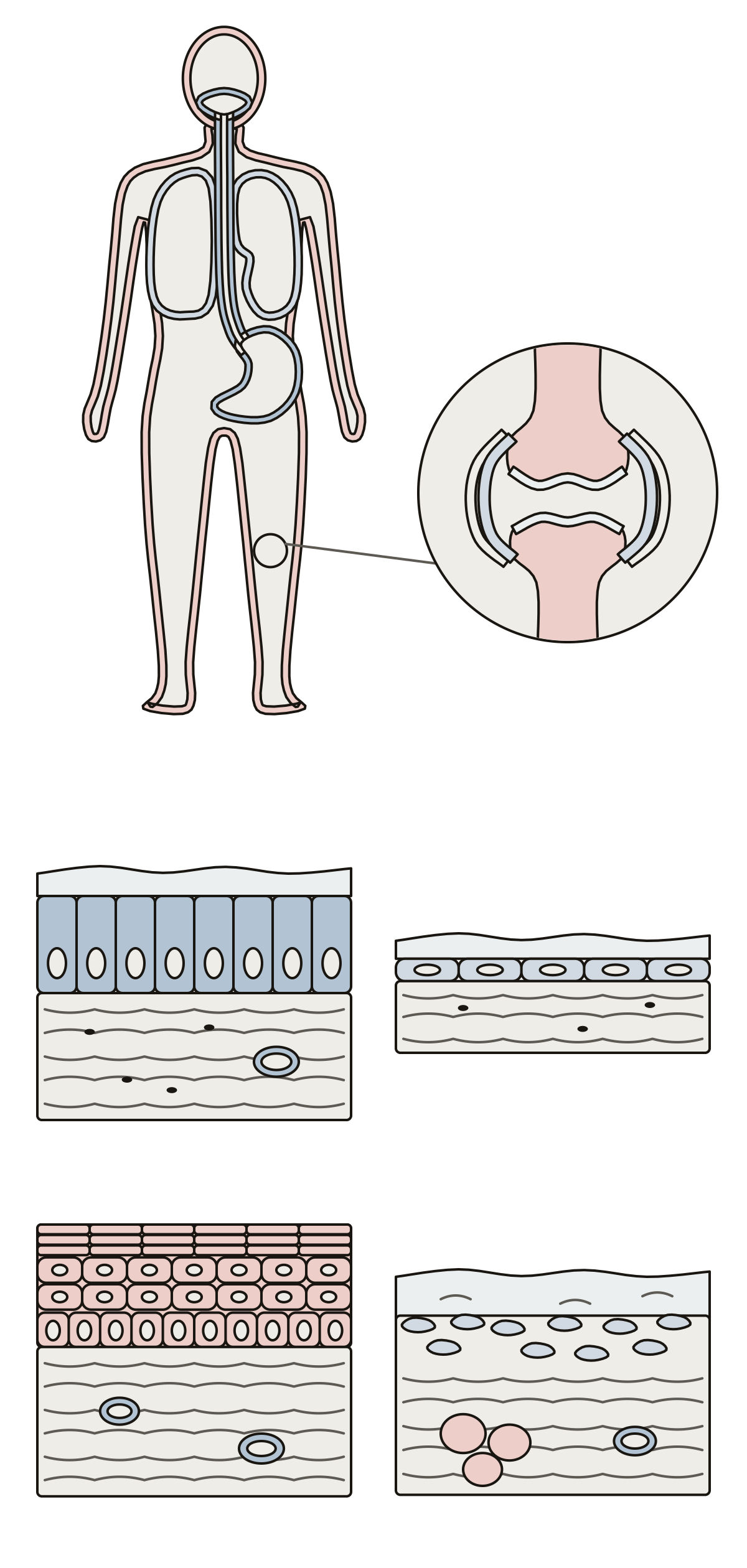 Tissue Membranes DiagramMucous, serous, cutaneous and synovial membranes on one figure, then epithelium, mucus, loose connective tissue, mesothelium, serous fluid, epidermis, dermis and synovial fluid in four cut blocks.
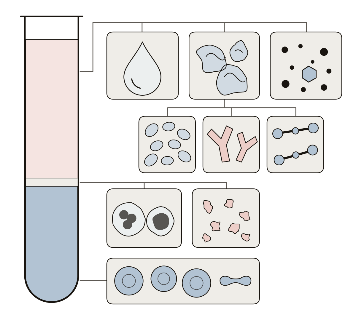 Components of Blood DiagramA spun tube of blood in three layers: plasma with its water, proteins and other solutes, the buffy coat of leukocytes and platelets, and the erythrocytes.
- Formed Elements of Blood Diagram UnlabeledThe formed elements drawing with nothing written on it, to print and fill in: seven kinds of cell and fragment on a smear, plasma in a spun tube, a word bank and an answer key.
Each page is one drawing of a body system, an organ or a tissue, with a numbered ring on every part. Start with the digestive system, 25 parts from mouth to anus with a simple set of 10 main organs for younger classes, or with the child figure and its 20 everyday words.
Each drawing prints blank or labeled. The pages teach names and places in the body and give no medical advice.
Keep exploring
- Animal body-part diagrams (94)
- Blank US maps (61)
- Blank maps (159)
- Bone labeling quizzes (49)
- Capitals quizzes (11)
- Car and engine part diagrams (54)
- Cell and organelle diagrams (63)
- Clothing, shoes and jewelry (45)
- Computer, phone and camera diagrams (43)
- Anatomy concept maps (1)
- Country map quizzes (28)
- Animal anatomy diagrams (100)
- Human Body Quizzes
- All quizzes